Abstract
Background
Multi-drug resistant bacteria hazards to the health of humans could be an agent in the destruction of human generation. Natural products of Bacillus species are the main source to access progressive antibiotics that can be a good candidate for the discovery of novel antibiotics. Wild honey as a valuable food has been used in medicine with antimicrobial effects.
Objective
Bacillus strains isolated from wild honey were evaluated for the potential antimicrobial activity against human and plant bacterial and fungal pathogens.
Methods
Three bacterial isolates were identified as strain Khuz-1 (98.27% similarity with Bacillus safensis subsp. Safensis strain FO-36bT), strain Khuz-2 (99.18% similarity with Bacillus rugosus strain SPB7T), and strain Khuz-3 (99.78% similarity with Bacillus velezensis strain CR-502 T) by 16S rRNA gene sequences. The strains were characterized by their ability to inhibit the growth of human and phytopathogenic fungi.
Results
The results indicated that B. rugosus strain Khuz-2 inhibited the growth of phytopathogenic and human fungal more effective than other ones. It seems that the strain Khuz-2 has a suitable antimicrobial and antifungal potential as a good candidate for further pharmaceutical research.
Conclusion
Based on the results of GC–MS, Pyrrolo [1,2-a] pyrazine-1,4-dion, hexahydro-3-(2-methylpropyle) (PPDHM) was the major compound for all strains which have a various pharmacological effect. Isolation and identification of beneficial bacteria from natural sources can play an important role in future pharmaceutical and industrial applications.
Supplementary Information
The online version contains supplementary material available at 10.1186/s12906-022-03551-y.
Keywords: Bacillus sp., Antimicrobial activity, Human pathogens, Phytopathogenic pathogenes, GC–MS
Introduction
Honey as a valuable food has been used in ancient medicine. It has a remarkable effect on the remediation of wound healing, bedsore treatment, and ulcers [1]. Recently, the progression of multi-drug resistant bacteria hazards to the health of humans which could be an agent in the destruction of human generation. Natural products of bacteria are the main source to access progressive antibiotics that can be a good candidate for the discovery of novel antibiotics [2]. However, the finding of new antibiotics is very scarce at the industrial level and needs to examine by new methods [3]. In 1966 Burkholder et al. extracted a pyrrole antibiotic from marine bacteria for the first time [4]. Consequently, various antimicrobial compounds were introduced and produced by different micro-biome, such as archaea, bacteria and fungi [5]. It seems that the isolation and identification of novel bacteria can be a new approach in the achievement of unknown natural sources which leads us to find new antibiotics for the accumulation of our starved pharmaceutical business [6]. Natural products discovery efforts began in pharmaceutical companies, mainly in the United States, Europe and Japan, and modest efforts fol-lowed in isolated academic laboratories worldwide [7]. Since 2013, around 1453 new chemical entities have been accepted by US Food and Drug administration [7]. The Most important natural product used in the anti-infective area, especially anti- bacterial therapy. Around twelve natural product is used to treat Gram-positive or Gram-negative bacterial infections in humans and animals [8]. In this research, the antimicrobial ability of Bacillus sp. strains were discussed which was isolated and identified for the first time from wild honey collected from Khuzestan Province, Iran.
Materials and methods
Sample collection and growth conditions
A literature search was directed up to 2018, on the electronic databases of Scopus, PubMed, and Web of Science. The search was accomplished by using the following search strings in the title/abstract/keywords: “Antimicrobial Activity of environmental bacteria” AND “Wild Honey*” OR both of them. Obtained articles were imported to EndNoteX9 reference management software. All articles were separately screened for, duplicity, and eligibility by two authors individually [9]. According to our search, it seemed that there is few research about the investigation of the bacterial population in Khuzestan wild honey and the Identification of these bacteria can be helpful in both basic and applied research. Therefore, for this aim, honey samples were collected from Shushtar city, Khuzestan Province, Iran. Khuzestan Province as the historical Iranian province is situated in the southwest of Iran, in the neighborhood of the Persian Gulf and the Iran-Iraq border (32°02′44″N 48°51′24″E). The samples were transported to the laboratory in sterile condition. The microbial population of samples was enriched using specific media [10] and the colonies were isolated from 18 to 24 h. Isolates were cultured in the nutrient broth, containing (g/L), NaCl 9, MgSO4.7H2O 9.7, MgCl2.H2O 7.0, CaCl2 3.6, KCl 2.0, NaHCO3 0.06 and NaBr 0.026, where pH was adjusted to 7.3 ± 0.2 before autoclaving. Cultures were incubated at 30 °C in an orbital shaker, at 150 rpm min−1 for 72 h. To culture on solid media, 12–15 gl−1 agar was added to the new nutrient broth, then it was incubated at 30 °C for 48 h. Cell morphology and biochemical tests were carried out to identify isolates [6, 11].
Physiological characteristics
The principal tests used for strains Khuzestan1 (Khuz-1), Khuzestan2 (Khuz-2) and Khuzestan3 (Khuz-3). These purposes are Hydrogen Sulphide Production (H2S), potassium hydroxide (KOH), Urease Test(URE), Catalase Test (CAT), Tween20,40 and 80 test, starch hydrolysis test, Gelatin hydrolysis test, L-Tyrosine test [12, 13].
Bacterial isolates were tested for growth at different pH (4- 11), (using increments of 1 pH unit) on lab made nutrient broth. The pH values were adjusted using buffer system including 0.1 M citric acid/0.1 M sodium citrate for pH 4.0–5.0; 0.1 M KH2PO4/0.1 M NaOH for pH 6.0–8.0; 0.1 M NaHCO3/0.1 M Na2CO3 for pH 9.0–10.0; 0.05 M and Na2HPO4/0.1 M NaOH for pH 11.0 with different temperature ranges (5, 10, 20, 30, 40 and 50 °C). Samples were measured by UV absorbance at 600 nm wave length (OD: 600) after 48 h.
Following identification, susceptibility test of the isolated bacteria was performed using the disk diffusion sensitivity method employing paper disks impregnated with seventeen different types of antibiotics (Padtan Teb Co., Iran): cefalexin (30 μg/mL−1), chloramphenicol (30 μg/mL−1), azithromycin (15 μg/mL−1), amikacin (30 μg/mL−1), ampicillin (10 μg/mL-1), penicillin (10 μg/mL−1), rifampicin (5 μg/mL−1), erythromycin (15 μg/mL−1), ciprofloxacin (5 μg/mL−1), cefoxitin (30 μg/mL−1), ceftriaxone (30 μg/mL−1), nitrofurantoin (300 μg/mL−1), doxycycline (5 μg/mL−1), tetracycline (30 μg/mL−1), amoxicillin (10 μg/mL−1), gentamicin (10 μg/mL−1) and erythromycin (15 μg/mL−1). Each bacterium spread over the surface of NA medium in petri dishes followed by the distribution of paper disks impregnated with the antibiotics. Then, the cultures remained under incubation at 30 °C.
Molecular Identification
Genomic DNA extraction was done by DNA extraction Kit (Cinagene DNa plus, South Korea), according to manufacturer’s protocol. Universal used primers were 16F 5’- AGAGTTTGATCCTGGCTCAG-3’, and reverse 16R; 5’- TACCTTGTTAGGACTTCACC-3’ primers [14]. Each amplification reaction contained 1µL of each primer, dNTP (10 mM) 0.5 µm, PCR buffer 2.5 µL, MgCl2 (50 mM) 0.75 µL, template DNA 1 µL, smartaq DNA polymerase 0.2 µL, and dH2O 19.05 µL, in a final volume of 25µL. The 16S rRNA gene amplification protocol was 95 °C for 5 min, followed by 40 cycles of 95 °C for 1 min, 55 °C for 55 s and 72 °C for 1 min and 25 s, with final 10 min extension at 72 °C [15]. High-Pure PCR Product Purification kit (Roche) was used for the amplified fragment purification and then sequenced by utilizing forward and reverse primers by Macrogen (Korea) [16]. The phylogenic relationship of the isolates was determined by comparing the sequencing data with the related 16S rRNA gene sequences in the GenBank database of the National Center for Biotechnology Information (https://www.ncbi.nlm.nih.gov/), via BLAST search. In addition, the sequences were conducted in the EzBioCloud identity service led to retrieving its closest phylogenetic neighbors [17]. The 16S rRNA gene reads were assembled using Chromas pro software and aligned using the multiple sequence alignment program CLUSTAL_X (version 1.83) [18] Phylogenetic analysis was performed using the neighbor-joining method in MEGA version 7 software package [14, 19].
Preparation of pathogenic microorganisms
The antimicrobial properties of isolates were assayed by the agar well diffusion method. Based on the previous studies, we selected the serious pathogenic microorganisms that are involved in globally important human or plant diseases [20, 21]. In this study, Bacillus cereus ATCC 11,778, Escherichia coli O157 PTCC 1276, Klebsiella pneumonia PTCC 10,031, Shigella flexneri PTCC 1234, Pseudomonas aeroginosa ATCC 10,231, Streptococcus mutams ATCC 35,668 and Candida albicans ATCC 10,231 were studied as human pathogenic organisms and Fusarum oxysparum (IBRC-M 30,067), Aspergillus flavus (IBRC-M 30,029), Neurospora crassa (IBRC-M 30,138), Botrytis cinerea (IBRC-M 30,162), Erwinia amylovora (IBRC-M 10,748), Psudomonas syringea pv. Syringae (IBRC-M 10,702), Xanthomonas campestris (IBRC-M 10,644) and, Rhizobium radiobacter (IBRC-M 10,701) were used as plant pathogenic. According to the agar well diffusion method, described by Ennahar et al., 2000 the bacterial extracts were filtered through a 0.22 µm pore filter membrane after centrifugation of bacterial culture at 5000 rpm for 10 min. The pathogenic microorganisms (107 CFU/mL) were inoculated into a sterile plate with 20 mL of their selective media. Subsequently, the plate was gently shacked for even spread and suitable mixing of the microorganisms and media. It was permitted to solidify. Then, 5 wells of about 6 mm in diameter were prepared on the surface of the agar plates using a sterile cylinder and the plates were turned upside down. The wells were labeled with a marker. Each well was occupied with 0.1 mL of the bacterial extracts. Then, the plates were incubated at 37 °C for 24 h and the inhibition zone was measured. Finally, the results were organized [22].
Antimicrobial assays
First, each colony of bacteria was cultured in 50 mL of liquid nutrient medium and incubated at 30 °C with shaking for 72 h. Then, to separate the extract from the medium, the culture was centrifuged at 4000 rpm for 20 min. Subsequently, the supernatant was collected and filtered through a 0.22 µm membrane filter [6, 23].
Minimum inhibitory concentration (MIC) and Minimum bactericidal concentration (MBC)
The MIC of the extract against bacteria was determined using the micro broth dilution method as recommended by the National Committee for Clinical Laboratory Standards (NCCLS). The MIC value is considered as the lowest concentration that completely inhibited bacterial growth after 48 h of incubation at 30 °C. The various concentrations (10–1 g / ml to 10–8 g / ml) of the extract were used to detect the MIC value against tested pathogens. 16 μl suitable medium was added into each well and 20 μl of the 0.5 McFarland suspension of pathogen bacterial (1:10 diluted) was added into the wells. Finally, different concentrations of the extract (20 μl) was added into the same wells and sterile broth was added into the wells to makeup to 300 μL. The positive control wells contain 0.5 McFarlend (OD: 600) of the pathogen and culture medium and the negative control wells contained an antimicrobial extract and culture media. After cultivation, the microplates were mixed wells to make the mixture quite uniform and placed in an incubator for 24 h at 30° C. The MIC value was determined by the first well in which the visible growth of microorganisms was inhibited. For the determination of MBC, a portion of liquid (5 μl) from each well that did not exhibit any growth was taken and then incubated at 30 oC for 24 h. The lowest concentration that revealed no visible bacterial growth after sub-culturing was taken as MBC [6].
Minimum inhibitory concentration (MIC) and (MFC)
To determine the MIC value for bacterial extract against fungal pathogens, the macro broth dilution method was used. In brief, serial dilutions of bacterial extract ranging from 10–1 g/ml to 10–8 g/ml were prepared in 96 well microtitration plates [24]. Then, 100 µL suitable medium was added to the well and 100 µL of fungal inoculums at a concentration of 106 CFU/mL. This serial dilution was repeated and the positive control (medium with fungal) and culture medium and the negative control (medium only) contained an antimicrobial extract and culture media. the plate was incubated at a suitable temperature. After 48 h, tubular opacity and growth of fungi were evaluated in comparison with the control plate. To obtain the MFC, 10 ml of each dilution was taken from each well and spread on suitable medium. Plates were incubated at 28 ℃ for 72 h. The MFC was defined as the lowest concentration capable of inhibiting 99% fungal growth or fewer colonies [23, 24].
Gas Chromatography–Mass Spectroscopyanalysis (GC–MS)
GC/MS analysis as a suitable method for identification of unknown and different substances within a test sample was carried out using Gas Hewlett PackardHP-5890 series II equipped with split/splitless injector and a capillary column (30 m, 0.25 mm, 0.25 lm) fused with phenylpolysilphenylene siloxane. The injector and detector temperatures were set at 280 and 300 °C, respectively, and the oven temperature was kept at 80 °C for 1 min, rose to 300°Cat20°C/min. Helium was used as carrier gas at a constant flow of 1.0 ml/min. A volume of 2ll was injected in the splitless mode and the purge time was 1 min. The MS (Hewlett–Pack-ard 5889B MS Engine) with selected ion monitoring (SIM) was used. The mass spectrometer was operated at 70 eV and scan fragments from 50 to 650 m/z. Peak identification of crude algal extracts was performed based on comparing the obtained mass spectra with those available in NIST library [25].
Statistical analysis
Statistical analysis was performed by using SPSS software version 16 to calculate the mean of zones of inhibition on tested microorganisms.
Result
Isolation and Identification of the Bacillus Strain
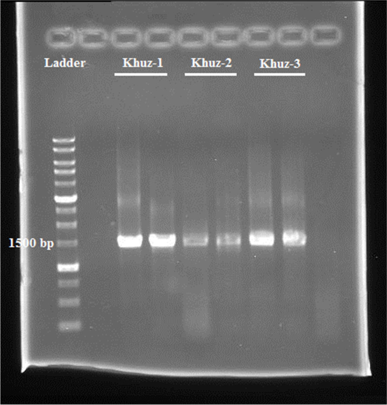
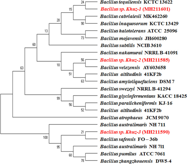
In the present study, three different Bacillus strains were isolated from Shushtar city, Khuzestan Province from wild honey, which is named Khuz- 1, 2 and 3 (abbreviation of Khuzestan). The isolates were purified on NA medium and preserved in a refrigerator maintained at-70 °C [16]. We doubted that the bacteria isolated belonged to genus Bacillus. To confirm this hypothesis, we isolated DNA for the usual PCR of the 16S rRNA gene. For this target, we amplified a 1.5 kb DNA sub-fragment of the 16S rRNA gene, which is extremely maintained within the genus Bacillus (Fig. 1). The 16S rRNA gene sequences of mentioned strains Khuz- 1, 2 and 3 were submitted in gene bank with accession numbers; MH211590, MH211601 and MH211585, respectively. In addition, the phylogenetic tree of 16S rRNA genome sequencing of the isolates showed that they are closely related to Bacillus safensis, Bacillus rugosus, Bacillus velezensis (Fig. 2).
Fig. 1.

The gel electrophoresis of 16S rRNA gene sequences of isolated strains. The length of fragments were approximately ≈ 1500 bp
Fig. 2.

Neighbor-joining phylogenetic tree [26] derived from 16S rRNA gene sequence data [14] showing the positions of seven isolated bacteria including strains Khuz-1, Khuz-2 and Khuz-3 among related bacteria. Numbers at branch points are bootstrap percentages based on 500 replicates. Bar, 0.02 changes per nucleotide position [19]
Biochemical characterization of bacteria
Bacteria were identified based on the colony, pigment and microscopy shape. All strains were selected for further assays and their biochemical characteristics are summarized in Table 1.
Table 1.
The isolated strains and their biochemical characteristics
| ID | KoH | H2O2 | Tween20 | Tween40 | Tween80 | Starch | Gelatin | Tyrosine | Urease | H2S |
|---|---|---|---|---|---|---|---|---|---|---|
| Strain Khuz- 1 | + | + | + | + | - | - | - | + | + | + |
| Strain Khuz- 2 | + | + | + | + | + | + | + | + | + | + |
| Strain Khuz- 3 | + | + | + | + | - | + | + | + | + | + |
+ Positive reaction,—negative reaction
All isolates were Gram-positive. The growth was observed in the range of pH 4 to 10 with the ability to grow up to 10 pH. The optimum pH for all strains were 7 and 8 (Table 2). Additionally, all bacteria can grow the widespread range of temperature at 5 to 50 0C. The optimum temperature for all strains was between 20 to 30 0C, which is recorded in Table 2. As shown in Table 3, the antibiotic resistance of isolated bacteria towards different antibiotics was also determined for phenotypical characterization of isolates. According to our results, all strains were highly resistant to Cefazolin and Cefalexin.
Table 2.
The range of temperature (°C) and pH of isolated strains and their optimized ranges
| ID | |||||||||
|---|---|---|---|---|---|---|---|---|---|
| Temperature °C | 5 | 10 | 15 | 20 | 30 | 40 | 50 | 55 | 60 |
| Strain Khuz-1 | - | + | + | + + | + | + | + | - | - |
| Strain Khuz-2 | - | + | + | + + | + | + | + | - | - |
| Strain Khuz-3 | - | + | + | + + | + | + | + | - | - |
| pH | 4 | 5 | 6 | 7 | 8 | 9 | 10 | 11 | 12 |
| Strain Khuz-1 | - | + | + | + + | + | + | + | + | - |
| Strain Khuz-2 | - | + | + | + + | + | + | + | + | - |
| Strain Khuz-3 | - | + | + | + + | + | + | + | + | - |
+ Positive reaction, + + strongly positive reaction,—negative reaction
Table 3.
Culture susceptibility test of the identified bacterial contaminants to different antibiotics. Data are expressed as the average of three determinations ± standard deviations. NI Not Inhibited
| Property | Strain Khuz-1 | Strain Khuz-2 | Strain Khuz-3 |
|---|---|---|---|
| Azithromycin | 26 ± 0.4 s | 19 ± 0.2 s | 20 ± 0.5 s |
| Rifampicin | 17 ± 0.4 s | 27 ± 0.2 s | 3 ± 0.2r |
| Erythromycin | 26 ± 0.2 s | 26 ± 0.1 s | 25 ± 0.3 s |
| Amikacin | 32 ± 0.4 s | 26 ± 0.3 s | 21 ± 0.2 s |
| Ciprofloxacin | 32 ± 0.2 s | 32 ± 0.1 s | 31 ± 0.3 s |
| Tetracyclin | 25 ± 0.1 s | 37 ± 0.4 s | 15 ± 0.2 s |
| Cefaxitin | 26 ± 0.3 s | 32 ± 0.4 s | 29 ± 0.3 s |
| Gentamicin | 24 ± 0.1 s | 25 ± 0.2 s | 26 ± 0.3 s |
| Cefalexin | 40 ± 0.4 s | 39 ± 0.1 s | 38 ± 0.4 s |
| Ampicilin | 31 ± 0.2 s | 30 ± 0.1 s | 26 ± 0.3 s |
| Chloramphenicol | 5 ± 0.2r | 25 ± 0.3 s | 29 ± 0.2 s |
| Ceferiaxon | 17 ± 0.4 s | 36 ± 0.2 s | 31 ± 0.3 s |
| Nitrofurntion | 22 ± 0.3 s | 26 ± 0.3 s | 20 ± 0.1 s |
| Cefazolin | 42 ± 0.2 s | 39 ± 0.1 s | 40 ± 0.3 s |
| Pencillin | 36 ± 0.1 s | 34 ± 0.2 s | 27 ± 0.3 s |
| Amoxicillin | 33 ± 0.2 s | 31 ± 0.3 s | 25 ± 0.3 s |
| Doxycycline | 31 ± 0.3 s | 27 ± 0.1 s | 26 ± 0.2 s |
Antimicrobial and Antifungal activity of bacteria
These two isolates (Bacillus safensis strain Khuz-1 and Bacillus velezensis strain Khuz-3) were able to produce inhibition zones against the pathogenic microorganisms. On the other hand, all of the isolates were not active against human bacterial pathogens like Bacillus cereus, Echerchia coli, Klesbsiella pneumonia, Shigella aureus and streptococcus aureus.
Our results indicated that Bacillus rugosus strain Khuz-2 (MH211601) have the ability to produce inhibition zones against human pathogen, C. albicans (21 mm) and depicted antimicrobial effect against plant pathogen F. oxysparum, N. crassa, B. cinerea,, E. amylovora. R. radiobacter and A. flavus. However, B. rugosus wasn’t active against X. campestris and P. syringe. This isolate showed the highest inhibition zone against E. amylovora (30 mm) (Table 4, 5). B. velezensis was not active against human pathogen; unlike being active against F.oxysparum and N. crassa (20 and 16 mm). On the other hand, B. safensis wasn’t active against any pathogen. The MIC and MBC values showed that B. velezensis extract against Plants microbial display antimicrobial activity just against N. crassa and F. oxysparum. The activity of B. velezensis against N. crassa was considerable in the highest values (MIC = 25 μg/ml). However, the extraction of B. velezensis biomass did not show any activity against human and the others pathogens (Table 6). The activity of B. rugosus against plant and human tested fungi were considerable, with the highest values for R. radiobacter (MIC = 50 μg/ml). The extract was inactive against human bacteria. (Table 7).
Table 4.
Inhibition zones (mm) against different human pathogenic microorganisms. Data are expressed as the average of three determinations ± standard deviations. NI Not Inhibited
| Human pathogen | Strain Khuz-1 | Strain Khuz-2 | Strain Khuz-3 |
|---|---|---|---|
| Candida albicans | NI | 21 | 21 |
Table 5.
Inhibition zones (mm) against different plant pathogens microorganisms
| Plant Pathogens a | Strain Khuz-1 | Strain Khuz-2 | Strain Khuz-3 |
|---|---|---|---|
| Psudomonas syringe pv. Syringae | NI | NI | NI |
| Xanthomonas campestris | NI | NI | NI |
| Erwinia amylovora | NI | 30 | NI |
| Rhizobium radiobacter | NI | 21 | NI |
| Fusarum oxysparum | NI | 19 | 20 |
| Aspergillus flavus | NI | 12 | NI |
| Neurospora crassa | NI | 15 | 16 |
| Botrytis cinerea | NI | 20 | NI |
aValues are mean of three different tests. Means followed by the same letters are not significantly different in each row for each isolates (p < 0.05),—> 100 μg/ml
Table 6.
minimum inhibitory concentration (MIC) and minimum fungicidal concentration (MFC) B. velezensis extract against Plants microbial of the most effective (values in μg/ml)
| Pathogensa | MIC | MFC |
|---|---|---|
| Fusarum oxysparum | 12.5 μg/ml | 12.5 μg/ml |
| Neurospora crassa | 25 μg/ml | 25 μg/ml |
aValues are mean of three different tests. Means followed by the same letters are not significantly different in each row for each isolates (p < 0.05),—> 100 μg/ml
Table 7.
minimum inhibitory concentration (MIC) and minimum fungicidal concentration (MFC) B. rugosus extract against Human and Plants microbial of the most effective (values in μg/ml)
| Pathogensa | MIC | MBC |
|---|---|---|
| Erwinia amylovora | 25 μg/ml | 25 μg/ml |
| Rhizobium radiobacter | 50 μg/ml | 50 μg/ml |
| Fusarum oxysparum | 12.5 μg/ml | 12.5 μg/ml |
| Aspergillus flavus | 25 μg/ml | 25 μg/ml |
| Neurospora crassa | 25 μg/ml | 25 μg/ml |
| Botrytis cinerea | 25 μg/ml | 25 μg/ml |
| Candida albicans | 25 μg/ml | 25 μg/ml |
aValues are mean of three different tests. Means followed by the same letters are not significantly different in each row for each isolates (p < 0.05),—> 100 μg/ml
Chemical composition of extracts
A gas chromatography-mass spectrometry (GC–MS) analysis of the extract was used for the identification of the components [25]. Following the GC–MS analysis, the identified compounds with their percentages, formula, retention time and molecular weight have been represented in Table 8, 9 and 10 for three strains. The data were available in supplementary PDF S1, 2 and 3 files.
Table 8.
Chemical composition of Khuz-1 strain using GC/MS analysis
| No | Compound name | Percentage | Rt a | Formula | Molecular weight |
|---|---|---|---|---|---|
| 1 | methylcyclohexane | 3.69 | 11.65 | C7H14 | 98 |
| 2 | 2-Piperidinone | 3.37 | 20.65 | C5H9NO | 99 |
| 3 | Acetamide, 2-amino-N-(1-methylethyl) | 4.81 | 20.93 | c5h12N2O | 116 |
| 4 | Acetamidoacetic acid-Glycine, N-acetyl | 2.40 | 24.35 | C4H7NO3 | 117 |
| 5 | Decane-2-methyl | 1.12 | 25.46 | C11H24 | 156 |
| 6 | 1- Propanamine-2-methyl | 13.30 | 29.42 | C4H11N | 73 |
| 7 | Phenol, 2,4-bis(1, 1-dimethylethyl) | 1.60 | 31.30 | c14h22O | 206 |
| 8 | Heptadecane | 1.44 | 31.61 | C17H36 | 240 |
| 9 | 1-Octanamine | 8.17 | 32.31 | C8H19N | 129 |
| 10 | Piperidine, 3-methyl | 1.76 | 34.99 | C6H13N | 99 |
| 11 | Octadecane | 0.80 | 37.01 | C18H38 | 254 |
| 12 | 2- Decene, 3-methyl- (Z) | 9.94 | 37.82 | c11h22 | 154 |
| 13 | 2- Decene, 3-methyl- (Z) | 1.92 | 38.31 | c11H22 | 154 |
| 14 | Pyrrolo[1,2-a] pyrazine-1,4-dion,hexahydro-3-(2-methylpropyle) | 5.29 | 40.01 | C11H18N2O2 | 210 |
| 15 | Pyrrolo[1,2-a] pyrazine-1,4-dion,hexahydro-3-(2-methylpropyle) | 20.51 | 40.46 | C11H18N2O2 | 210 |
| 16 | Pyrrolo[1,2-a] pyrazine-1,4-dion,hexahydro-3-(2-methylpropyle) | 5.77 | 40.62 | C11H18N2O2 | 210 |
| 17 | Pyrrolo[1,2-a] pyrazine-1,4-dion,hexahydro-3-(2-methylpropyle) | 2.56 | 40.71 | C11H18N2O2 | 210 |
| 18 | Ergotaman-3'.6'.18-trione, 9,10-dihydro-12'hydroxy-2'-methyl-5'-(phenylmethyl)-,(5'.alpha.,10 alpha) | 1.76 | 48.20 | C33H37N5O5 | 583 |
| 19 | Pyrrolo[1,2-a] pyrazine-1,4-dion,hexahydro-3-(Phenylmethyl) | 7.85 | 49.05 | C14H16N2O2 | 244 |
| 20 | 9-Octadecenamide (Z)$$ Oleamide | 1.92 | 49.23 | C18H35NO | 281 |
aRt Retention time (minutes)
Table 9.
Chemical composition of Khuz-2 strain using GC/MS analysis
| No | Compound name | Percentage | Rt a | Formula | Molecular weight |
|---|---|---|---|---|---|
| 1 | Pirrolidine, 1-[8-(3-octyloxiranyl)-1-oxooctyl] | 24.17 | 11.654 | C2H2Cl2O2 | 351 |
| 2 | Pirrolidine, 1-[8-(3-octyloxiranyl)-1-oxooctyl] | 1.26 | 18.249 | C22H41NO2 | 351 |
| 3 | 2-Piperidinone$$ alpha-Piperidone | 2.69 | 20.615 | C22H41NO2 | 99 |
| 4 | Undecane, 3,7-dimethyl- | 1.11 | 25.451 | C5H9NO | 184 |
| 5 | Undecane,2,4-dimethyl- | 0.95 | 31.306 | C13H28 | 184 |
| 6 | n-nonadecane | 1.26 | 31.6 | C13H28 | 268 |
| 7 | Hexadecane | 0.79 | 36.999 | C19H40 | 226 |
| 8 | 2-Decene,3-methyl-,(Z) | 12.01 | 37.802 | C16H34 | 154 |
| 9 | Heptadecane 4-methyl | 0.63 | 37.975 | C11H22 | 254 |
| 10 | Pyrrolo[1,2-a] pyrazine-1,4-dion,hexahydro-3-(2-methylpropyle) | 39.9 | 38.921 | C18H38 | 210 |
| 11 | Nonadecane | 0.63 | 41.807 | C19H40 | 268 |
| 12 | Ergotaman-3'.6'.18-trione, 9,10-dihydro-12'hydroxy-2'-methyl-5'-(phenylmethyl)-,(5'.alpha.,10 alpha) | 1.90 | 48.197 | C33H37N5O5 | 583 |
| 13 | Pyrrolo[1,2-a] pyrazine-1,4-dion,hexahydro-3-(phenylmethyl) | 10.74 | 49.033 | C14H16N2O2 | 244 |
| 14 | 9-Octadecenamide (Z)$$ Oleamide | 1.90 | 49.224 | C18H35NO | 281 |
aRt Retention time (minutes)
Table 10.
Chemical composition of Khuz-3 strain using GC/MS analysis
| No | Compound name | Percentage | Rt a | Formula | Molecular weight |
|---|---|---|---|---|---|
| 1 | Trans-2-hexenal | 4.40 | 11.729 | C6H10O | 98 |
| 2 | Pyrazin, 2,6-dimethyl | 0.94 | 12.404 | C6H8N2 | 108 |
| 3 | Propan, 1,1-dichloro | 0.37 | 13.891 | C3H6Cl2 | 112 |
| 4 | Dodecane | 0.32 | 18.27 | C12H26 | 170 |
| 5 | 2-Piperidone | 2.51 | 21.226 | C5H9NO | 99 |
| 6 | Dodecane-5-methyl | 0.17 | 24.919 | C13H28 | 184 |
| 7 | Tetradecane | 0.45 | 25.48 | C14H30 | 198 |
| 8 | Tetradecane | 0.17 | 26.799 | C14H30 | 198 |
| 9 | Tetradecane | 0.00 | C15H32 | 212 | |
| 10 | 1-dodecene | 0.37 | 30.405 | C12H24 | 168 |
| 11 | 1-Dodecanol | 0.00 | C12H26O | 186 | |
| 12 | Phenol, 2,4-bis(1, 1-dimethylethyl) | 0.52 | 31.312 | C14H22O | 206 |
| 13 | Tetradecane, 4-ethyl- | 0.57 | 31.631 | C16H34 | 226 |
| 14 | Tetradecane | 0.25 | 32.756 | C14H30 | 198 |
| 15 | Tetradecane | 0.30 | 34.083 | C14H30 | 198 |
| 16 | Hexadecane | 0.51 | 36.516 | C16H34 | 226 |
| 17 | 1- octanol, 2-butyl- | 0.44 | 36.748 | C12H26O | 186 |
| 18 | Pentacosane | 0.89 | 37.025 | C25H52 | 352 |
| 19 | 2- Decene, 3-methyl- (Z) | 9.27 | 38.411 | C11h22 | 154 |
| 20 | Pyrrolo[1,2-a] pyrazine-1,4-dion,hexahydro-3-(2-methylpropyle) | 1.03 | 38.747 | C11H18N2O2 | 210 |
| 21 | Pyrrolo[1,2-a] pyrazine-1,4-dion,hexahydro-3-(2-methylpropyle) | 37.23 | 41.32 | C11H18N2O2 | 210 |
| 22 | Tetradecane | 0.83 | 43.141 | C14H30 | 198 |
| 23 | Nonadecanamide | 1.72 | 46.058 | C19H39NO | 297 |
| 24 | Pyrrolidine, 1-(1,6-dooxooctadecyl)- | 2.80 | 46.907 | C22H41NO2 | 351 |
| 25 | 9-Octadecenamide (Z)$$ Oleamide | 33.95 | 49.59 | C18H35NO | 281 |
| 26 | Trans-2-hexenal | 4.40 | 11.729 | C6H10O | 98 |
| 27 | Pyrazin, 2,6-dimethyl | 0.94 | 12.404 | C6H8N2 | 108 |
aRt Retention time (minutes)
Based on the results for strain khuz1, three compounds were detected in higher concentration compared to other found compounds including Pyrrolo[1,2-a] pyrazine-1,4-dion,hexahydro-3-(2-methylpropyle) (PPDHM) (20.51%), 1- Propanamine-2-methyl (13.30%), and 2- Decene, 3-methyl- (Z) (9.93%%). According to the results for strain khuz 2, four compounds were detected in higher concentration compared to other found compounds including Pyrrolo[1,2-a] pyrazine-1,4-dion, hexahydro-3-(2-methylpropyle) (39.9%), Pirrolidine, 1-[8-(3-octyloxiranyl)-1-oxooctyl] (24.1%), 2-Decene,3-methyl-,(Z) (12%), and Pyrrolo[1,2-a] pyrazine-1,4-dion,hexahydro-3-(phenylmethyl) (10.7%). In addition, for strain khuz 3, three compounds were detected in higher concentration compared to other found compounds including: Pyrrolo[1,2-a] pyrazine-1,4-dion,hexahydro-3-(2-methylpropyle) (37.23%), 9-Octadecenamide (Z)$$ Oleamide (33.9%), and 2- Decene, 3-methyl- (Z) (9.26%).
Discussion
In recent decades, the incidence of resistance to antimicrobial agents has increased greatly due to the overuse of antibiotics worldwide [27]. Today, resistance to antimicrobial agents is one of the greatest health problems and threatens human health worldwide. Therefore, the findings of new antibiotics are an important point for the dissolving of the problems [2]. Recently, the researchers have found that a variety of natural sources, such as essential oil and extraction of herbs [24, 28, 29], environmental bacteria [23] and unhazardous fungi [30] can be used as novel antibiotics agents. Scientifics information on the safety and effectiveness of natural antibiotics’ remedies have shown that these agents have good anti-pathogenic activity with very low side effects [7, 8]. The among microorganisms, environmental bacteria have various beneficial potations in medical and pharmaceutical applications such as bio-absorption of heavy metals [31], bio-remediation of toxics [14], production of antimicrobial peptides and antibiotics [32–34]. One of the most important methods for strain identification and distribution is 16S rRNA sequencing method [11]. In this study, the results of 16S rRNA sequencing determined that the isolates belong to Bacillus sp. Isolated strains based on their high anti-fungal activity was closely related to B. safensis and B. velezensis. Several Bacillus have a suppressive effect on the growth of certain phytopathogens and could be used as biocontrol and bioremedial agents [12]. Some members of Bacillus genus have high potentials for usage as control agents against various fungal diseases [35]. In the current study, the sensitivity of Bacillus sp. was investigated for various used antibiotics. All isolated were highly resistant to Cefazolin and Cefalexin. Strain Khuz 3 did not show any sensitivity to Rafampin which is in contrast to B. velezensis. On the other hand, among the isolates, B. safensis strain Khuz1 did not show any inhibition zone. Strain Khuz 2 belonging to B. rugosus had antifungal and anti-bacterial effects against human and plant pathogen including C. albicans, E. amylovera, R. radiobacter, F. axysparum, A. flavus, N. crassa and B. cinera.
Considering the GC–MS results of three strains (Khuz1, Khuz 2, and Khuz 3), PPDHM was the most dominant chemical found in all strains. PPDHM is a heterocyclic compound belonging to diketopiperazine group, which has various pharmacological effects. In addition, chemicals of diketopiperazine group are produced as secondary metabolites by microorganisms with a wide range of bioactivity [36]. The compounds of diketopiperazine group are one of the smallest classes of cyclic peptides with various pharmaceutical effects such as antimicrobial, antiviral, and antitumor activities [36]. Kiran et al., 2018 showed that pyrrolo[1,2-a]pyrazine-1,4-dione,hexahydro isolated from Bacillus tequilensis strain MSI45 could effectively control multi-drug resistant Staphylococcus aureus [37]. Furthermore, pyrrolo[1,2-a]pyrazine-1,4-dione, hexahydro from sponge associated Bacillus sp. was reported to reduce oxidative damage by radicals [38]. PPDHM was currently identified as a major chemical with antimicrobial effects in the metabolites of an endophytic Mortierella alpina strain isolated from the Antarctic moss Schistidium antarctici [39]. This compound was also previously isolated from sponge-associated bacteria and showed bioactivity including antibiofilm and antilarval activity against Vibrio halioticoli [40].
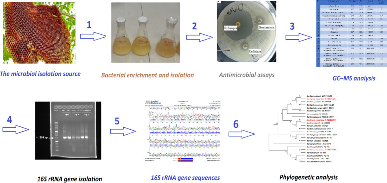
The other most dominant chemical found in Khuz 2 which belongs to diketopiperazine group was Pyrrolo [1,2-a] pyrazine-1,4-dion, hexahydro-3-(phenylmethyl) (PPDHP). Sanjenbam and coworkers reported anticandidal activities of PPDHP from Streptomyces sp. VITPK9 (isolated from a brine spring soil sediment sample) against Candida albicans, Candida krusei, and Candida tropicalis [22]. In addition, the bioactivity of PPDHP has been investigated by [41]. Based on the results, PPDHP was less toxic to the normal cell and is nonhemolytic at low concentrations of < 100 μg/mL. In addition, genotoxicity study showed that PPDHP had minimal chromosomal aberrations relative to streptomycin. On the whole, the GC–MS analysis showed the presence of potential antimicrobial agents belonging to diketopiperazine group PPDHM and PPDHP as the major compounds. Therefore, Bacillus sp. which was isolated from wild honey is a promising bacteria for the biotechnological production of antibiotics. All the procedures are summarized in Fig. 3 as a schematic flow chart.
Fig. 3.
A schematic flow chart of all the procedures
Conclusion
It has been considered the critical lack of new antimicrobials is the clinical pipeline. In 2019 WHO identified only six innovative antibiotics, which were used in the clinical development of priority pathogens. However, a lack of access to quality antimicrobials remained a major issue. We have isolated three strain of Bacillus genus from honey that differs in their antagonistic activity against a number from phytopathogenic and human fungi. Only B. rugosus strain Khuz 2 was able to protect C. albicans human fungi. Thus, B. rugosus stain Khuz 2 can find application as antibacterial and antifungus in agriculture as bioinoculant for agriculturally important plants. The major compounds of strain Khuz 1, 2 and 3 obtained from GC–MS are Pyrrolo[1,2-a] pyrazine-1,4-dion, hexahydro-3-(2-methylpropyl).
Supplementary Information
Acknowledgements
This study was supported by Tabriz University of Medical Sciences for Pharmacy. Student’s Project (grant numbers 60598 and 58766).
Abbreviations
- GC–MS
Gas chromatography–mass spectrometry
- PPDHM
Pyrrolo [1,2-a] pyrazine-1,4-dion, hexahydro-3-(2-methylpropyle)
- Khuz 1
Khuzestan1
- Khuz 2
Khuzestan2
- Khuz 3
Khuzestan3
- DNA
Deoxyribonucleic Acid
- OD
Optical Density
- MIC
Minimum inhibitory concentration
- MBC
Minimum bactericidal concentration
Authors’ contributions
BE, MS, RH and RZ carried out the experiments. SE analyzed GR mass results. SH, FL, AD and AD designed the project and prepared the manuscript together. All authors reviewed the manuscript. All authors read and approved the final manuscript.
Funding
This work was supported by Tabriz University of Medical Sciences, (grant NO: 60598 and 58766), Tabriz University of Medical Sciences. The funders had no role in study design, data collection and analysis, decision to publish, or preparation of the manuscript.
Availability of data and materials
The datasets used and/or analyzed during the current study are available from the corresponding author on reasonable request.
Declarations
Ethics approval and consent to participate
Not applicable.
Consent for publication
Not applicable.
Competing interests
There are neither ethical nor financial conflicts of interest involved in the manuscript. The manuscript contains only original unpublished data and is not submitted for publication elsewhere.
Footnotes
Publisher’s Note
Springer Nature remains neutral with regard to jurisdictional claims in published maps and institutional affiliations.
References
- 1.Saranraj P, Sivasakthi S. Comprehensive review on honey: Biochemical and medicinal properties. J Acad Ind Res. 2018;6(10):165. [Google Scholar]
- 2.Anand TP, Bhat AW, Shouche YS, Roy U, Siddharth J, Sarma SP. Antimicrobial activity of marine bacteria associated with sponges from the waters off the coast of South East India. Microbiol Res. 2006;161(3):252–262. doi: 10.1016/j.micres.2005.09.002. [DOI] [PubMed] [Google Scholar]
- 3.Singh LS, Sharma H, Talukdar NC. Production of potent antimicrobial agent by actinomycete, Streptomyces sannanensis strain SU118 isolated from phoomdi in Loktak Lake of Manipur. India BMC microbiology. 2014;14(1):1–13. doi: 10.1186/1471-2180-14-1. [DOI] [PMC free article] [PubMed] [Google Scholar]
- 4.Burkholder PR, Pfister RM, Leitz FH. Production of a pyrrole antibiotic by a marine bacterium. Appl Microbiol. 1966;14(4):649–653. doi: 10.1128/am.14.4.649-653.1966. [DOI] [PMC free article] [PubMed] [Google Scholar]
- 5.Beesoo R, Neergheen-Bhujun V, Bhagooli R, Bahorun T. Apoptosis inducing lead compounds isolated from marine organisms of potential relevance in cancer treatment. Mutat Res. 2014;768:84–97. doi: 10.1016/j.mrfmmm.2014.03.005. [DOI] [PubMed] [Google Scholar]
- 6.Elyasifar B, Jafari S, Hallaj-Nezhadi S, Chapeland-leclerc F, Ruprich-Robert G, Dilmaghani A. Isolation and identification of antibiotic-producing halophilic bacteria from dagh biarjmand and haj aligholi salt deserts, Iran. Pharm Sci. 2019;25(1):70–77. doi: 10.15171/PS.2019.11. [DOI] [Google Scholar]
- 7.Katz L, Baltz RH. Natural product discovery: past, present, and future. J Ind Microbiol Biotechnol. 2016;43(2–3):155–176. doi: 10.1007/s10295-015-1723-5. [DOI] [PubMed] [Google Scholar]
- 8.Gatadi S, Gour J, Nanduri S. Natural product derived promising anti-MRSA drug leads: a review. Bioorg Med Chem. 2019;27(17):3760–3774. doi: 10.1016/j.bmc.2019.07.023. [DOI] [PubMed] [Google Scholar]
- 9.Behzadi P, Gajdács M. Writing a strong scientific paper in medicine and the biomedical sciences: a checklist and recommendations for early career researchers. Biologia Futura. 2021;72(4):395–407. doi: 10.1007/s42977-021-00095-z. [DOI] [PubMed] [Google Scholar]
- 10.Elyasi Far B, Yari Khosroushahi A, Dilmaghani A. In silico study of alkaline serine protease and production optimization in Bacillus sp. khoz1 closed bacillus safensis isolated from honey. Int J Pept Res Ther. 2020;26(4):2241–51.
- 11.Tarhriz V, Mohammadzadeh F, Hejazi MS, Nematzadeh G, Rahimi E. Isolation and characterization of some aquatic bacteria from Qurugol lake in Azerbaijan under aerobic conditions. Adv Environ Biol. 2011;5(10):3173–8. [Google Scholar]
- 12.Tarhriz V, Hamidi A, Rahimi E, Eramabadi M, Eramabadi P, Yahaghi E, et al. Isolation and characterization of naphthalene-degradation bacteria from Qurugol Lake located at Azerbaijan. Biosci Biotechnol Res Asia. 2014;11(2):715–722. doi: 10.13005/bbra/1326. [DOI] [Google Scholar]
- 13.MacFaddin JF. Biochemical tests for identification of medical bacteria. 2000. [Google Scholar]
- 14.Ebrahimi V, Eyvazi S, Montazersaheb S, Yazdani P, Hejazi MA, Tarhriz V, et al. Polycyclic aromatic hydrocarbons degradation by aquatic bacteria isolated from Khazar Sea, the World’s Largest Lake. Pharm Sci. 2020;27(1):121–130. doi: 10.34172/PS.2020.28. [DOI] [Google Scholar]
- 15.Tarhriz V, Hirose S, Fukushima S-i, Hejazi MA, Imhoff JF, Thiel V, et al. Emended description of the genus Tabrizicola and the species Tabrizicola aquatica as aerobic anoxygenic phototrophic bacteria. Antonie van Leeuwenhoek. 2019;112(8):1169–75. doi: 10.1007/s10482-019-01249-9. [DOI] [PubMed] [Google Scholar]
- 16.Tarhriz V, Nematzadeh G, Vahed SZ, Hejazi MA, Hejazi MS. Alishewanella tabrizica sp. nov., isolated from Qurugöl Lake. Int J Syst Evol Microbiol. 2012;62(Pt_8):1986–91. doi: 10.1099/ijs.0.031567-0. [DOI] [PubMed] [Google Scholar]
- 17.Yoon S-H, Ha S-M, Kwon S, Lim J, Kim Y, Seo H, et al. Introducing EzBioCloud: a taxonomically united database of 16S rRNA gene sequences and whole-genome assemblies. Int J Syst Evol Microbiol. 2017;67(5):1613. doi: 10.1099/ijsem.0.001755. [DOI] [PMC free article] [PubMed] [Google Scholar]
- 18.Thompson JD, Gibson TJ, Plewniak F, Jeanmougin F, Higgins DG. The CLUSTAL_X windows interface: flexible strategies for multiple sequence alignment aided by quality analysis tools. Nucleic Acids Res. 1997;25(24):4876–4882. doi: 10.1093/nar/25.24.4876. [DOI] [PMC free article] [PubMed] [Google Scholar]
- 19.Kumar S, Stecher G, Tamura K. MEGA7: molecular evolutionary genetics analysis version 7.0 for bigger datasets. Mol Biol Evol. 2016;33(7):1870–4. doi: 10.1093/molbev/msw054. [DOI] [PMC free article] [PubMed] [Google Scholar]
- 20.Eyvazi S, Vostakolaei MA, Dilmaghani A, Borumandi O, Hejazi MS, Kahroba H, et al. The oncogenic roles of bacterial infections in development of cancer. Microb Pathog. 2020;141:104019. doi: 10.1016/j.micpath.2020.104019. [DOI] [PubMed] [Google Scholar]
- 21.Sundin GW, Wang N. Antibiotic resistance in plant-pathogenic bacteria. Annu Rev Phytopathol. 2018;56:161–180. doi: 10.1146/annurev-phyto-080417-045946. [DOI] [PubMed] [Google Scholar]
- 22.Ennahar S, Deschamps N. Anti-Listeria effect of enterocin A, produced by cheese-isolated Enterococcus faecium EFM01, relative to other bacteriocins from lactic acid bacteria. J Appl Microbiol. 2000;88(3):449–457. doi: 10.1046/j.1365-2672.2000.00985.x. [DOI] [PubMed] [Google Scholar]
- 23.Tarhriz V, Eyvazi S, Shakeri E, Hejazi MS, Dilmaghani A. Antibacterial and antifungal activity of novel freshwater bacterium Tabrizicola aquatica as a prominent natural antibiotic available in Qurugol Lake. Pharm Sci. 2020;26(1):88–92. doi: 10.34172/PS.2019.56. [DOI] [Google Scholar]
- 24.Donadu MG, Peralta-Ruiz Y, Usai D, Maggio F, Molina-Hernandez JB, Rizzo D, et al. Colombian essential oil of ruta graveolens against nosocomial antifungal resistant Candida strains. J Fungi. 2021;7(5):383. doi: 10.3390/jof7050383. [DOI] [PMC free article] [PubMed] [Google Scholar]
- 25.Afshar FH, Zadehkamand M, Rezaei Z, Delazar A, Tarhriz V, Asgharian P. Chemical compositions, antimicrobial effects, and cytotoxicity of Asia minor wormwood (Artemisia splendens Willd.) growing in Iran. BMC Chem. 2021;15(1):1–9. doi: 10.1186/s13065-020-00727-w. [DOI] [PMC free article] [PubMed] [Google Scholar]
- 26.Saitou N, Nei M. The neighbor-joining method: a new method for reconstructing phylogenetic trees. Mol Biol Evol. 1987;4(4):406–425. doi: 10.1093/oxfordjournals.molbev.a040454. [DOI] [PubMed] [Google Scholar]
- 27.Prestinaci F, Pezzotti P, Pantosti A. Antimicrobial resistance: a global multifaceted phenomenon. Pathog Glob Health. 2015;109(7):309–318. doi: 10.1179/2047773215Y.0000000030. [DOI] [PMC free article] [PubMed] [Google Scholar]
- 28.Hosseini K, Jasori S, Delazar A, Asgharian P, Tarhriz V. Phytochemical analysis and anticancer activity of Falcaria vulgaris Bernh growing in Moghan plain, northwest of Iran. BMC Complement Med Ther. 2021;21(1):1–10. doi: 10.1186/s12906-021-03464-2. [DOI] [PMC free article] [PubMed] [Google Scholar]
- 29.Tarhriz V, Yari Khosroushahi A, Ebrahimi Ghasor L, Elyasifar B, Dilmaghani A. Effect of essential oils of zingiber officinale, cinnamomum verum, trachyspermum ammi, cuminum cyminum, and carum carvi on bacteria inducing clonal dysbiosis in vitro. J Mazandaran Univ Med Sci. 2021;31(201):16–27. [Google Scholar]
- 30.Hosseini K, Ahangari H, Chapeland-leclerc F, Ruprich-Robert G, Tarhriz V, Dilmaghani A. Role of fungal infections in carcinogenesis and cancer development: a literature. [DOI] [PMC free article] [PubMed]
- 31.Tarhriz V, Akbari Z, Dilmaghani A, Hamidi A, Hejazi MA, Hejazi MS. Bioreduction of iron and biosorption of heavy metals (Ni 2+, Co 2+, Pb 2+) by a novel environmental bacterium, Tabrizicola aquatica RCRI19 t. Asian J Water Environ Pollut. 2019;16(3):73–81. doi: 10.3233/AJW190035. [DOI] [Google Scholar]
- 32.Clardy J, Fischbach M, Currie C. The natural history of antibiotics. Current biology: CB. 2009;19(11):R437. doi: 10.1016/j.cub.2009.04.001. [DOI] [PMC free article] [PubMed] [Google Scholar]
- 33.Seyfi R, Kahaki FA, Ebrahimi T, Montazersaheb S, Eyvazi S, Babaeipour V, et al. Antimicrobial peptides (AMPs): roles, functions and mechanism of action. Int J Pept Res Ther. 2020;26(3):1451–1463. doi: 10.1007/s10989-019-09946-9. [DOI] [Google Scholar]
- 34.Ebrahimzadeh S, Ahangari H, Soleimanian A, Hosseini K, Ebrahimi V, Ghasemnejad T, et al. Colorectal cancer treatment using bacteria: focus on molecular mechanisms. BMC Microbiol. 2021;21(1):1–12. doi: 10.1186/s12866-021-02274-3. [DOI] [PMC free article] [PubMed] [Google Scholar]
- 35.Roy A, Mahata D, Paul D, Korpole S, Franco OL, Mandal SM. Purification, biochemical characterization and self-assembled structure of a fengycin-like antifungal peptide from Bacillus thuringiensis strain SM1. Front Microbiol. 2013;4:332. doi: 10.3389/fmicb.2013.00332. [DOI] [PMC free article] [PubMed] [Google Scholar]
- 36.Sanjenbam P, Gopal JV, Kannabiran K. Isolation and identification of anticandidal compound from Streptomyces sp. VITPK9. Appl Biochem Microbiol. 2014;50(5):492–9. doi: 10.1134/S0003683814050081. [DOI] [Google Scholar]
- 37.Kiran GS, Priyadharsini S, Sajayan A, Ravindran A, Selvin J. An antibiotic agent pyrrolo [1, 2-a] pyrazine-1, 4-dione, hexahydro isolated from a marine bacteria Bacillus tequilensis MSI45 effectively controls multi-drug resistant Staphylococcus aureus. RSC Adv. 2018;8(32):17837–17846. doi: 10.1039/C8RA00820E. [DOI] [PMC free article] [PubMed] [Google Scholar]
- 38.Tan LT-H, Chan K-G, Pusparajah P, Yin W-F, Khan TM, Lee L-H, et al. Mangrove derived Streptomyces sp. MUM265 as a potential source of antioxidant and anticolon-cancer agents. BMC Microbiol. 2019;19(1):1–16. doi: 10.1186/s12866-019-1409-7. [DOI] [PMC free article] [PubMed] [Google Scholar]
- 39.Durai S, Vigneshwari L, Balamurugan K. C aenorhabditis elegans-based in vivo screening of bioactives from marine sponge-associated bacteria against V ibrio alginolyticus. J Appl Microbiol. 2013;115(6):1329–1342. doi: 10.1111/jam.12335. [DOI] [PubMed] [Google Scholar]
- 40.Sheoran N, Nadakkakath AV, Munjal V, Kundu A, Subaharan K, Venugopal V, et al. Genetic analysis of plant endophytic Pseudomonas putida BP25 and chemo-profiling of its antimicrobial volatile organic compounds. Microbiol Res. 2015;173:66–78. doi: 10.1016/j.micres.2015.02.001. [DOI] [PubMed] [Google Scholar]
- 41.Kannabiran K. Bioactivity of Pyrrolo [1, 2-a] pyrazine-1, 4-dione, hexahydro-3-(phenylmethyl)-Extracted from Streptomyces sp. VITPK9 Isolated from the Salt Spring Habitat of Manipur, India. Asian Journal of Pharmaceutics (AJP): Free full text articles from Asian J Pharm. 2016;10(04).
Associated Data
This section collects any data citations, data availability statements, or supplementary materials included in this article.
Supplementary Materials
Data Availability Statement
The datasets used and/or analyzed during the current study are available from the corresponding author on reasonable request.


