Abstract
SARS-CoV-2 mRNA-based vaccines are about 95% effective in preventing COVID-191–5. The dynamics of antibody-secreting plasmablasts and germinal centre B cells induced by these vaccines in humans remain unclear. Here we examined antigen-specific B cell responses in peripheral blood (n = 41) and draining lymph nodes in 14 individuals who had received 2 doses of BNT162b2, an mRNA-based vaccine that encodes the full-length SARS-CoV-2 spike (S) gene1. Circulating IgG- and IgA-secreting plasmablasts that target the S protein peaked one week after the second immunization and then declined, becoming undetectable three weeks later. These plasmablast responses preceded maximal levels of serum anti-S binding and neutralizing antibodies to an early circulating SARS-CoV-2 strain as well as emerging variants, especially in individuals who had previously been infected with SARS-CoV-2 (who produced the most robust serological responses). By examining fine needle aspirates of draining axillary lymph nodes, we identified germinal centre B cells that bound S protein in all participants who were sampled after primary immunization. High frequencies of S-binding germinal centre B cells and plasmablasts were sustained in these draining lymph nodes for at least 12 weeks after the booster immunization. S-binding monoclonal antibodies derived from germinal centre B cells predominantly targeted the receptor-binding domain of the S protein, and fewer clones bound to the N-terminal domain or to epitopes shared with the S proteins of the human betacoronaviruses OC43 and HKU1. These latter cross-reactive B cell clones had higher levels of somatic hypermutation as compared to those that recognized only the SARS-CoV-2 S protein, which suggests a memory B cell origin. Our studies demonstrate that SARS-CoV-2 mRNA-based vaccination of humans induces a persistent germinal centre B cell response, which enables the generation of robust humoral immunity.
The concept of using mRNAs as vaccines was introduced over 30 years ago6,7. Key refinements that improved the biological stability and translation capacity of exogenous mRNA enabled development of these molecules as vaccines8,9. The emergence of SARS-CoV-2 in December 2019, and the ensuing pandemic, has revealed the potential of this platform9–11. Hundreds of millions of people have received one of the two SARS-CoV-2 mRNA-based vaccines that were granted emergency use authorization by the US Food and Drug Administration in December 2020. Both of these vaccines demonstrated notable immunogenicity in phase-I/II studies and efficacy in phase-III studies1–4,12–14. Whether these vaccines induce the robust and persistent germinal centre reactions that are critical for generating high-affinity and durable antibody responses has not been examined in humans. To address this question, we conducted an observational study of 41 healthy adults (8 of whom had a history of confirmed SARS-CoV-2 infection) who received the Pfizer–BioNTech SARS-CoV-2 mRNA vaccine BNT162b2 (Extended Data Tables 1, 2). Blood samples were collected at baseline, and at weeks 3 (pre-boost), 4, 5, 7 and 15 after the first immunization. Fine needle aspirates (FNAs) of the draining axillary lymph nodes were collected from 14 participants (none with history of SARS-CoV-2 infection) at weeks 3 (pre-boost), 4, 5, 7, and 15 after the first immunization (Fig. 1a).
Fig. 1|. Plasmablast and antibody response to SARS-CoV-2 immunization.
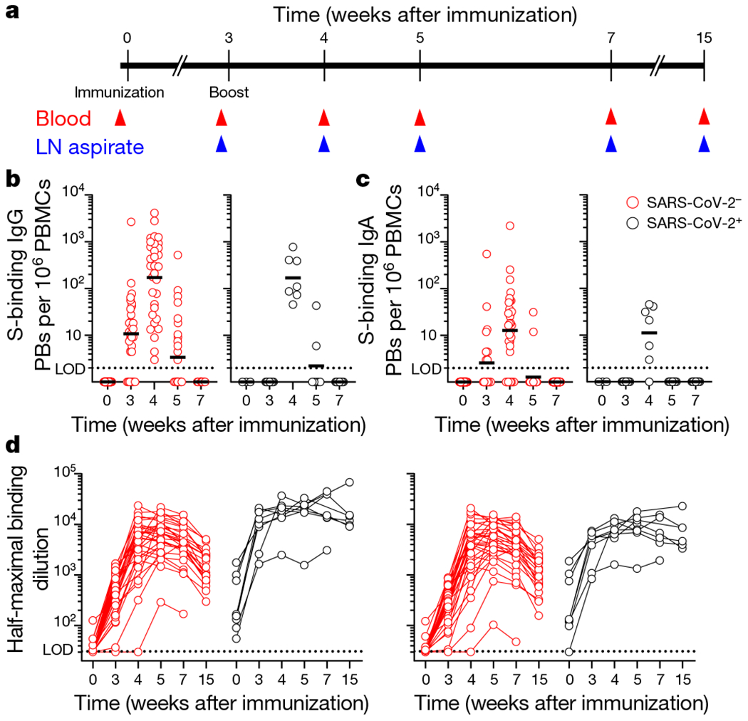
a, Study design. Forty-one healthy adult volunteers (ages 28–73, 8 with a history of SARS-CoV-2 infection) were enrolled and received the BNT162b2 mRNA SARS-CoV-2 vaccine. Blood was collected before immunization, and at 3, 4, 5, 7 and 15 weeks after immunization. For 14 participants (ages 28–52, none with a history of SARS-CoV-2 infection), FNAs of ipsilateral axillary lymph nodes (LNs) were collected at 3, 4, 5, 7 and 15 weeks after immunization. b, c, ELISpot quantification of S-binding IgG- (b) and IgA- (c) secreting plasmablasts (PBs) in blood at baseline, and at 3, 4, 5 and 7 weeks after immunization in participants without (red) and with (black) a history of SARS-CoV-2 infection. d, Plasma IgG titres against SARS-CoV-2 S (left) and the RBD of S (right) measured by ELISA in participants without (red) and with (black) a history of SARS-CoV-2 infection at baseline, and at 3, 4, 5, 7 and 15 weeks after immunization. Dotted lines indicate limits of detection. Symbols at each time point in b–d represent one sample (n = 41). Results are from one experiment performed in duplicate.
We used an enzyme-linked immune absorbent spot (ELISpot) assay to measure antibody-secreting plasmablasts in blood that bound SARS-CoV-2 S protein. We detected SARS-CoV-2-S-specfic IgG- and IgA-secreting plasmablasts 3 weeks after primary immunization in 24 of 33 participants with no history of SARS-CoV-2 infection, but in 0 of 8 participants who had previously been infected with SARS-CoV-2. Plasmablasts peaked in blood during the first week after boosting (week 4 after primary immunization), with frequencies that varied widely from 3 to 4,100 S-binding plasmablasts per 106 peripheral blood mononuclear cells (PBMCs) (Fig. 1b, c). We found that plasma IgG antibody titres against S, measured by enzyme-linked immunosorbent assay (ELISA), increased in all participants over time, and reached peak geometric mean half-maximal binding titres of 5,567 and 15,850 at 5 weeks after immunization among participants without and with history of SARS-CoV-2 infection, respectively, with a subsequent decline by 15 weeks after immunization. Anti-S IgA titres and IgG titres against the receptor-binding domain (RBD) of S showed similar kinetics, and reached peak geometric mean half-maximal binding titres of 172 and 739 for anti-S IgA and 4,501 and 7,965 for anti-RBD IgG among participants without and with history of SARS-CoV-2 infection, respectively, before declining. IgM responses were weaker and more transient, peaking 4 weeks after immunization among participants without history of SARS-CoV-2 infection with a geometric mean half-maximal binding titre of 78 and were undetectable in all but 2 previously infected participants (Fig. 1d, Extended Data Fig. 1a).
The functional quality of serum antibody was measured using high-throughput focus reduction neutralization tests15 on Vero cells expressing TMPRSS2 against three authentic infectious SARS-CoV-2 strains with sequence variations in the S gene16,17: (1) a Washington strain (2019n-CoV/USA) with a prevailing D614G substitution (WA1/2020 D614G); (2) a B.1.1.7 isolate with signature changes in the S gene18, including mutations resulting in the deletion of residues 69, 70,144 and 145 as well as N501Y, A570D, D614G and P681H substitutions; and (3) a chimeric SARS-CoV-2 with a B.1.351 S gene in the Washington strain background (Wash-B.1.351) that contained the following changes: D80A, deletion of residues 242–244, R246I, K417N, E484K, N501Y, D614G and A701V. Serum neutralizing titres increased markedly in participants without a history of SARS-CoV-2 infection after boosting, with geometric mean neutralization titres against WA1/2020 D614G of 58 at 3 weeks after primary immunization and 572 at 2 or 4 weeks after boost (5 or 7 weeks after primary immunization). Neutralizing titres against the B.1.1.7 and B.1.351 variants were lower, with geometric mean neutralization titres of 49 and 373 against B.1.1.7 and 36 and 137 against B.1.351 after primary and secondary immunization, respectively. In participants with a history of previous SARS-CoV-2 infection, neutralizing titres against all three viruses were detected at baseline (geometric mean neutralization titres of 241.8, 201.8 and 136.7 against WA1/2020 D614G, B.1.1.7 and B.1.351, respectively). In these participants, neutralizing titres increased more rapidly and to higher levels after immunization, with geometric mean neutralization titres of 4,544, 3,584 and 1,897 against WA1/2020 D614G, B.1.1.7 and B.1.351, respectively, after primary immunization, and 9,381, 9,351 and 2,749 against WA1/2020 D614G, B.1.1.7 and B.1.351, respectively, after secondary immunization. These geometric mean neutralization titres were 78-, 73- and 53-fold higher after primary immunization and 16-, 25- and 20-fold higher after boosting against WA1/2020 D614G, B.1.1.7 and B.1.351, respectively, than in participants without a history of SARS-CoV-2 infection (Extended Data Fig. 1b).
The BNT162b2 vaccine is injected into the deltoid muscle, which drains primarily to the lateral axillary lymph nodes. We used ultrasonography to identify and guide FNA of accessible axillary nodes on the side of immunization approximately 3 weeks after primary immunization. In 5 of the 14 participants, a second draining lymph node was identified and sampled after secondary immunization (Fig. 2a). Germinal centre B cells (defined as CD19+CD3−IgDlowBCL6+CD38int lymphocytes) were detected in all lymph nodes (Fig. 2b, d, Extended Data Fig. 2a, Extended Data Table 3). We co-stained FNA samples with two fluorescently labelled S probes to detect S-binding germinal centre B cells. A control tonsillectomy sample with a high frequency of germinal centre B cells that was collected before the COV1D-19 pandemic from an unrelated donor was stained as a negative control. S-binding germinal centre B cells were detected in FNAs from all 14 participants following primary immunization. The kinetics of the germinal centre response varied among participants, but S-binding germinal centre B cell frequencies increased at least transiently in all participants after boosting and persisted at high frequency in most individuals for at least 7 weeks. Notably, S-binding germinal centre B cells remained at or near their peak frequency 15 weeks after immunization in 8 of the 10 participants sampled at that time point, and these prolonged germinal centre responses had high proportions of S-binding cells (Fig. 2c–e, Extended Data Fig. 2b).
Fig. 2|. Germinal centre B cell response to SARS-CoV-2 immunization.
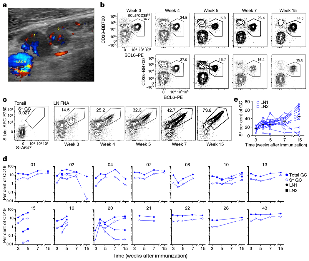
a, Representative colour Doppler ultrasound image of two draining lymph nodes (‘1’ and ‘2’) adjacent to the axillary vein ‘LAX V’ 5 weeks after immunization. b, c, Representative flow cytometry plots of BCL6 and CD38 staining on IgDlowCD19+CD3− live singlet lymphocytes in FNA samples (b; LN1, top row; LN2, bottom row) and S staining on BCL6+CD38int germinal centre B cells in tonsil and FNA samples (c) at the indicated times after immunization. d, e, Kinetics of total (blue) and S+ (white) germinal centre (GC) B cells as gated in b and c (d) and S-binding per cent of germinal centre B cells (e) from FNA of draining lymph nodes. Symbols at each time point represent one FNA sample; square symbols denote the second lymph node sampled (n = 14). Horizontal lines indicate the median.
To evaluate the domains targeted by the S-protein-specific germinal centre response after vaccination, we generated recombinant monoclonal antibodies from single-cell-sorted S-binding germinal centre B cells (defined by the surface-marker phenotype CD19+CD3−IgDlowCD20highCD38intCD71+CXCR5+ lymphocytes) from three of the participants one week after boosting (Extended Data Fig. 2a). Fifteen, five and seventeen S-binding, clonally distinct monoclonal antibodies were generated from participants 07, 20 (lymph node 1) and 22, respectively (Extended Data Table 4). Of the 37 S-binding monoclonal antibodies, 17 bound the RBD, 6 recognized the N-terminal domain and 3 were cross-reactive with S proteins from seasonal betacoronavirus OC43; 2 of these monoclonal antibodies also bound S from seasonal betacoronavirus HKU1 (Fig. 3a). Clonal relatives of 14 out of 15, 1 out of 5 and 12 out of 17 of the S-binding monoclonal antibodies were identified among bulk-sorted total plasmablasts from PBMCs and germinal centre B cells at 4 weeks after immunization from participants 07, 20 and 22, respectively (Fig. 3b, Extended Data Figs. 2c, 3a, b, Extended Data Tables 5, 6). Clones related to S-binding monoclonal antibodies had significantly increased mutation frequencies in their immunoglobulin heavy chain variable region (IGHV) genes compared to previously published naive B cells19, particularly those related to monoclonal antibodies that cross-reacted with seasonal betacoronaviruses (Fig. 3c, d).
Fig. 3|. Clonal analysis of germinal centre response to SARS-CoV-2 immunization.
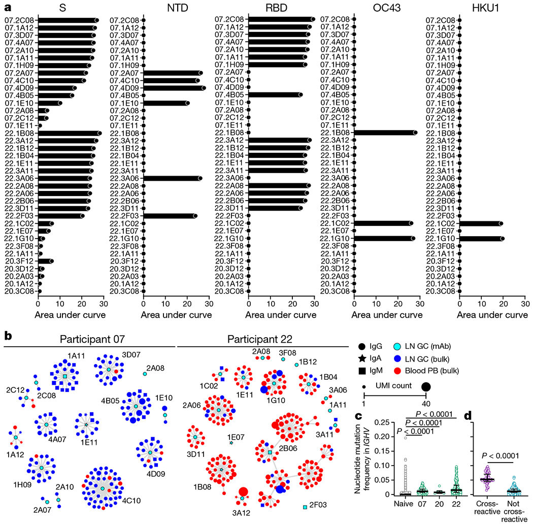
a, Binding of monoclonal antibodies (mAbs) generated from germinal centre B cells to SARS-CoV-2 S, N-terminal domain (NTD) of S, RBD, or S proteins of betacoronavirus OC43 or HKU1, measured by ELISA. Results are from one experiment performed in duplicate. Baseline for area under the curve was set to the mean + three times the s.d. of background binding to bovine serum albumin. b, Clonal relationship of sequences from S-binding germinal centre-derived monoclonal antibodies (cyan) to sequences from bulk repertoire analysis of plasmablasts from PBMCs (red) and germinal centre B cells (blue) sorted 4 weeks after immunization. Each clone is visualized as a network in which each node represents a sequence and sequences are linked as a minimum spanning tree of the network. Symbol shape indicates sequence isotype: IgG (circle), IgA (star) and IgM (square); symbol size corresponds to sequence count. c, d, Comparison of nucleotide mutation frequency in IGHV genes of naive B cells sorted from individuals vaccinated with influenza virus vaccine19 (grey) to clonal relatives of S-binding monoclonal antibodies among plasmablasts sorted from PBMCs and germinal centre B cells 4 weeks after immunization (green) in indicated participants (c) and between clonal relatives of S-binding monoclonal antibodies cross-reactive (purple) or not (teal) to seasonal coronavirus S proteins among plasmablasts sorted from PBMCs and germinal centre B cells 4 weeks after immunization (d). Horizontal lines and error bars indicate the median and interquartile range. Sequence counts were 2,553 (naive), 199 (participant 07), 6 (participant 20), 240 (participant 22), 54 (cross-reactive) and 391 (not cross-reactive). P values from two-sided Kruskal–Wallis test with Dunn’s post-test between naive B cells and S-binding clones (c) or two-sided Mann–Whitney U test (d).
In addition to germinal centre B cells, we detected robust plasmablast responses in the draining lymph nodes of all 14 participants in the FNA cohort. S-binding plasmablasts (defined as CD19+CD3−IgDlowCD20lowCD38+CD71+BLIMP1+ lymphocytes) were detected in all of the lymph nodes that we sampled, and increased in frequency after boosting (Extended Data Fig. 4a, b). The detected plasmablasts were unlikely to be a contaminant of blood, because CD14+ monocyte and/or granulocyte frequencies were below 1% in all FNA samples (well below the 10% threshold that was previously established19) (Extended Data Table 3). Moreover, S-binding plasmablasts were detected in FNA samples at 5, 7 and 15 weeks after immunization, when they had become undetectable in blood from all participants in the cohort. The vast majority of S-binding lymph node plasmablasts were isotype-switched at 4 weeks after primary immunization, and IgA-switched cells accounted for 25% or more of the plasmablasts in 6 out of 14 participants (Extended Data Fig. 4c, d).
This study evaluated whether SARS-CoV-2 mRNA-based vaccines induce antigen-specific plasmablast and germinal centre B cell responses in humans. The vaccine induced a strong IgG-dominated plasmablast response in blood that peaked one week after the booster immunization. In the draining lymph nodes, we detected robust SARS-CoV-2 S-binding germinal centre B cell and plasmablast responses in aspirates from all 14 of the participants. These responses were detectable after the first immunization but greatly expanded after the booster injection. Notably, S-binding germinal centre B cells and plasmablasts persisted for at least 15 weeks after the first immunization (12 weeks after secondary immunization) in 8 of the 10 participants who were sampled at that time point. These responses to mRNA vaccination are superior to those seen after seasonal influenza virus vaccination in humans19, in whom haemagglutinin-binding germinal centre B cells were detected in only three out of eight participants. More robust germinal centre responses are consistent with antigen dissemination to multiple lymph nodes and the self-adjuvating characteristics of the mRNA–lipid nanoparticle vaccine platform compared to nonadjuvanted inactivated vaccines used for seasonal influenza virus vaccination7,20,21. Our data in humans corroborate reports that demonstrate the induction of potent germinal centre responses by SARS-CoV-2 mRNA-based vaccines in mice22,23.
To our knowledge, this is the first study to provide direct evidence for the induction of a persistent antigen-specific germinal centre B cell response after vaccination in humans. Dynamics of germinal centre B cell responses vary widely depending on the model system in which they are studied, although the most active period of the response usually occurs over the course of a few weeks. Primary alum-adjuvanted protein immunization of mice typically leads to germinal centre responses that peak 1–2 weeks after immunization and contract at least 10-fold within 5–7 weeks24–26. Germinal centre responses induced by immunization with more robust adjuvants such as sheep red blood cells, complete Freund’s adjuvant or saponin-based adjuvants tend to peak slightly later, at 2–4 weeks after vaccination, and can persist at low frequencies for several months27–33. Although studies of extended durability are rare, antigen-specific germinal centre B cells have been found to persist for at least one year, albeit at very low levels28,30. In this study, we show SARS-CoV-2 mRNA vaccine-induced germinal centre B cells are maintained at or near peak frequencies for at least 12 weeks after secondary immunization.
The persistence of S-binding germinal centre B cells and plasmablasts in draining lymph nodes is a positive indicator for induction of long-lived plasma cell responses25. Future studies will be needed to examine whether mRNA vaccination induces a robust S-specific long-lived plasma cell compartment in the bone marrow. As part of such studies, it will be critical to generate a comprehensive set of monoclonal antibodies derived from plasmablasts and germinal centre B cells isolated from several time points to define the breadth of the B cell response elicited by this vaccine. None of the 14 participants in our study who underwent FNA of draining lymph nodes had a history of SARS-CoV-2 infection. Thus, further comparison of vaccine-induced germinal centre responses from naive and previously infected individuals will be informative. Finally, the work presented here focuses on the B cell component of the germinal centre reaction. A robust T follicular helper response sustains the germinal centre reaction34,35. As such, studies are planned to investigate the magnitude, specificity and durability of the T follicular helper cell response after SARS-CoV-2 mRNA vaccination in humans.
A preliminary observation from our study is the dominance of RBD-targeting clones among responding germinal centre B cells. A more detailed analysis36 of these RBD-binding monoclonal antibodies assessed their in vitro inhibitory capacity against the WA1/2020 D614G strain using an authentic SARS-CoV-2 neutralization assay: five showed high neutralization potency, with 80% neutralization values of less than 100 ng ml−1. For the most part, RBD-binding clones contained few (<3) nonsynonymous nucleotide substitutions in their IGHV genes, which indicates that they originated from recently engaged naive B cells. This contrasts with the three cross-reactive germinal centre B cell clones that recognized conserved epitopes within the S proteins of betacoronaviruses. These cross-reactive clones had significantly higher mutation frequencies, which suggests a memory B cell origin. These data are consistent with previous findings from seasonal influenza virus vaccination in humans that show that the germinal centre reaction can engage pre-existing memory B cells directed against conserved epitopes as well as naive clones targeting novel epitopes19. However, these cross-reactive clones were not identified in all individuals and comprised a small fraction of responding B cells, consistent with a similar analysis of SARS-CoV-2 mRNA vaccine-induced plasmablasts37. Overall, our data demonstrate the capacity of SARS-CoV-2 mRNA-based vaccines to induce robust and prolonged germinal centre reactions. The induced germinal centre reaction recruited cross-reactive memory B cells as well as newly engaged clones that target unique epitopes within SARS-CoV-2 S protein. Elicitation of high affinity and durable protective antibody responses is a hallmark of a successful humoral immune response to vaccination. By inducing robust germinal centre reactions, SARS-CoV-2 mRNA-based vaccines are on track for achieving this outcome.
Methods
No statistical methods were used to predetermine sample size.
Sample collection, preparation, and storage
All studies were approved by the Institutional Review Board of Washington University in St Louis. Written consent was obtained from all participants. Forty-one healthy volunteers were enrolled, of whom 14 provided axillary lymph node samples (Extended Data Table 1). In 5 of the 14 participants, a second draining lymph node was identified and sampled following secondary immunization. One participant (15) received the second immunization in the contralateral arm; draining lymph nodes were identified and sampled on both sides. Blood samples were collected in EDTA tubes, and PBMCs were enriched by density gradient centrifugation over Ficoll 1077 (GE) or Lymphopure (BioLegend). The residual red blood cells were lysed with ammonium chloride lysis buffer, and cells were immediately used or cryopreserved in 10% dimethylsulfoxide in fetal bovine serum (FBS). Ultrasound-guided FNA of axillary lymph nodes was performed by a radiologist or a qualified physician’s assistant under the supervision of a radiologist. Lymph node dimensions and cortical thickness were measured, and the presence and degree of cortical vascularity and location of the lymph node relative to the axillary vein were determined before each FNA. For each FNA sample, six passes were made under continuous real-time ultrasound guidance using 25-gauge needles, each of which was flushed with 3 ml of RPMI 1640 supplemented with 10% FBS and 100 U ml−1 penicillin–streptomycin, followed by three 1-ml rinses. Red blood cells were lysed with ammonium chloride buffer (Lonza), washed with phosphate-buffered saline (PBS) supplemented with 2% FBS and 2 mM EDTA, and immediately used or cryopreserved in 10% dimethylsulfoxide in FBS. Participants reported no adverse effects from phlebotomies or serial FNAs.
Cell lines
Expi293F cells were cultured in Expi293 Expression Medium (Gibco). Vero E6 (CRL-1586, American Type Culture Collection), Vero cells expressing TMPRSS2 (Vero-TMPRSS2 cells)38 (a gift from S. Ding), and Vero cells expressing human ACE2 and TMPRSS2 (Vero-hACE2-TMPRSS2) (a gift of A. Creanga and B. Graham) cells were cultured at 37°C in Dulbecco’s modified Eagle medium (DMEM) supplemented with 10% FBS, 10 mM HEPES (pH 7.3), 1 mM sodium pyruvate, 1× nonessential amino acids and 100 U ml−1 of penicillin–streptomycin. Vero-TMPRSS2 cell cultures were supplemented with 5 μg ml−1 of blasticidin. Vero-hACE2-TMPRSS2 cell cultures were supplemented with 10 μg ml−1 of puromycin.
Viruses
The 2019n-CoV/USA_WA1/2020 isolate of SARS-CoV-2 was obtained from the US Centers for Disease Control. The B.1.1.7 isolate from the UK was obtained from an infected individual. The point mutation D614G in the S gene was introduced into an infectious complementary DNA clone of the 2019n-CoV/USA_WA1/2020 strain as previously described39. Nucleotide substitutions were introduced into a subclone puc57-CoV-2-F5-7 containing the S gene of the SARS-CoV-2 wild-type infectious clone40. The S gene of the B.1.351 variant (first identified in South Africa) was produced synthetically by Gibson assembly. The full-length infectious cDNA clones of the variant SARS-CoV-2 viruses were assembled by in vitro ligation of seven contiguous cDNA fragments following a previously described protocol40. In vitro transcription was then performed to synthesize full-length genomic RNA. To recover the mutant viruses, the RNA transcripts were electroporated into Vero E6 cells. The viruses from the supernatant of cells were collected 40 h later and served as p0 stocks. All viruses were passaged once in Vero-hACE2-TMPRSS2 cells and subjected to deep sequencing after RNA extraction to confirm the introduction and stability of substitutions17. All virus preparation and experiments were performed in an approved biosafety level 3 facility.
Antigens
Recombinant soluble SARS-CoV-2 S protein, recombinant RBD of S, human coronavirus OC43 S, and human coronavirus HKU1 S were expressed as previously described41. In brief, mammalian cell codon-optimized nucleotide sequences coding for the soluble ectodomain of the S protein of SARS-CoV-2 (GenBank: MN908947.3, amino acids 1–1213) including a C-terminal thrombin cleavage site, T4 foldon trimerization domain and hexahistidine tag, and for the RBD (amino acids 319–541) along with the signal peptide (amino acids 1–14) plus a hexahistidine tag were cloned into mammalian expression vector pCAGGS. The S protein sequence was modified to remove the polybasic cleavage site (RRAR to A), and two pre-fusion stabilizing proline mutations were introduced (K986P and V987P, wild-type numbering). Expression plasmids encoding for the S of common human coronaviruses OC43 and HKU1 were provided by B. Graham42. Recombinant proteins were produced in Expi293F cells (ThermoFisher) by transfection with purified DNA using the ExpiFectamine 293 Transfection Kit (ThermoFisher). Supernatants from transfected cells were collected 3 days after transfection, and recombinant proteins were purified using Ni-NTA agarose (ThermoFisher), then buffer-exchanged into PBS and concentrated using Amicon Ultracel centrifugal filters (EMD Millipore). For flow cytometry staining, recombinant S was labelled with Alexa Fluor 647–NHS ester or biotinylated using the EZ-Link Micro NHS-PEG4-Biotinylation Kit (Thermo Fisher); excess Alexa Fluor 647 and biotin were removed using 7-kDa Zeba desalting columns (Pierce).
ELISpot assay
Plates were coated with Flucelvax Quadrivalent 2019/2020 seasonal influenza virus vaccine (Sequiris), S or RBD. A direct ex vivo ELISpot assay was performed to determine the number of total, vaccine-binding or recombinant S-binding IgG- and IgA-secreting cells present in PBMC samples using IgG/IgA double-colour ELISpot Kits (Cellular Technology) according to the manufacturer’s instructions. ELISpot plates were analysed using an ELISpot counter (Cellular Technology).
ELISAs
Assays were performed in 96-well plates (MaxiSorp; Thermo) coated with 100 μl of recombinant S, RBD, N-terminal domain of S (SinoBiological), OC43 S, HKU1 S or bovine serum albumin diluted to 1 μg ml−1 in PBS, and plates were incubated at 4 °C overnight. Plates then were blocked with 10% FBS and 0.05% Tween 20 in PBS. Plasma or purified monoclonal antibodies were serially diluted in blocking buffer and added to the plates. Plates were incubated for 90 min at room temperature and then washed 3 times with 0.05% Tween 20 in PBS. Goat anti-human IgG-HRP (goat polyclonal, Jackson ImmunoResearch, 1:2,500), IgA (goat polyclonal, Jackson ImmuoResearch, 1:2,500) or IgM (goat polyclonal, Caltag, 1:4,000) were diluted in blocking buffer before adding to wells and incubating for 60 min at room temperature. Plates were washed 3 times with 0.05% Tween 20 in PBS and 3 times with PBS before the addition of o-phenylenediamine dihydrochloride peroxidase substrate (Sigma-Aldrich). Reactions were stopped by the addition of 1 M hydrochloric acid. Optical density measurements were taken at 490 nm. The area under the curve for each monoclonal antibody and half-maximal binding dilution for each plasma sample were calculated using Graphpad Prism v.8.
Focus reduction neutralization test
Plasma samples were declotted by diluting 1:10 in DMEM supplemented with 2% FBS, 10 mM HEPES and 100 U ml−1 penicillin–streptomycin and incubating for 3 h at 37 °C. Serial dilutions of resulting serum were incubated with 102 focus-forming units of different strains or variants of SARS-CoV-2 for 1 h at 37 °C. Antibody–virus complexes were added to Vero-TMPRSS2 cell monolayers in 96-well plates and incubated at 37 °C for 1 h. Subsequently, cells were overlaid with 1% (w/v) methylcellulose in MEM supplemented with 2% FBS. Plates were collected 30 h later by removing overlays and fixed with 4% PFA in PBS for 20 min at room temperature. Plates were washed and sequentially incubated with an oligoclonal pool of mouse anti-S monoclonal antibodies (SARS2-2, SARS2-11, SARS2-16, SARS2-31, SARS2-38, SARS2-57 and SARS2-71) (ref. 43) and HRP-conjugated goat anti-mouse IgG (polyclonal, Sigma, 1:500) in PBS supplemented with 0.1% saponin and 0.1% bovine serum albumin. SARS-CoV-2-infected cell foci were visualized using TrueBlue peroxidase substrate (KPL) and quantified on an ImmunoSpot microanalyser (Cellular Technology).
Flow cytometry and cell sorting
Staining for flow cytometry analysis and sorting was performed using freshly isolated or cryo-preserved FNA, PBMC or tonsil samples. For analysis, cells were incubated for 30 min on ice with biotinylated and Alexa Fluor 647 conjugated recombinant soluble S and PD-1-BB515 (EH12.1, BD Horizon, 1:100) in 2% FBS and 2 mM EDTA in PBS (P2), washed twice, then stained for 30 min on ice with IgG–BV480 (goat polyclonal, Jackson ImmunoResearch, 1:100), IgA–FITC (M24A, Millipore, 1:500), CD45–A532 (HI30, Thermo, 1:50), CD38–BB700 (HIT2, BD Horizon, 1:500), CD20–Pacific Blue (2H7, 1:400), CD27–BV510 (O323, 1:50), CD8–BV570 (RPA-T8, 1:200), IgM–BV605 (MHM-88, 1:100), HLA-DR–BV650 (L243, 1:100), CD19–BV750 (HIB19, 1:100), CXCR5–PE–Dazzle 594 (J252D4, 1:50), IgD–PE–Cy5 (IA6-2, 1:200), CD14–PerCP (HCD14, 1:50), CD71–PE–Cy7 (CY1G4, 1:400), CD4–Spark685 (SK3, 1:200), streptavidin–APC–Fire750, CD3–APC–Fire810 (SK7, 1:50) and Zombie NIR (all BioLegend) diluted in Brilliant Staining buffer (BD Horizon). Cells were washed twice with P2, fixed for 1 h at 25 °C using the True Nuclear fixation kit (BioLegend), washed twice with True Nuclear Permeabilization/Wash buffer, stained with FOXP3–BV421 (206D, BioLegend, 1:15), Ki-67–BV711 (Ki-67, BioLegend, 1:200), Tbet–BV785 (4B10, BioLegend, 1:400), BCL6–PE (K112-91, BD Pharmingen, 1:25), and BLIMP1–A700 (646702, R&D, 1:50) for 1 h at 25 °C, washed twice with True Nuclear Permeabilization/Wash buffer, and acquired on an Aurora using SpectroFlo v.2.2 (Cytek). Flow cytometry data were analysed using FlowJo v.10 (Treestar).
For sorting germinal centre B cells, FNA single-cell suspensions were stained for 30 min on ice with CD19–BV421 (HIB19, 1:100), CD3–FITC (HIT3a, 1:200), IgD–PerCP–Cy5.5 (IA6-2, 1:200), CD71–PE (CY1G4, 1:400), CXCR5–PE–Dazzle 594 (J252D4, 1:50), CD38–PE–Cy7 (HIT2, 1:200), CD20–APC–Fire750 (2H7, 1:100), Zombie Aqua (all BioLegend), and Alexa Fluor 647 conjugated recombinant soluble S. For sorting plasmablasts, PBMCs were stained for 30 min on ice with CD20–PB (2H7, 1:400), CD71–FITC (CY1G4, 1:200), CD4–PerCP (OKT4, 1:100), IgD–PE (IA6-2, 1:200), CD38–PE–Cy7 (HIT2, 1:200), CD19–APC (HIB19, 1:200) and Zombie Aqua (all BioLegend). Cells were washed twice, and single S-binding germinal centre B cells (live singlet CD3−CD19+IgDlowCD20highCD38intCD71+CXCR5+S+) were sorted using a FACSAria II into 96-well plates containing 2 μl Lysis Buffer (Clontech) supplemented with 1 U μl−1 RNase inhibitor (NEB), or total germinal centre B cells or plasmablasts (live singlet CD3−CD19+IgDlowCD20lowCD38+CD71+) were bulk-sorted into buffer RLT Plus (Qiagen) and immediately frozen on dry ice.
Monoclonal antibody generation
Antibodies were cloned as previously described44. In brief, VH, Vκ and Vλ genes were amplified by reverse transcription PCR and nested PCR reactions from singly sorted germinal centre B cells using primer combinations specific for IgG, IgM, IgA, Igκ and Igλ from previously described primer sets45, and then sequenced. To generate recombinant antibodies, restriction sites were incorporated via PCR with primers to the corresponding heavy and light chain V and J genes. The amplified VH, Vκ and Vλ genes were cloned into IgG1 and Igκ or Igλ expression vectors, respectively, as previously described45–47. Heavy and light chain plasmids were co-transfected into Expi293F cells (Gibco) for expression, and antibody was purified using protein A agarose chromatography (Goldbio). Sequences were obtained from PCR reaction products and annotated using the ImMunoGeneTics (IMGT)/V-QUEST database (http://www.imgt.org/IMGT_vquest/)48,49. Mutation frequency was calculated by counting the number of nonsynonymous nucleotide mismatches from the germline sequence in the heavy chain variable segment leading up to the CDR3, while excluding the 5′ primer sequences that could be error-prone.
Bulk B cell receptor sequencing
RNA was purified from sorted plasmablasts from PBMCs and germinal centre B cells from lymph nodes from participants 07, 20 (lymph node 1) and 22 using the RNeasy Plus Micro kit (Qiagen). Reverse transcription, unique molecular identifier (UMI) barcoding, cDNA amplification, and Illumina linker addition to B cell heavy chain transcripts were performed using the human NEBNext Immune Sequencing Kit (New England Biolabs) according to the manufacturer’s instructions. High-throughput 2× 300-bp paired-end sequencing was performed on the Illumina MiSeq platform with a 30% PhiX spike-in according to manufacturer’s recommendations, except for performing 325 cycles for read 1 and 275 cycles for read 2.
Processing of B cell receptor bulk-sequencing reads
Demultiplexed pair-end reads were BLAST’ed using blastn v.2.11.0 (ref. 50) for PhiX removal and subsequently preprocessed using pRESTO v.0.6.2 (ref. 51) as follows. (1) Reads with a mean Phred quality score below 20 were filtered. (2) Reads were aligned against template switch sequences and constant region primers (Extended Data Table 5), with a maximum mismatch rate of 0.5 and 0.2 respectively. (3) A UMI was assigned to each read by extracting the first 17 nucleotides preceding the template switch site. (4) Sequencing and multiplexing errors in the UMI region were then corrected using a previously published approach52. In brief, reads with similar UMIs were clustered using cd-hit-est v.4.8.1 (ref. 53) on the basis of the pairwise distance of their UMIs with a similarity threshold of 0.83 that was estimated from 10,000 reads. The UMI-based read groups were further clustered within themselves on the basis of the pairwise distance of the non-UMI region of their reads with a similarity threshold of 0.8. Read clusters spanning multiple multiplexed samples were assigned to the majority sample. (5) Separate consensus sequences for the forward and reverse reads within each read cluster were constructed with a maximum error score of 0.1 and minimum constant region primer frequency of 0.6. If multiple constant region primers were associated with a particular read cluster, the majority primer was used. (6) Forward and reverse consensus sequence pairs were assembled by first attempting de novo assembly with a minimum overlap of 8 nucleotides and a maximum mismatch rate of 0.3. If unsuccessful, this was followed by reference-guided assembly using blastn v.2.11.0 (ref. 50) with a minimum identity of 0.5 and an E-value threshold of 1 × 10⁻⁵. (7) Isotypes were assigned by local alignment of the 3′ end of each consensus sequence to isotype-specific internal constant region sequences (Extended Data Table 5) with a maximum mismatch rate of 0.3. Sequences with inconsistent isotype assignment and constant region primer alignment were removed. (8) Duplicate consensus sequences, except those with different isotype assignments, were collapsed into unique sequences. Only unique consensus sequences with at least two contributing reads were used subsequently (Extended Data Table 6).
B cell receptor genotyping
Initial germline V(D)J gene annotation was performed using IgBLAST v.1.17.1 (ref. 54) with IMGT/GENE-DB release 202113-2 (ref. 55). IgBLAST output was parsed using Change-O v.1.0.2 (ref. 56). Quality control was performed, requiring each sequence to have non-empty V and J gene annotations; exhibit chain consistency in all annotations; bear fewer than 10 non-informative (non-A/T/G/C, such as N or –) positions; and carry a CDR3 with no N and a nucleotide length that is a multiple of 3. Individualized genotypes were inferred using TIgGER v.1.0.0 (ref. 57) and used to finalize V(D)J annotations. Sequences annotated as non-productively rearranged by IgBLAST were removed from further analysis.
Clonal lineage analysis
B cell clonal lineages were inferred on the basis of productively rearranged heavy chain sequences using hierarchical clustering with single linkage58,59. Sequences were first partitioned based on common V and J gene annotations and CDR3 lengths. Within each partition, sequences with CDR3s that were within 0.15 normalized Hamming distance from each other were clustered as clones. This distance threshold was determined by manual inspection in conjunction with kernel density estimates to identify the local minimum between the two modes of the within-participant bimodal distance-to-nearest distribution (Extended Data Fig. 3a). Following clonal clustering, full-length clonal consensus germline sequences were reconstructed for each clone with D-segment and N/P regions masked with Ns, resolving any ambiguous gene assignments by majority rule. Within each clone, duplicate IMGT-aligned V(D)J sequences from bulk sequencing were collapsed with the exception of duplicates derived from different B cell compartments or isotypes. Clones were visualized as networks60 using igraph v.1.2.5 (ref. 61). First, a full network was calculated for each clone, in which an edge was drawn between every pair of sequences with CDR3s that were within 0.15 normalized Hamming distance from each other. Then, a minimum spanning tree was derived from the full network, in which only edges essential for ensuring that all sequences connected in the full network remain connected in the minimum spanning tree either directly or indirectly were retained. The minimum spanning tree was then visualized for each clone.
Calculation of somatic hypermutation frequency
Mutation frequency was calculated by counting the number of nucleotide mismatches from the germline sequence in the observed heavy chain variable segment leading up to the CDR3, while excluding the first 18 positions that could be error-prone owing to the primers used for generating the monoclonal antibody sequences. Calculation was performed using the calcObservedMutations function from SHazaM v.1.0.2 (ref. 56).
Extended Data
Extended Data Fig. 1|. Antibody response to SARS-CoV-2 immunization.

a, Plasma IgA (left) and IgM (right) titres against SARS-CoV-2 S measured by ELISA in participants without (red) and with (black) a history of SARS-CoV-2 infection at baseline, and 3, 4, 5, 7 and 15 weeks after immunization. b, Neutralizing activity of serum against WA1/2020 D614G (left), B.1.1.7 (middle) and a chimeric virus expressing B.1.351 S (right) in Vero-TMPRSS2 cells at baseline, 3, and 5 or 7 weeks after immunization in participants without (red) and with (black) a history of SARS-CoV-2 infection. P values from two-sided Mann-Whitney tests. Dotted lines indicate limits of detection. Horizontal lines indicate the geometric mean. Symbols at each time point represent one sample (n = 41). Results are from one experiment performed in duplicate.
Extended Data Fig. 2|. Gating strategies for analysis of germinal centre response to SARS-CoV-2 immunization.
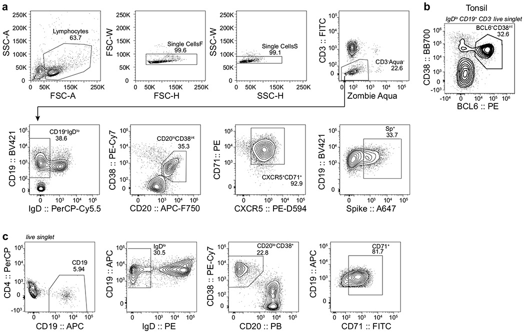
a, c, Sorting gating strategies for S-binding germinal centre B cells from FNAs (a) and total plasmablasts from PBMCs (c). b, Representative plot of germinal centre B cells in tonsil.
Extended Data Fig. 3|. Clonal analysis of germinal centre response to SARS-CoV-2 immunization.
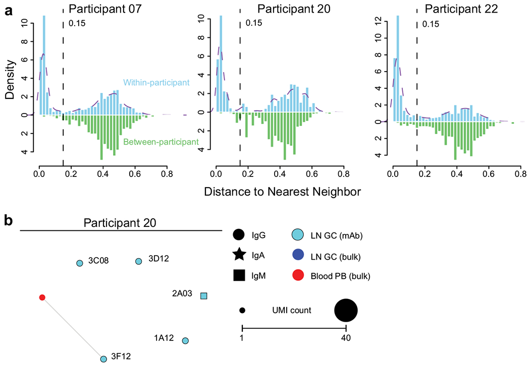
a, Distance-to-nearest-neighbour plots for choosing a distance threshold for inferring clones via hierarchical clustering. After partitioning sequences based on common V and J genes and CDR3 length, the nucleotide Hamming distance of a CDR3 to its nearest nonidentical neighbour from the same participant within its partition was calculated and normalized by CDR3 length (blue histogram). For reference, the distance to the nearest nonidentical neighbour from other participants was calculated (green histogram). A clustering threshold of 0.15 (dashed black line) was chosen via manual inspection and kernel density estimate (dashed purple line) to separate the two modes of the within-participant distance distribution representing, respectively, sequences that were probably clonally related and unrelated. b, Clonal relationship of sequences from S-binding germinal centre-derived monoclonal antibodies (cyan) to sequences from bulk repertoire analysis of plasmablasts sorted from PBMCs (red) and germinal centre B cells (blue) 4 weeks after immunization. Each clone is visualized as a network in which each node represents a sequence and sequences are linked as a minimum spanning tree of the network. Symbol shape indicates sequence isotype: IgG (circle), IgA (star) and IgM (square); symbol size corresponds to sequence count.
Extended Data Fig. 4|. Lymph node plasmablast response to SARS-CoV-2 immunization.
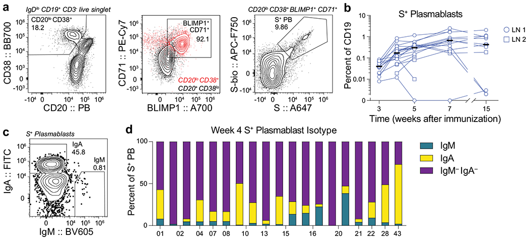
a, c, Representative flow cytometry plots showing gating of CD20lowCD38+ CD71+BLIMP1+S+ plasmablasts from IgDlowCD19+CD3− live singlet lymphocytes (a) and IgA and IgM staining on S+ plasmablasts (c) in FNA samples. b, Kinetics of S+ plasmablasts gated as in a from FNA of draining lymph nodes. Symbols at each time point represent one FNA sample; square symbols denote second lymph node sampled (n = 14). Horizontal lines indicate the median. d, Percentages of IgM+ (teal), IgA+ (yellow) or IgM−IgA− (purple) S+ plasmablasts gated as in c in FNA of draining lymph nodes 4 weeks after primary immunization. Each bar represents one sample (n = 14).
Extended Data Table 1|.
Participant demographics
| Variable | Total N=41 N (%) |
Lymph node N=14 N (%) |
|---|---|---|
| Age (median [range]) | 37 (28-73) | 37 (28-52) |
| Sex | ||
| Female | 18 (43.9) | 8 (57.1) |
| Male | 23 (56.1) | 6 (42.9) |
| Race | ||
| White | 30 (73.2) | 11 (78.6) |
| Asian | 9 (22) | 2 (14.3) |
| Black | 1 (2.4) | 1 (7.1) |
| Other | 1 (2.4) | 0 (0) |
| Ethnicity | ||
| Not of Hispanic, Latinx, or Spanish origin | 39 (95.1) | 13 (92.9) |
| Hispanic, Latinx, Spanish origin | 2 (4.9) | 1 (7.1) |
| BMI (median [range]) | 25.4 (21.4-40) | 23.9 (21.4-40) |
| Comorbidities | ||
| Lung disease | 2 (4.9) | 1 (7.1) |
| Diabetes mellitus | 0 (0) | 0 (0) |
| Hypertension | 7 (17.1) | 2 (14.3) |
| Cardiovascular | 0 (0) | 0 (0) |
| Liver disease | 0 (0) | 0 (0) |
| Chronic kidney disease | 0 (0) | 0 (0) |
| Cancer on chemotherapy | 0 (0) | 0 (0) |
| Hematological malignancy | 0 (0) | 0 (0) |
| Pregnancy | 0 (0) | 0 (0) |
| Neurological | 0 (0) | 0 (0) |
| HIV | 0 (0) | 0 (0) |
| Solid organ transplant recipient | 0 (0) | 0 (0) |
| Bone marrow transplant recipient | 0 (0) | 0 (0) |
| Hyperlipidemia | 1 (2.4) | 0 (0) |
| Confirmed SARS-CoV-2 infection | 8 (19.5) | 0 (0) |
| Time from SARS-CoV-2 infection to baseline visit in days (median [range]) | 122 (50-307) | -- |
Extended Data Table 2|.
Vaccine side effects
| Variable | Total N=41 N (%) |
Total N=41 N (%) |
|
|---|---|---|---|
| First dose | Second dose | ||
| None | 3 (7.3) | None | 1 (2.4) |
| Chills | 5 (12.2) | Chills | 15 (36.6) |
| Fever | 2 (4.9) | Fever | 6 (14.6) |
| Headache | 6 (14.6) | Headache | 11 (26.8) |
| Injection site pain | 33 (80.5) | Injection site pain | 36 (87.8) |
| Muscle or joint pain | 9 (21.9) | Muscle or joint pain | 32 (78) |
| Fatigue | 8 (19.5) | Fatigue | 23 (56.1) |
| Sweating | 0 (0) | Sweating | 2 (4.8) |
|
Duration of side effects in hours (median [range])
| |||
| Chills | 48 (6-72) | Chills | 24 (4-48) |
| Fever | 9 (6-12) | Fever | 24 (1-48) |
| Headache | 9 (3-48) | Headache | 24 (4-48) |
| Injection site pain | 36 (2-120) | Injection site pain | 36 (2-120) |
| Muscle or joint pain | 36 (1-48) | Muscle or joint pain | 24 (1-48) |
| Fatigue | 30 (3-48) | Fatigue | 24 (3-144) |
| Sweating | 0 (0) | Sweating | 18 (18) |
Extended Data Table 3|.
Lymph node population frequencies
| Participant | Week post immunization | LN # | Total GC (%CD19) | S+ GC (%CD19) | S+ PB (%CD19) | CD14 (%live singlet) |
|---|---|---|---|---|---|---|
| 01 | 3 | 1 | 15.1744 | 3.2617 | 0.0718 | 0.2134 |
| 01 | 4 | 1 | 13.7195 | 5.4700 | 0.8436 | 0.0588 |
| 01 | 5 | 1 | 11.1280 | 4.0445 | 1.1611 | 0.1461 |
| 01 | 7 | 1 | 31.1819 | 13.5866 | 1.6436 | 0.1469 |
| 01 | 15 | 1 | 29.0930 | 18.1334 | 2.0446 | 0.1628 |
| 02 | 3 | 1 | 8.2782 | 1.3827 | 0.0132 | 0.1590 |
| 02 | 4 | 1 | 34.1504 | 9.7890 | 0.2102 | 0.1705 |
| 02 | 4 | 2 | 14.0743 | 4.7121 | 0.2521 | 0.1371 |
| 02 | 5 | 1 | 44.6608 | 11.6465 | 0.4086 | 0.0743 |
| 02 | 5 | 2 | 13.5353 | 3.8936 | 0.2893 | 0.1361 |
| 02 | 7 | 1 | 21.8959 | 6.0492 | 0.3725 | 0.2486 |
| 02 | 7 | 2 | 23.1123 | 9.3883 | 0.5557 | 0.7063 |
| 02 | 15 | 1 | 7.1063 | 0.0197 | 0.0020 | 0.1649 |
| 04 | 3 | 1 | 4.6727 | 0.5706 | 0.0538 | 0.1934 |
| 04 | 4 | 1 | 13.9308 | 2.8621 | 0.0885 | 0.3733 |
| 04 | 5 | 1 | 11.3856 | 5.9721 | 0.4296 | 0.7237 |
| 04 | 7 | 1 | 43.8266 | 6.4556 | 0.2019 | 0.1436 |
| 04 | 15 | 1 | 3.7193 | 0.7127 | 0.0025 | 0.9901 |
| 07 | 3 | 1 | 21.0403 | 4.6411 | 0.0697 | 0.1927 |
| 07 | 4 | 1 | 19.6634 | 4.9771 | 0.2599 | 0.1507 |
| 07 | 5 | 1 | 11.3504 | 3.8557 | 0.2576 | 0.4177 |
| 07 | 7 | 1 | 39.2049 | 14.7032 | 0.6765 | 0.6582 |
| 07 | 15 | 1 | 28.9957 | 20.1221 | 0.3248 | 0.0349 |
| 08 | 3 | 1 | 9.9010 | 1.2181 | 0.0651 | 0.2738 |
| 08 | 4 | 1 | 1.3233 | 0.4150 | 0.0904 | 0.1670 |
| 08 | 5 | 1 | 3.9913 | 0.9238 | 0.1507 | 0.3377 |
| 08 | 7 | 1 | 12.1411 | 4.2393 | 0.7559 | 0.8097 |
| 10 | 3 | 1 | 7.7130 | 1.1146 | 0.0399 | 0.2216 |
| 10 | 4 | 1 | 5.9172 | 1.4892 | 0.3494 | 0.0676 |
| 10 | 4 | 2 | 5.7036 | 1.3733 | 0.1867 | 0.0860 |
| 10 | 5 | 1 | 2.8213 | 0.9125 | 0.3428 | 0.0772 |
| 10 | 5 | 2 | 3.4006 | 1.0486 | 0.4156 | 0.0517 |
| 10 | 7 | 1 | 7.3456 | 3.1376 | 0.8776 | 0.2061 |
| 10 | 7 | 2 | 4.2626 | 1.0628 | 0.1663 | 0.1314 |
| 10 | 15 | 1 | 19.2991 | 14.2480 | 1.0759 | 0.0340 |
| 10 | 15 | 2 | 7.0114 | 2.7615 | 0.1849 | 0.0292 |
| 13 | 3 | 1 | 14.8994 | 3.5681 | 0.0199 | 0.5633 |
| 13 | 4 | 1 | 8.5564 | 3.0085 | 0.2116 | 0.3352 |
| 13 | 5 | 1 | 7.1981 | 2.1647 | 0.3036 | 0.4254 |
| 13 | 7 | 1 | 15.9621 | 3.7382 | 0.6532 | 0.3253 |
| 13 | 15 | 1 | 20.1410 | 10.1032 | 0.4304 | 1.6315 |
| 15 | 3 | 1 | 13.0526 | 2.9882 | 0.0838 | 0.0715 |
| 15 | 4 | 1 | 26.8834 | 8.1055 | 0.1196 | 0.1948 |
| 15 | 4 | 2 | 0.3157 | 0.0169 | 0.1039 | 0.1379 |
| 15 | 5 | 1 | 44.5687 | 9.7412 | 0.3016 | 0.0786 |
| 15 | 5 | 2 | 0.6237 | 0.0247 | 0.0098 | 0.2388 |
| 16 | 3 | 1 | 5.1983 | 0.7456 | 0.0195 | 0.2619 |
| 16 | 4 | 1 | 7.6114 | 1.5173 | 0.0510 | 0.1431 |
| 16 | 4 | 2 | 1.2226 | 0.2419 | 0.0190 | 0.1142 |
| 16 | 5 | 1 | 17.0381 | 3.3806 | 0.1261 | 0.0570 |
| 16 | 5 | 2 | 7.4397 | 1.8530 | 0.1101 | 0.1405 |
| 20 | 3 | 1 | 0.4058 | 0.0257 | 0.0076 | 0.2140 |
| 20 | 4 | 1 | 7.4714 | 3.0853 | 0.0115 | 0.1478 |
| 20 | 4 | 2 | 7.8030 | 1.2875 | 0.0505 | 0.1149 |
| 20 | 5 | 1 | 2.0994 | 0.3146 | 0.0000 | 0.2934 |
| 20 | 5 | 2 | 3.3579 | 0.7320 | 0.0811 | 0.1607 |
| 20 | 7 | 1 | 0.5222 | 0.0378 | 0.0000 | 0.4082 |
| 20 | 7 | 2 | 13.6571 | 4.4611 | 0.2402 | 0.1100 |
| 20 | 15 | 1 | 0.4687 | 0.0391 | 0.0033 | 0.8360 |
| 20 | 15 | 2 | 16.9158 | 11.1431 | 0.4222 | 0.2930 |
| 21 | 4 | 1 | 20.1451 | 5.9981 | 0.3831 | 0.1274 |
| 21 | 7 | 1 | 14.4498 | 5.2617 | 1.7381 | 0.1088 |
| 22 | 3 | 1 | 24.7057 | 4.4770 | 0.0352 | 0.1818 |
| 22 | 4 | 1 | 20.6325 | 5.4021 | 0.1707 | 0.1613 |
| 22 | 5 | 1 | 19.6620 | 4.9759 | 0.3147 | 0.2005 |
| 22 | 7 | 1 | 25.8583 | 7.3746 | 0.7954 | 0.3576 |
| 22 | 15 | 1 | 22.0049 | 9.7484 | 0.8297 | 0.2798 |
| 28 | 3 | 1 | 6.3630 | 1.1301 | 0.0372 | 0.1303 |
| 28 | 4 | 1 | 6.2272 | 1.1718 | 0.1281 | 0.2226 |
| 28 | 15 | 1 | 12.7274 | 7.2239 | 1.1391 | 0.6584 |
| 43 | 3 | 1 | 29.0401 | 4.5319 | 0.0505 | 0.5172 |
| 43 | 4 | 1 | 29.3971 | 9.5485 | 0.6432 | 0.3236 |
| 43 | 5 | 1 | 26.0545 | 6.3729 | 0.5631 | 0.9658 |
| 43 | 7 | 1 | 29.4153 | 15.3730 | 2.4781 | 0.0902 |
| 43 | 15 | 1 | 35.1837 | 15.5455 | 0.9446 | 0.0106 |
Extended Data Table 4|.
Immunoglobulin gene usage of S-binding monoclonal antibodies
| Heavy Chain |
Light Chain |
||||||||
|---|---|---|---|---|---|---|---|---|---|
| Name | Native isotype | Gene usage | Nucleotide mutations* | AA mutations | HCDR3 | Gene usage | Nucleotide mutations* | AA mutations | LCDR3 |
| 07.1A11 | IgM | VH3-15 DH1-7 JH4 | 4/283=0.0141 | 4 | TTGWFTGTYGDYFDY | VK1-33 JK4 | 1/267=0.0037 | 1 | QQYDNLPPT |
| 07.1A12 | IgA1 | VH3-49 D2-15 JH4 | 3/281=0.0107 | 3 | TRVKYCSGGSCYGYHFDH | VK3-15 JK3 | 4/262=0/0.0153 | 3 | QQYNNWFT |
| 07.1E10 | IgG1 | VH4-34 D6-19 JH6 | 5/272=0.0184 | 5 | ARVVIAVAGTYPIQVYYYYGMDV | VK1-27 JK1 | 2/266=0.0075 | 2 | QKYNSAPRT |
| 07.1E11 | IgA1 | VH3-30 D3-22 JH6 | 1/278=0.0036 | 1 | AKEEMIEDWGMDV | VL3-1 JL2 | 2/265=0.0075 | 2 | QAWDRSTVV |
| 07.1H09 | IgG1 | VH3-66 DH3-10 JH3 | 3/275=0.0109 | 3 | ARDFREGAFDI | VK1-9 JK4 | 0/266=0 | 0 | QQLNSYPPT |
| 07.2A07 | IgG1 | VH3-21 DH2-21 JH4 | 6/277=0.0217 | 6 | ARAGFVPKRAYCGGDCWYYFDY | VK3-11 JK4 | 0/263=0 | 0 | QQRSNWLT |
| 07.2A08 | IgG1 | VH4-4 DH6-19 JH4 | 2/275=0.0073 | 2 | ATDGGWYTFDH | VL3-1 JL2 | 3/265=0.0113 | 3 | QAWGSSTVV |
| 07.2A10 | IgG1 | VH4-31 DH3-16 JH3 | 1/278=0.0036 | 1 | ARYPVWGAFDI | VK1-33 JK3 | 3/267=0.0112 | 3 | QHYDNLPPT |
| 07.2C08 | IgG1 | VH1-58 DH2-15 JH3 | 2/275=0.0073 | 2 | AAAYCSGGSCSDGFDI | VK3-20 JK1 | 5/266=0.0188 | 5 | QQYGSSPWT |
| 07.2C12 | IgG1 | VH3-30-3 DH1-26 JH3 | 3/277=0.0108 | 3 | ARARGGSYSGAFDI | VK3-20 JK2 | 1/267=0.0037 | 1 | QQYGSSPMYT |
| 07.3D07 | IgG1 | VH3-30-3 DH5-18 JH4 | 3/277=0.0108 | 3 | ARVLWLRGMFDY | VL6-57 JL3 | 2/278=0.0072 | 1 | QSYDISNHWV |
| 07.4A07 | IgG1 | VH3-30-3 DH3-10 JH4 | 3/277=0.0108 | 2 | ARGDYYGSGSYPGKTFDY | VK1-33 JK4 | 1/266=0.0038 | 1 | QQYDNLPLT |
| 07.4B05 | IgG1 | VH1-69 DH1-26 JH5 | 1/277=0.0036 | 1 | ARGRLDSYSGSYYSWFDP | VK4-1 JK2 | 2/283=0.0071 | 2 | QQYYSTPYT |
| 07.4C10 | IgG1 | VH3-23 DH3-22 JH4 | 1/276=0.0036 | 1 | AKNEMAMIVVVITLFDY | VL1-51 JL3 | 2/277=0.0072 | 2 | GTWDRSLSAWV |
| 07.4D09 | IgG1 | VH4-4 DH2-15 JH4 | 1/274=0.0036 | 1 | ATKYCSGGSCSYFGY | VL2-23 JL3 | 0/277=0 | 0 | CSYAGSSTWV |
| 20.1A12 | IgG1 | VH3-30 DH1-26 JH4 | 2/277=0.0072 | 2 | AKGHSGSYRAPFDY | VK3-20 JK2 | 0/263=0 | 0 | QQYGSSYT |
| 20.2A03 | IgM | VH3-33 DH3-10 JH4 | 1/278=0.0036 | 1 | AREAYFGSGSSPDY | VL3-10 JL2 | 2/272=0.0074 | 2 | YSTDSSDNHRRV |
| 20.3C08 | IgG1 | VH3-7 DH3-22 JH4 | 1/278=0.0036 | 1 | AREGTYYYDSSAYYNGGLDY | VL3-10 JL2 | 2/268=0.0075 | 2 | YSTDSGGNPQGV |
| 20.3D12 | IgG1 | VH3-33 DH1-1 JH4 | 7/278=0.0252 | 7 | ATEPVQLEPEVRLDY | VL3-10 JL1 | 1/272=0.0037 | 1 | YSTDSSGNHRRL |
| 20.3F12 | IgG1 | VH3-7 DH4-11 JH4 | 0/278=0 | 0 | ARDQGVTTGPFDY | VL3-1 JL2 | 1/265=0.0038 | 1 | QAWDSSTVV |
| 22.1A11 | IgG3 | VH3-30-3 DH3-16 JH4 | 0/278=0 | 0 | ARDLVVWEELAGGY | VL3-10 JL3 | 2/270=0.0074 | 2 | YSTDSSGNHGV |
| 22.1B04 | IgG1 | VH3-30 DH2-15 JH4 | 4/279=0.0143 | 3 | AKQGGGTYCGGGSCYRGYFDY | VK1-33 JK4 | 2/266=0.0075 | 2 | QQYDNLPLT |
| 22.1B08 | IgA1 | VH1-46 DH4-17 JH3 | 16/278=0.0576 | 15 | ARDPRVPAVTNVNDAFDL | VK3-11 JK2 | 5/267=0.0187 | 5 | QQRSNRPPRWT |
| 22.1B12 | IgG1 | VH3-53 DH3-10 JH4 | 8/273=0.0293 | 5 | ARSHLEVRGVFDN | VK4-1 JK2 | 1/282=0.0035 | 1 | QQYYSTPCS |
| 22.1C02 | IgG1 | VH3-20 DH7-27 JH4 | 5/275=0.0182 | 4 | ARGTGAADY | VK3-20 JK2 | 6/266=0.0226 | 6 | QQYGRSPYT |
| 22.1E07 | IgA1 | VH3-33 DH4-17 JH4 | 3/278=0.0108 | 3 | AREGVYGDIGGAGLDY | VL3-10 JL1 | 4/272=0.0147 | 2 | YSTDSSVNGRV |
| 22.1E11 | IgG1 | VH3-30 DH2-15 JH4 | 2/274=0.0073 | 2 | AKMGGVYCSAGNCYSGRLEY | VK1-33 JK3 | 0/263=0 | 0 | QQYDNLLT |
| 22.1G10 | IgG1 | VH4-59 DH2-21 JH5 | 12/275=0.0436 | 11 | ARETVNNWVDP | VK4-1 JK1 | 10/282=0.0355 | 8 | QQYFTTPWT |
| 22.2A06 | IgG3 | VH5-51 DH3-3 JH4 | 1/277=0.0036 | 1 | ARREWGGSLGHIDY | VL6-57 JL2 | 4/276=0.0145 | 4 | QSFDSSNVV |
| 22.2A08 | IgG1 | VH4-59 DH2-2 JH6 | 4/274=0.0146 | 4 | ARGQGVPAALYGMDV | VL1-40 JL2 | 3/281=0.0107 | 3 | QSYDGSLSGSV |
| 22.2B06 | IgM | VH3-53 DH1-1 JH6 | 2/275=0.0073 | 2 | ARDLQLYGMDV | VL3-21 JL2 | 2/268=0.0075 | 2 | QVWDSSSDPVV |
| 22.2F03 | IgM | VH1-18 DH6-13 JH6 | 1/277=0.0036 | 1 | ARVPGLVGYSSSWYDNEKNYYYYYYGMDV | VL3-25 JL1 | 1/270=0.0037 | 1 | QSADSSGTYV |
| 22.3A06 | IgG1 | VH3-23 DH5-18 JH5 | 2/277=0.0072 | 2 | AKADTAMAWYNWFDP | VK3-11 JK4 | 3/264=0.0114 | 3 | QHRSNWPLT |
| 22.3A11 | IgG1 | VH4-34 DH7-27 JH2 | 1/270=0.0037 | 1 | ARVWVRWWYFDL | VL3-21 JL1 | 4/272=0.0147 | 4 | QVWDNSSDQPNYV |
| 22.3A12 | IgG1 | VH1-46 DH2-21 JH4 | 4/275=0.0145 | 4 | ASSLPARGGVPGRLNY | VL1-51 JL2 | 3/277=0.0108 | 3 | GTWDSSLSVVV |
| 22.3D11 | IgG1 | VH5-51 D4-17 JH5 | 1/278=0.0036 | 1 | ARHHLDYDDYVGHWFDP | VL1-40 JL3 | 0/281=0 | 0 | QSYDSSLSGSGV |
| 22.3F08 | IgG1 | VH1-18 DH2-2 JH6 | 4/275=0.0145 | 4 | ASCPRRPAAIGDYYGMDV | VL3-21 JL7 | 1/271=0.0037 | 1 | QVWDSSSYHAV |
V-region nonsynonymous nucleotide substitutions
Extended Data Table 5|.
Template switch sequences, constant region primers and isotype-specific internal constant region sequences for bulk BCR sequencing and processing
| Template switch sequences | |
|---|---|
|
| |
| TS-shift0 | TACGGG |
| TS-shift1 | ATACGGG |
| TS-shift2 | TCTACGGG |
| TS-shift3 | CGATACGGG |
| TS-shift4 | GATCTACGGG |
|
| |
| Constant region primers | |
|
| |
| Human-IGHM | GAATTCTCACAGGAGACGAGG |
| Human-IGHD | TGTCTGCACCCTGATATGATGG |
| Human-IGHA | GGGTGCTGYMGAGGCTCAG |
| Human-IGHE | TTGCAGCAGCGGGTCAAGG |
| Human-IGHG | CCAGGGGGAAGACSGATG |
| Human-IGK | GACAGATGGTGCAGCCACAG |
| Human-IGL | AGGGYGGGAACAGAGTGAC |
|
| |
| Isotype-specific internal constant region sequences | |
|
| |
| Human-IGHA-InternalC | GGCTGGTCGGGGATGC |
| Human-IGHD-InternalC | GAGCCTTGGTGGGTGC |
| Human-IGHE-InternalC | GGCTCTGTGTGGAGGC |
| Human-IGHG-InternalC | GGCCCTTGGTGGARGC |
| Human-IGHM-InternalC | GGGCGGATGCACTCCC |
| Human-IGKC-IGKJ-InternalC | TTCGTTTRATHTCCAS |
| Human-IGLC-1-InternalC | TGGGGTTGGCCTTGGG |
| Human-IGLC-2-InternalC | AGGGGGCAGCCTTGGG |
| Human-IGLC-3-InternalC | YRGCCTTGGGCTGACC |
| Human-IGLC-4-InternalC | GCTGCCAAACATGTGC |
Extended Data Table 6|.
Processing of bulk sequencing BCR reads
| Participant | Sample | Cell Count | Sequence Count |
|||
|---|---|---|---|---|---|---|
| Input | Preprocessed | Post-QC Productive Heavy Chains | Unique Heavy Chain VDJs | |||
| 07 | d28 blood plasmablast | 8361 | 2307288 | 8294 | 6031 | 3014 |
| 20 | d28 blood plasmablast | 6136 | 2068139 | 2320 | 1453 | 951 |
| 22 | d28 blood plasmablast | 15496 | 1801330 | 16126 | 13733 | 6266 |
| 07 | d28 lymph node germinal centre | 25754 | 1104539 | 2429 | 1700 | 1211 |
| 22 | d28 lymph node germinal centre | 10236 | 2117620 | 552 | 364 | 268 |
Supplementary Material
Acknowledgements
We thank the donors for providing specimens, L. Kessels, A. J. Winingham, K. Safi and the staff of the Infectious Diseases Clinical Research Unit at Washington University School of Medicine for assistance with scheduling participants and sample collection, and E. Lantelme for facilitating sorting. The Vero-TMPRSS2 cells were provided by S. Ding. The expression plasmids encoding the S of coronaviruses OC43 and HKU1 were provided by K. Corbett and B. Graham. The laboratory of A.H.E. was supported by National Institute of Allergy and Infectious Diseases (NIAID) grants U01AI141990 and 1U01AI150747 and NIAID Centers of Excellence for Influenza Research and Surveillance (CEIRS) contract HHSN272201400006C. The laboratories of A.H.E. and F.K. were supported by NIAID CEIRS contract HHSN272201400008C and NIAID Collaborative Influenza Vaccine Innovation Centers contract 75N93019C00051. The laboratory of M.S.D. was supported by R01 AI157155. The laboratory of P.Y.-S. was supported by NIH grants AI134907 and UL1TR001439, and awards from the Sealy & Smith Foundation, Kleberg Foundation, the John S. Dunn Foundation, the Amon G. Carter Foundation, the Gilson Longenbaugh Foundation and the Summerfield Robert Foundation. J.S.T. was supported by NIAID 5T32CA009547. J.B.C. was supported by a Helen Hay Whitney Foundation postdoctoral fellowship. The WU368 study was reviewed and approved by the Washington University Institutional Review Board (approval no. 202012081).
Competing interests
The laboratory of A.H.E. received funding under sponsored research agreements that are unrelated to the data presented in the current study from Emergent BioSolutions and from AbbVie. A.H.E. is a consultant for Mubadala Investment Company and the founder of ImmuneBio Consulting. J.S.T., A.J.S., M.S.D. and A.H.E. are recipients of a licensing agreement with Abbvie that is unrelated to the data presented in the current study. M.S.D. is a consultant for Inbios, Vir Biotechnology, Fortress Biotech and Carnival Corporation, and on the Scientific Advisory Boards of Moderna and Immunome. The laboratory of M.S.D. has received unrelated funding support in sponsored research agreements from Moderna, Vir Biotechnology and Emergent BioSolutions. A patent application related to this work has been filed by Washington University School of Medicine. The Icahn School of Medicine at Mount Sinai has filed patent applications relating to SARS-CoV-2 serological assays and NDV-based SARS-CoV-2 vaccines which list F.K. as co-inventor. Mount Sinai has spun out a company, Kantaro, to market serological tests for SARS-CoV-2. F.K. has consulted for Merck and Pfizer (before 2020), and is currently consulting for Pfizer, Seqirus and Avimex. The Laboratory of F.K. is also collaborating with Pfizer on animal models of SARS-CoV-2. The laboratory of P.-Y.S. has received sponsored research agreements from Pfizer, Gilead, Merck and IGM Sciences Inc. The content of this manuscript is solely the responsibility of the authors and does not necessarily represent the official view of NIAID or NIH.
Footnotes
Online content
Any methods, additional references, Nature Research reporting summaries, source data, extended data, supplementary information, acknowledgements, peer review information; details of author contributions and competing interests; and statements of data and code availability are available at https://doi.org/10.1038/s41586-021-03738-2.
Reporting summary
Further information on research design is available in the Nature Research Reporting Summary linked to this paper.
Supplementary information The online version contains supplementary material available at https://doi.org/10.1038/s41586-021-03738-2.
Peer review information Nature thanks the anonymous reviewers for their contribution to the peer review of this work. Peer reviewer reports are available.
Data availability
Antibody sequences are deposited on GenBank under the following accession numbers: MW926396–MW926407, MW926409–MW926430, MW926432–MW926441 and MZ292481–MZ292510, available from Gen-Bank/EMBL/DDBJ. Bulk sequencing reads are deposited on Sequence Read Archive under BioProject PRJNA731610. Processed B cell receptor data are deposited at https://doi.org/10.5281/zenodo.5042252. The IMGT/V-QUEST database is accessible at http://www.imgt.org/IMGT_vquest/. Other relevant data are available from the corresponding authors upon request.
References
- 1.Mulligan MJ et al. Phase I/II study of COVID-19 RNA vaccine BNT162b1 in adults. Nature 586, 589–593 (2020). [DOI] [PubMed] [Google Scholar]
- 2.Jackson LA et al. An mRNA vaccine against SARS-CoV-2 – preliminary report. N. Engl. J. Med 383, 1920–1931 (2020). [DOI] [PMC free article] [PubMed] [Google Scholar]
- 3.Sahin U et al. COVID-19 vaccine BNT162b1 elicits human antibody and TH1 T cell responses. Nature 586, 594–599 (2020). [DOI] [PubMed] [Google Scholar]
- 4.Polack FP et al. Safety and efficacy of the BNT162b2 mRNA Covid-19 vaccine. N. Engl. J. Med 383, 2603–2615 (2020). [DOI] [PMC free article] [PubMed] [Google Scholar]
- 5.Baden LR et al. Efficacy and safety of the mRNA-1273 SARS-CoV-2 vaccine. N. Engl. J. Med 384, 403–416 (2021). [DOI] [PMC free article] [PubMed] [Google Scholar]
- 6.Zhou X et al. Self-replicating Semliki forest virus RNA as recombinant vaccine. Vaccine 12, 1510–1514 (1994). [DOI] [PubMed] [Google Scholar]
- 7.Cagigi A & Loré K Immune responses induced by mRNA vaccination in mice, monkeys and humans. Vaccines 9, 61 (2021). [DOI] [PMC free article] [PubMed] [Google Scholar]
- 8.Karikó K et al. Incorporation of pseudouridine into mRNA yields superior nonimmunogenic vector with increased translational capacity and biological stability. Mol. Ther 16, 1833–1840 (2008). [DOI] [PMC free article] [PubMed] [Google Scholar]
- 9.Schlake T, Thess A, Fotin-Mleczek M & Kallen K-J Developing mRNA-vaccine technologies. RNA Biol. 9, 1319–1330 (2012). [DOI] [PMC free article] [PubMed] [Google Scholar]
- 10.Graham BS, Mascola JR & Fauci AS Novel vaccine technologies: essential components of an adequate response to emerging viral diseases. J. Am. Med. Assoc 319, 1431–1432 (2018). [DOI] [PubMed] [Google Scholar]
- 11.Bettini E & Locci M SARS-CoV-2 mRNA vaccines: immunological mechanism and beyond. Vaccines 9, 147 (2021). [DOI] [PMC free article] [PubMed] [Google Scholar]
- 12.Amit S, Regev-Yochay G, Afek A, Kreiss Y & Leshem E Early rate reductions of SARS-CoV-2 infection and COVID-19 in BNT162b2 vaccine recipients. Lancet 397, 875–877 (2021). [DOI] [PMC free article] [PubMed] [Google Scholar]
- 13.Dagan N et al. BNT162b2 mRNA Covid-19 vaccine in a nationwide mass vaccination setting. N. Engl. J. Med 384, 1412–1423 (2021). [DOI] [PMC free article] [PubMed] [Google Scholar]
- 14.Vasileiou E et al. Interim findings from first-dose mass COVID-19 vaccination roll-out and COVID-19 hospital admissions in Scotland: a national prospective cohort study. Lancet 397, 1646–1657 (2021). [DOI] [PMC free article] [PubMed] [Google Scholar]
- 15.Case JB et al. Neutralizing antibody and soluble ACE2 inhibition of a replication-competent VSV-SARS-CoV-2 and a clinical isolate of SARS-CoV-2. Cell Host Microbe 28, 475–485.e5 (2020). [DOI] [PMC free article] [PubMed] [Google Scholar]
- 16.Liu Y et al. Neutralizing activity of BNT162b2-elicited serum. N. Engl. J. Med 384, 1466–1468 (2021). [DOI] [PMC free article] [PubMed] [Google Scholar]
- 17.Chen RE et al. Resistance of SARS-CoV-2 variants to neutralization by monoclonal and serum-derived polyclonal antibodies. Nat. Med 27, 717–726 (2021). [DOI] [PMC free article] [PubMed] [Google Scholar]
- 18.Leung K, Shum MH, Leung GM, Lam TT & Wu JT Early transmissibility assessment of the N501Y mutant strains of SARS-CoV-2 in the United Kingdom, October to November 2020. Euro Surveill. 26, 2002106 (2021). [DOI] [PMC free article] [PubMed] [Google Scholar]
- 19.Turner JS et al. Human germinal centres engage memory and naive B cells after influenza vaccination. Nature 586, 127–132 (2020). [DOI] [PMC free article] [PubMed] [Google Scholar]
- 20.Pardi N et al. Expression kinetics of nucleoside-modified mRNA delivered in lipid nanoparticles to mice by various routes. J. Control. Release 217, 345–351 (2015). [DOI] [PMC free article] [PubMed] [Google Scholar]
- 21.Liang F et al. Efficient targeting and activation of antigen-presenting cells in vivo after modified mRNA vaccine administration in rhesus macaques. Mol. Ther 25, 2635–2647 (2017). [DOI] [PMC free article] [PubMed] [Google Scholar]
- 22.Tai W et al. A novel receptor-binding domain (RBD)-based mRNA vaccine against SARS-CoV-2. Cell Res. 30, 932–935 (2020). [DOI] [PMC free article] [PubMed] [Google Scholar]
- 23.Lederer K et al. SARS-CoV-2 mRNA vaccines foster potent antigen-specific germinal center responses associated with neutralizing antibody generation. Immunity 53, 1281–1295.e5 (2020). [DOI] [PMC free article] [PubMed] [Google Scholar]
- 24.Kaji T et al. Distinct cellular pathways select germline-encoded and somatically mutated antibodies into immunological memory. J. Exp. Med 209, 2079–2097 (2012). [DOI] [PMC free article] [PubMed] [Google Scholar]
- 25.Weisel FJ, Zuccarino-Catania GV, Chikina M & Shlomchik MJ A Temporal switch in the germinal center determines differential output of memory B and plasma cells. Immunity 44, 116–130 (2016). [DOI] [PMC free article] [PubMed] [Google Scholar]
- 26.Good-Jacobson KL et al. Regulation of germinal center responses and B-cell memory by the chromatin modifier MOZ. Proc. Natl Acad. Sci. USA 111, 9585–9590 (2014). [DOI] [PMC free article] [PubMed] [Google Scholar]
- 27.Bachmann MF, Odermatt B, Hengartner H & Zinkernagel RM Induction of long-lived germinal centers associated with persisting antigen after viral infection. J. Exp. Med 183, 2259–2269 (1996). [DOI] [PMC free article] [PubMed] [Google Scholar]
- 28.Dogan I et al. Multiple layers of B cell memory with different effector functions. Nat. Immunol 10, 1292–1299 (2009). [DOI] [PubMed] [Google Scholar]
- 29.Rothaeusler K & Baumgarth N B-cell fate decisions following influenza virus infection. Eur. J. Immunol 40, 366–377 (2010). [DOI] [PMC free article] [PubMed] [Google Scholar]
- 30.Pape KA, Taylor JJ, Maul RW, Gearhart PJ & Jenkins MK Different B cell populations mediate early and late memory during an endogenous immune response. Science 331, 1203–1207 (2011). [DOI] [PMC free article] [PubMed] [Google Scholar]
- 31.Turner JS, Benet ZL & Grigorova I Transiently antigen primed B cells can generate multiple subsets of memory cells. PLoS ONE 12, e0183877 (2017). [DOI] [PMC free article] [PubMed] [Google Scholar]
- 32.Havenar-Daughton C et al. Rapid germinal center and antibody responses in non-human primates after a single nanoparticle vaccine immunization. Cell Rep. 29, 1756–1766.e8 (2019). [DOI] [PMC free article] [PubMed] [Google Scholar]
- 33.Cirelli KM et al. Slow delivery immunization enhances HIV neutralizing antibody and germinal center responses via modulation of immunodominance. Cell 177, 1153–1171.e28 (2019). [DOI] [PMC free article] [PubMed] [Google Scholar]
- 34.Qi H, Cannons JL, Klauschen F, Schwartzberg PL & Germain RN SAP-controlled T-B cell interactions underlie germinal centre formation. Nature 455, 764–769 (2008). [DOI] [PMC free article] [PubMed] [Google Scholar]
- 35.Johnston RJ et al. Bcl6 and Blimp-1 are reciprocal and antagonistic regulators of T follicular helper cell differentiation. Science 325, 1006–1010 (2009). [DOI] [PMC free article] [PubMed] [Google Scholar]
- 36.Schmitz AJ et al. A public vaccine-induced human antibody protects against SARS-CoV-2 and emerging variants. Preprint at 10.1101/2021.03.24.436864 (2021). [DOI] [PMC free article] [PubMed]
- 37.Amanat F et al. SARS-CoV-2 mRNA vaccination induces functionally diverse antibodies to NTD, RBD and S2. Cell 10.1016/j.cell.2021.06.005 (2021 [DOI] [PMC free article] [PubMed] [Google Scholar]
- 38.Zang R et al. TMPRSS2 and TMPRSS4 promote SARS-CoV-2 infection of human small intestinal enterocytes. Sci. Immunol 5, eabc3582 (2020). [DOI] [PMC free article] [PubMed] [Google Scholar]
- 39.Plante JA et al. Spike mutation D614G alters SARS-CoV-2 fitness. Nature 592, 116–121 (2021). [DOI] [PMC free article] [PubMed] [Google Scholar]
- 40.Xie X et al. An infectious cDNA clone of SARS-CoV-2. Cell Host Microbe 27, 841–848.e3 (2020). [DOI] [PMC free article] [PubMed] [Google Scholar]
- 41.Stadlbauer D et al. SARS-CoV-2 seroconversion in humans: a detailed protocol for a serological assay, antigen production, and test setup. Curr. Protoc. Microbiol 57, e100 (2020). [DOI] [PMC free article] [PubMed] [Google Scholar]
- 42.Pallesen J et al. Immunogenicity and structures of a rationally designed prefusion MERS-CoV spike antigen. Proc. Natl Acad. Sci. USA 114, E7348–E7357 (2017). [DOI] [PMC free article] [PubMed] [Google Scholar]
- 43.Liu Z et al. Identification of SARS-CoV-2 spike mutations that attenuate monoclonal and serum antibody neutralization. Cell Host Microbe 29, 477–488.e4 (2021). [DOI] [PMC free article] [PubMed] [Google Scholar]
- 44.Wrammert J et al. Broadly cross-reactive antibodies dominate the human B cell response against 2009 pandemic H1N1 influenza virus infection. J. Exp. Med 208, 181–193 (2011). [DOI] [PMC free article] [PubMed] [Google Scholar]
- 45.Smith K et al. Rapid generation of fully human monoclonal antibodies specific to a vaccinating antigen. Nat. Protoc 4, 372–384 (2009). [DOI] [PMC free article] [PubMed] [Google Scholar]
- 46.Wrammert J et al. Rapid cloning of high-affinity human monoclonal antibodies against influenza virus. Nature 453, 667–671 (2008). [DOI] [PMC free article] [PubMed] [Google Scholar]
- 47.Nachbagauer R et al. broadly reactive human monoclonal antibodies elicited following pandemic H1N1 influenza virus exposure protect mice against highly pathogenic H5N1 challenge. J. Virol 92, 1–17 (2018). [DOI] [PMC free article] [PubMed] [Google Scholar]
- 48.Brochet X, Lefranc M-P & Giudicelli V IMGT/V-QUEST: the highly customized and integrated system for IG and TR standardized V-J and V-D-J sequence analysis. Nucleic Acids Res. 36, W503–W508 (2008). [DOI] [PMC free article] [PubMed] [Google Scholar]
- 49.Giudicelli V, Brochet X & Lefranc M-P IMGT/V-QUEST: IMGT standardized analysis of the immunoglobulin (IG) and T cell receptor (TR) nucleotide sequences. Cold Spring Harb. Protoc 2011, pdb.prot5633 (2011). [DOI] [PubMed] [Google Scholar]
- 50.Camacho C et al. BLAST+: architecture and applications. BMC Bioinformatics 10, 421 (2009). [DOI] [PMC free article] [PubMed] [Google Scholar]
- 51.Vander Heiden JA et al. pRESTO: a toolkit for processing high-throughput sequencing raw reads of lymphocyte receptor repertoires. Bioinformatics 30, 1930–1932 (2014). [DOI] [PMC free article] [PubMed] [Google Scholar]
- 52.Jiang R et al. Thymus-derived B cell clones persist in the circulation after thymectomy in myasthenia gravis. Proc. Natl Acad. Sci. USA 117, 30649–30660 (2020). [DOI] [PMC free article] [PubMed] [Google Scholar]
- 53.Li W & Godzik A Cd-hit: a fast program for clustering and comparing large sets of protein or nucleotide sequences. Bioinformatics 22, 1658–1659 (2006). [DOI] [PubMed] [Google Scholar]
- 54.Ye J, Ma N, Madden TL & Ostell JM IgBLAST: an immunoglobulin variable domain sequence analysis tool. Nucleic Acids Res. 41, W34–W40 (2013). [DOI] [PMC free article] [PubMed] [Google Scholar]
- 55.Giudicelli V, Chaume D & Lefranc MP IMGT/GENE-DB: a comprehensive database for human and mouse immunoglobulin and T cell receptor genes. Nucleic Acids Res. 33, D256–D261 (2005). [DOI] [PMC free article] [PubMed] [Google Scholar]
- 56.Gupta NT et al. Change-O: a toolkit for analyzing large-scale B cell immunoglobulin repertoire sequencing data. Bioinformatics 31, 3356–3358 (2015). [DOI] [PMC free article] [PubMed] [Google Scholar]
- 57.Gadala-Maria D, Yaari G, Uduman M & Kleinstein SH Automated analysis of high-throughput B-cell sequencing data reveals a high frequency of novel immunoglobulin V gene segment alleles. Proc. Natl Acad. Sci. USA 112, E862–E870 (2015). [DOI] [PMC free article] [PubMed] [Google Scholar]
- 58.Gupta NT et al. Hierarchical clustering can identify B cell clones with high confidence in Ig repertoire sequencing data. J. Immunol 198, 2489–2499 (2017). [DOI] [PMC free article] [PubMed] [Google Scholar]
- 59.Zhou JQ & Kleinstein SH Cutting edge: Ig H chains are sufficient to determine most B cell clonal relationships. J. Immunol 203, 1687–1692 (2019). [DOI] [PMC free article] [PubMed] [Google Scholar]
- 60.Bashford-Rogers RJM et al. Network properties derived from deep sequencing of human B-cell receptor repertoires delineate B-cell populations. Genome Res. 23, 1874–1884 (2013). [DOI] [PMC free article] [PubMed] [Google Scholar]
- 61.Csardi G & Nepusz T The igraph software package for complex network research. InterJournal Complex Syst. 1695, 1–9 (2006). [Google Scholar]
Associated Data
This section collects any data citations, data availability statements, or supplementary materials included in this article.
Supplementary Materials
Data Availability Statement
Antibody sequences are deposited on GenBank under the following accession numbers: MW926396–MW926407, MW926409–MW926430, MW926432–MW926441 and MZ292481–MZ292510, available from Gen-Bank/EMBL/DDBJ. Bulk sequencing reads are deposited on Sequence Read Archive under BioProject PRJNA731610. Processed B cell receptor data are deposited at https://doi.org/10.5281/zenodo.5042252. The IMGT/V-QUEST database is accessible at http://www.imgt.org/IMGT_vquest/. Other relevant data are available from the corresponding authors upon request.


