Abstract
Bimetallic nanoparticles offer unique chemical, physical and optical properties that are not available for monometallic nanoparticles. Bimetallic nanoparticles play a major role in various therapeutic, industrial and energy fields. Recently, nanoparticles of Copper/Zinc bimetallic nanoparticles have attracted attention in various fields, especially medicine. In this study, bimetallic CuO/ZnO nanostructures were biosynthesized using plant extracts. The plant-mediated synthesis nanoparticles were characterized by Transmission electron microscopy (TEM), X-ray diffraction analysis (XRD), Field Emission Scanning Electron Microscopy (FESEM) and Energy-Dispersive Spectroscopy (EDAX). The cytotoxicity of plant-mediated synthesis bimetallic nanoparticles and the synergistic effects of these nanoparticles in combination with the anticancer drug doxorubicin on MCF-7 cancer cells were evaluated by MTT assay.
Keywords: Cytotoxicity, Environmentally friendly, Copper/Zinc bimetallic nanoparticle, MCF-7 cancer cells, MTT assay
Introduction
Nanotechnology is a novel technology that has spread rapidly due to its transformative, miraculous and incredible effects (Cao et al. 2021a; Chu et al. 2021). Nanotechnology has wide applications in various fields from food (Ningthoujam et al. 2022; Parimaladevi et al. 2018), medicine (Chu et al. 2022), medical diagnostics (Nazari-Vanani et al. 2019) and biotechnology (Kannan et al. 2021; Rabiee et al. 2021) to electronics (Song et al. 2015), computers (Zhao et al. 2021), communications (Zhao, Khan 2021), transportation (Mathew et al. 2019), energy (Dey et al. 2022), environment (Kannan et al. 2020; Liu et al. 2022), materials (Cao et al. 2021b; Wang et al. 2022), aerospace (Haynes and Asmatulu 2013), and national security (Zha 2021). Currently, the special properties of these nanomaterials such as magnetic properties, quantum size, optical and biological properties have been considered by scientists (Luo et al. 2021; Wang et al. 2022; Rahdar et al. 2021a; Rahdar et al. 2021b). Nanomaterials are one of the promising areas of medical research and have been used to diagnostic and treatment of many dangerous diseases, including COVID-19 (Tang et al. 2021). Among the types of nanoparticles, bimetallic nanostructure from the combination of two different metals have unique properties in the field of light, heat, catalytic, drug delivery and therapeutic effects (Alijani et al. 2020). The physical and chemical properties of bimetallic and tri-metal nanoparticles are different from the properties of individual metals due to the compositions and interactions between the individual components of these nanostructures (Khatami et al. 2018; Wang et al. 2018). Nanoparticle compounds lead to the search and design of new nanostructure with unique properties that attract the attention of scientists in various fields of medicine and academia. On the other hand, due to drug resistance, inefficiency of current treatments, including side effects and high costs for the treatment of various infectious diseases, autoimmunity and cancer, researchers have turned to the use of nanoparticles in medical treatments (Gao et al. 2019; Zhang 2018).
Scientists provide numerous examples of preparing bimetallic nanoparticles using physical and chemical methods (Yang et al. 2019; Gao et al. 2021; Öztürk et al. 2020). Physical and chemical methods are mainly dependent on expensive and toxic equipment and reagents (Benkai et al. 2016; Rahdar et al. 2022; Wang et al. 2021). Since there are major problems in the methods of physical and chemical synthesis of nanostructures, it is essential to use safe, cost-effective methods without applying harmful chemical compounds (Haghighat et al. 2021; Li et al. 2020; Zhang et al. 2022). In the meantime, the biosynthesis approach has been reported as an easy, environmentally friendly and cost-effective method for the synthesis of nanoparticles using plant extracts. Green synthesis of nanoparticles using plant extracts has some benefits such as lower cost, environmental friendliness and the possibility of cheap and easy production on an industrial scale. In addition, in green synthesis methods, unlike the main chemical methods, there is no need to use high temperature and pressure and several chemical compounds, so this approach as an economical, valuable and environmentally friendly method can be used to produce metal particles should be used on an industrial scale. In green synthesis of nanostructure using plant extracts, these extracts act as a reducing agent and also as a coating agent in the process of synthesis and stability of nanoparticles (Khatami et al. 2021).
Copper is a metallic element with high ductility and thermal and electrical conductivity (Peralta 1994). Copper is an essential mineral for living organs because it plays a key role in the production of the respiratory enzyme of cytochrome oxidase C (Kulandaivel et al. 2022; Stern 2010). Copper nanoparticles exhibit unique properties such as catalytic, antifungal (Asghar et al. 2018) and antibacterial activity (Parimaladevi et al. 2018) that are not found in commercial copper. Copper oxide nanoparticles with apoptotic effect (Chakraborty and Basu 2017) on cancer cells are desirable anticancer agents (Chakraborty and Basu 2017). Zinc is a rare trace mineral that has the highest levels in the body after iron (Allai et al. 2022). Zinc is found in Zinc Finger Proteins and enzymes such as Superoxide Dismutase (Kaneda-Nakashima et al. 2022; Khan and Malik 2022). Research shows that there is a direct relationship between the amount of zinc in the body and the inflammation of cells. Reducing the amount of zinc in the body stimulates immune cells. In this way, the lack of zinc in the body causes inflammation in the cells of the body. One of the severe side effects of zinc deficiency in the body is the stimulation of cancer cells and inflammation of some cells (Yang et al. 2017; Prasad 2008). Research has shown that zinc is an important therapeutic agent in the control and treatment of cancer, as high concentrations increase apoptosis in cancer cells. It is also known as a tissue and skin repairer and is widely used in cosmetics (Aflatoonian et al. 2017; Bisht and Rayamajhi 2016).
In this study, Copper/Zinc bimetallic nanostructures were biosynthesized using plant extracts. The characterization of the biosynthesized nanostructure was studied by XRD SEM EDS and TEM analyses. Finally, the anticancer properties of bimetallic nanoparticles synthesized against breast cancer cells were evaluated by MTT method.
Materials and methods
Preparation of Copper/Zinc bimetallic nanoparticles
Copper/Zinc bimetallic nanoparticles were produced with Lonicera caprifolium plant extract. 7 g of plant powder was mixed with 50 mL of deionized water (sterile) and stirred at 25 °C (24 h). It was then placed at 100 °C for 15 min. The prepared extract was finally filtered. 3.4 g of Zn (CH3COO)2 * 2H2O (Merck, ≤ 100%) and 1.4 g of CuCl2 * 2H2O (Merck, ≥ 99%) were added to 200 mL of the extract at 80 °C and dissolved; then, the pH of the mixture was increased to 7.5 by adding NaOH (Merck) ≥ 99%). The reaction mixture was sterilized for 3 h at 80 °C. After washing with deionized water, it was dried at 80 °C and calcined at 500 °C for 5 h (Chu et al. 2022).
Characterization of Copper/Zinc bimetallic nanoparticles
The chemical composition and morphology of the Copper/Zinc bimetallic nanoparticles synthesized were evaluated using the Sigma VP microscope (ZEISS German). The Copper/Zinc bimetallic nanoparticles morphology was studied by TEM microscope Tecnai G2 Spirit (Twin; FEI, Czech Republic) at voltage of 120 kV. XRD analysis was performed to determine the crystal phase and crystal size of Copper/Zinc bimetallic nanoparticles using Panalytical Holland X’PertPro at an angle of 2θ from 10° to 80°. This analysis was performed with a Cu Kα anode of 1.54 angstroms. The particle size was calculated using Debye–Scherrer formula:
In the Debye–Scherrer formula, the K components are the crystal shape coefficient (0.9), ʎ is the wavelength of X-ray (0.154 nm), θ is the diffraction angle, and the beta peak width at half of the maximum height.
MTT assay
MCF-7 cancer cells were prepared from Pasteur Institute in Tehran, Iran. Cells in Dulbecco’s Modified Eagle’s Medium (DMEM; GIBCO) medium containing 10% FBS; GIBCO and 1% penicillin/streptomycin antibiotic solution were cultured and stored at 37 °C and 5% carbon dioxide. After reaching a density of 90%, the cells were treated with different concentrations (0–600 µg/mL) of biogenic Copper/Zinc bimetallic. Then, the effect of inhibiting the growth of cancer cell line was measured by MTT method. The optical absorption of Formazan crystals after dissolution in DMSO: Dimethyl sulfoxide was recorded at 570 nm with the help of ELISA.
Results
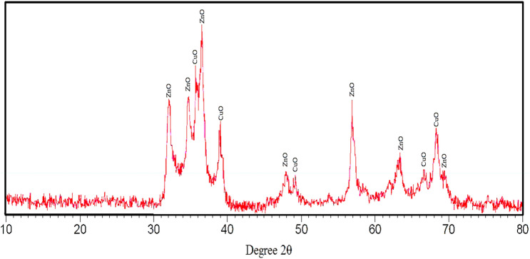
Figure 1 shows the XRD spectrum of the resulting synthesized Copper/Zinc bimetallic nanoparticles. In the XRD spectrum, distinct and sharp peaks are observed in the range of 31.9, 34.5, 35.5, 36.3, 38.8, 47.7, 56.7, 63.01, 66.5 and 68.1 degrees. Peaks of 35.5, 38.8, 66.5 and 68.4 degrees correspond to (002), (111), (311) and (220) Plane of pure CuO nanoparticles, respectively (Cao et al. 2021b). This single-phase with monoclinic structure confirms copper oxide nanoparticles. The strongest peak is at 2θ = 35.5 degrees, which according to the Debye–Scherrer formula, the average crystallite size of copper oxide nanoparticles is below that 50 nm. Peaks of 31.9, 34.5, 36.3, 47.7, 56.7, and 63.01 degrees correspond to (100), (002), (101), (102), and (110) planes of pure ZnO nanoparticles, respectively. This confirms the hexagonal wurtzite structure of zinc oxide nanoparticles (Bisht and Rayamajhi 2016). The strongest peak is at 2θ = 36.3 degrees, which according to the Debye–Scherrer formula, the average crystallite size of zinc oxide nanoparticles is below that 50 nm.
Fig. 1.
XRD patterns of synthesized Copper/Zinc bimetallic nanoparticles
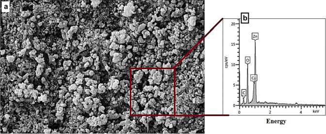
Figure 2 shows the results of the FESEM-EDS analysis. The surface morphology of bimetallic nanoparticles with a magnitude of 20.0 Kx is shown in Fig. 2a. Spherical like nanoparticles can be seen in the image. The synthesized biogenic nanoparticles contain zinc, copper, oxygen and carbon with weight percentages of 50.2, 16.7, 19.3 and 13.8 Wt%, respectively. (Fig. 2b) The presence of carbon element corresponds to the organic precursor present in the synthesis steps (plant extract).
Fig. 2.
FESEM-EDS micrographs of the Copper/Zinc bimetallic nanoparticles: a SEM image and EDS element (b)
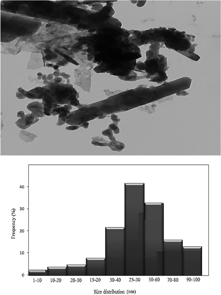
Figure 3 shows a TEM micrograph (a) and size distribution chart (b) of Copper/Zinc bimetallic nanoparticles with a bright-field background. Different types of rod, spherical and polyhedral Copper/Zinc bimetallic nanoparticles with different orientations are shown. This is consistent with the SEM results. As can be clearly seen in the picture, in some areas, the spherical nanoparticles are placed in order.
Fig. 3.
TEM micrograph of Copper/Zinc bimetallic nanoparticles (a) and size distribution chart (b)
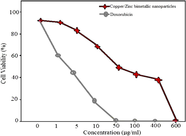
The cytotoxic effects of Copper/Zinc bimetallic nanoparticles on MCF-7 cell line were analyzed using MTT assay. The cytotoxic effects of anticancer drug (doxorubicin) were studied. The cytotoxic effects activity of Copper/Zinc bimetallic nanoparticles on MCF-7 cell line were studied. The IC50 was about 54 µg/mL. These NPs showed toxic effects against MCF-7 cell line, and at a concentration of more 600 µg/mL, the cytotoxic effects reached 100% (Fig. 4).
Fig. 4.

The MCF-7 cell viability: at various concentrations of Copper/Zinc bimetallic nanoparticles and anticancer drug
Discussion
In this study, Copper/Zinc bimetallic nanostructures were synthesized and characterized using plant extracts. The cytotoxicity of bimetallic nanostructures synthesized alone and the synergistic effects of these nanoparticles in combination with the anticancer drug doxorubicin on MCF-7 cancer cells were evaluated. Among nanoparticles, the effect of monometallic nanoparticles in various fields, including medical and industrial, is less than that of bimetallic and multimetallic nanoparticles. At present, in the treatment of disease, monometallic nanoparticles do not achieve the desired result. Therefore, according to the desired purpose of combining monometallic nanoparticles, they increase the effectiveness of bimetallic and trimetallic nanoparticles produced (Ali et al. 2021). In this study, Copper/Zinc bimetallic nanostructures were biosynthesized using green chemistry. Its physicochemical properties were evaluated using XRD SEM EDS TEM analysis. The synthesized bimetallic nanoparticles are completely pure. The structure of zinc oxide wurtzite and single-phase copper oxide was confirmed by XRD analysis. Copper/Zinc bimetallic nanoparticles have particles with different morphologies from spherical to rod and polyhedral. Bar and polyhedral structures increase the volume to the surface area of nanoparticles and increase the impact and contact surface.
Some synthesis of bimetallic nanoparticles was reported previously. Ismail et al. synthesized bimetallic Cu\Ni and Cu\Ag nanoparticles using ginger powders (Ismail et al. 2018). Kumari et al. (2015) obtained Ag–Au bimetallic nanoparticles using pomegranate juice (Meena Kumari et al. 2015). Dobrucka et al. (2019) produced CuO–ZnO and Au–CuO nanoparticles using Cnici benedicti. The synthesized CuO–ZnO and Au–CuO bimetallic nanoparticles were analyzed for antibacterial activity as well as their effect on cell viability, using two human brain glioma cell lines. The size of ZnO–CuO nanoparticles (28 nm) showed toxicity on brain cells, depending on the time and concentration. In the first stage, nanoparticles limited the ability of cell division. They then blocked the cell cycle (in the G2-M phase), eventually leading to cell death. Antimicrobial activity studies showed that CuO–Au nanoparticles inhibited the growth of tested microbes at lower concentrations than ZnO–CuO nanoparticles and both nanoparticles showed excellent lethality. Renata et al. (Dobrucka et al. 2021) reported the green synthesis of CuO/Au/ZnO nanostructures using Verbena officinalis extract. The toxic effect of CuO/Au/ZnO nanostructures on Jurkat cell line (ATCC® TIB-152™) was evaluated. The results showed that CuO/Au/ZnO nanostructures have a strong toxic effect on the cell line.
Conclusions
Copper/Zinc bimetallic nanoparticles were synthesized and characterized using plant extracts. The cytotoxicity of bimetallic nanoparticles synthesized alone and the synergistic effects of these nanoparticles in combination with the anticancer drug doxorubicin on MCF-7 cancer cells were evaluated by MTT method.
Acknowledgements
The author also thanks Hajar Q. Alijani (PhD) for valuable research assistance.
Author contributions
All the authors have read and approved the final manuscript.
Declarations
Conflict of interest
The authors confirm that the content of this article involves no competing interests.
Footnotes
Publisher's Note
Springer Nature remains neutral with regard to jurisdictional claims in published maps and institutional affiliations.
Contributor Information
Firoozeh Abolhasani Zadeh, Email: Firoozeh1981@gmail.com.
Dmitry Olegovich Bokov, Email: bokov_d_o@staff.sechenov.ru.
Omar Dheyauldeen Salahdin, Email: omer.dhia-aldeen@uoa.edu.iq.
Walid Kamal Abdelbasset, Email: walidkamal.wr@gmail.com.
Mohammed Abed Jawad, Email: mohammed.a.medical.lab@nuc.edu.iq.
Mustafa M. Kadhim, Email: mustafa_kut88@yahoo.com
Maytham T. Qasim, Email: Dr.maytham@alayen.edu.iq
Hamzah H. Kzar, Email: hamza.hashim@vet.uoqasim.edu.iq
Moaed E. Al-Gazally, Email: moaedalgazally@yahoo.com
Yasser Fakri Mustafa, Email: Dr.yassermustafa@uomosul.edu.iq.
M. Khatami, Email: mehrdad7khatami@gmail.com
References
- Aflatoonian M, Khatami M, Sharifi I, Pourseyedi S, Yaghobi H, Naderifar M. Evalution antimicrobial activity of biogenic zinc oxide nanoparticles on two standard gram positive and gram negative strains. Tehran Univ Med J. 2017;75:562–569. [Google Scholar]
- Ali S, et al. Noble metals based bimetallic and trimetallic nanoparticles: controlled synthesis, antimicrobial and anticancer applications. Crit Rev Anal Chem. 2021;51:454–481. doi: 10.1080/10408347.2020.1743964. [DOI] [PubMed] [Google Scholar]
- Alijani HQ, Pourseyedi S, Torkzadeh-Mahani M, Seifalian A, Khatami M. Bimetallic nickel-ferrite nanorod particles: greener synthesis using rosemary and its biomedical efficiency. Artif Cells Nanomed Biotechnol. 2020;48:242–251. doi: 10.1080/21691401.2019.1699830. [DOI] [PubMed] [Google Scholar]
- Allai FM, Gul K, Zahoor I, Ganaie TA, Nasir G, Azad Z (2022) Malnutrition: Impact of Zinc on Child Development. In: Microbial Biofertilizers and Micronutrient Availability. Springer, pp 83–100
- Asghar MA, Zahir E, Shahid SM, Khan MN, Asghar MA, Iqbal J, Walker G. Iron, copper and silver nanoparticles: green synthesis using green and black tea leaves extracts and evaluation of antibacterial, antifungal and aflatoxin B 1 adsorption activity. LWT. 2018;90:98–107. doi: 10.1016/j.lwt.2017.12.009. [DOI] [Google Scholar]
- Benkai L, et al. Grinding temperature and energy ratio coefficient in MQL grinding of high-temperature nickel-base alloy by using different vegetable oils as base oil. Chin J Aeron. 2016;29:1084–1095. doi: 10.1016/j.cja.2015.10.012. [DOI] [Google Scholar]
- Bisht G, Rayamajhi S. ZnO nanoparticles: a promising anticancer agent. Nanobiomedicine. 2016;3:3–9. doi: 10.5772/63437. [DOI] [PMC free article] [PubMed] [Google Scholar]
- Cao Y, et al. Ceramic magnetic ferrite nanoribbons: eco-friendly synthesis and their antifungal and parasiticidal activity. Ceram Int. 2021 doi: 10.1016/j.ceramint.2021.10.121. [DOI] [Google Scholar]
- Cao, et al. Green synthesis of bimetallic ZnO–CuO nanoparticles and their cytotoxicity properties. Sci Rep. 2021;11:1–8. doi: 10.1038/s41598-020-79139-8. [DOI] [PMC free article] [PubMed] [Google Scholar]
- Chu, et al. Enhancement in thermal energy and solute particles using hybrid nanoparticles by engaging activation energy and chemical reaction over a parabolic surface via finite element approach. Fractal Fract. 2021;5:119–124. doi: 10.3390/fractalfract5030119. [DOI] [Google Scholar]
- Chu Y, et al. Combined impact of Cattaneo-Christov double diffusion and radiative heat flux on bio-convective flow of Maxwell liquid configured by a stretched nano-material surface. Appl Math Comput. 2022;419:1–10. doi: 10.1016/j.amc.2021.126883. [DOI] [Google Scholar]
- Chakraborty R, Basu T. Metallic copper nanoparticles induce apoptosis in a human skin melanoma A-375 cell line. Nanotechnology. 2017;28:105101. doi: 10.1088/1361-6528/aa57b0. [DOI] [PubMed] [Google Scholar]
- Dey N, et al. Nanotechnology-assisted production of value-added biopotent energy-yielding products from lignocellulosic biomass refinery—a review. Bioresour Technol. 2022;344:126171. doi: 10.1016/j.biortech.2021.126171. [DOI] [PubMed] [Google Scholar]
- Dobrucka R, Kaczmarek M, Łagiedo M, Kielan A, Dlugaszewska J. Evaluation of biologically synthesized Au-CuO and CuO-ZnO nanoparticles against glioma cells and microorganisms. Saudi Pharmaceut J. 2019;27:373–383. doi: 10.1016/j.jsps.2018.12.006. [DOI] [PMC free article] [PubMed] [Google Scholar]
- Dobrucka R, Romaniuk-Drapała A, Kaczmarek M. Anti-leukemia activity of Au/CuO/ZnO nanoparticles synthesized used verbena officinalis extract. J Inorg Organomet Polym Mater. 2021;31:191–202. doi: 10.1007/s10904-020-01690-8. [DOI] [Google Scholar]
- Gao T et al (2019) Dispersing mechanism and tribological performance of vegetable oil-based CNT nanofluids with different surfactants. Tribolo Int. 131. 10.1016/j.triboint.2018.10.025
- Gao T. Grindability of carbon fiber reinforced polymer using CNT biological lubricant. Scient Rep. 2021;11:22535. doi: 10.1038/s41598-021-02071-y. [DOI] [PMC free article] [PubMed] [Google Scholar]
- Haghighat M, et al. Cytotoxicity properties of plant-mediated synthesized K-doped ZnO nanostructures. Bioprocess Biosyst Eng. 2021;45(1):97–105. doi: 10.1007/s00449-021-02643-2. [DOI] [PubMed] [Google Scholar]
- Haynes H, Asmatulu R (2013) Nanotechnology safety in the aerospace industry. In: Nanotechnology Safety. Elsevier, pp 85–97
- Ismail M, Khan MI, Khan SB, Khan MA, Akhtar K, Asiri AM. Green synthesis of plant supported CuAg and CuNi bimetallic nanoparticles in the reduction of nitrophenols and organic dyes for water treatment. J Mol Liq. 2018;260:78–91. doi: 10.1016/j.molliq.2018.03.058. [DOI] [Google Scholar]
- Kaneda-Nakashima K, et al. Role of Mel1/Prdm16 in bone differentiation and morphology. Exp Cell Res. 2022;410:112969. doi: 10.1016/j.yexcr.2021.112969. [DOI] [PubMed] [Google Scholar]
- Kannan K, Radhika D, Nesaraj AS, Kumar Sadasivuni K, Sivarama Krishna L. Facile synthesis of NiO-CYSO nanocomposite for photocatalytic and antibacterial applications. Inorg Chem Commun. 2020;122:108307. doi: 10.1016/j.inoche.2020.108307. [DOI] [Google Scholar]
- Kannan K, Radhika D, Gnanasangeetha D, Lakkaboyana SK, Sadasivuni KK, Gurushankar K, Hanafiah MM. Photocatalytic and antimicrobial properties of microwave synthesized mixed metal oxide nanocomposite. Inorg Chem Commun. 2021;125:108429. doi: 10.1016/j.inoche.2020.108429. [DOI] [Google Scholar]
- Khan ST, Malik A (2022) The enormity of zinc deficiency: an overview microbial biofertilizers and micronutrient availability 1–33
- Khatami M, Alijani H, Sharifi I. Biosynthesis of bimetallic and core shell nanoparticles: their biomedical applications: a review. IET Nanobiotechnol. 2018 doi: 10.1049/iet-nbt.2017.0308. [DOI] [PMC free article] [PubMed] [Google Scholar]
- Khatami M, et al. Simplification of gold nanoparticle synthesis with low cytotoxicity using a greener approach: opening up new possibilities. RSC Adv. 2021;11:3288–3294. doi: 10.1039/D0RA08822F. [DOI] [PMC free article] [PubMed] [Google Scholar]
- Kulandaivel S, Lin C-H, Yeh Y-C. The bi-metallic MOF-919 (Fe–Cu) nanozyme capable of bifunctional enzyme-mimicking catalytic activity. Chem Commun. 2022;58(4):569–572. doi: 10.1039/D1CC05908D. [DOI] [PubMed] [Google Scholar]
- Li Y, Macdonald DD, Yang J, Qiu J, Wang S. Point defect model for the corrosion of steels in supercritical water: Part I, film growth kinetics. Corrosion Sci. 2020;163:108280. doi: 10.1016/j.corsci.2019.108280. [DOI] [Google Scholar]
- Liu Y, et al. Detection and remediation of mercury contaminated environment by nanotechnology: Progress and challenges. Environ Pollut. 2022;293:118557. doi: 10.1016/j.envpol.2021.118557. [DOI] [PubMed] [Google Scholar]
- Luo S, Chen X, He Y, Gu Y, Zhu C, Yang G-H, Qu L-L. Recent advances on graphene nanoribbons for biosensing and biomedicine. J Mater Chem B. 2021;9(31):6129–6143. doi: 10.1039/D1TB00871D. [DOI] [PubMed] [Google Scholar]
- Mathew J, Joy J, George SC. Potential applications of nanotechnology in transportation: a review. J King Saud Univ-Sci. 2019;31:586–594. doi: 10.1016/j.jksus.2018.03.015. [DOI] [Google Scholar]
- Meena Kumari M, Jacob J, Philip D. Green synthesis and applications of Au–Ag bimetallic nanoparticles. Spectrochimica Acta Part A. 2015;137:185–192. doi: 10.1016/j.saa.2014.08.079. [DOI] [PubMed] [Google Scholar]
- Nazari-Vanani R, Sattarahmady N, Yadegari H, Khatami M, Heli H. Electrochemical biosensing of 16s rRNA gene sequence of Enterococcus faecalis. Biosens Bioelectron. 2019;142:111541. doi: 10.1016/j.bios.2019.111541. [DOI] [PubMed] [Google Scholar]
- Ningthoujam R et al. (2022) Nanotechnology in food science. In: Bio-Nano Interface. Springer, pp 59–73
- Yan J. Chiral protein supraparticles for tumor suppression and synergistic immunotherapy: an enabling strategy for bioactive supramolecular chirality construction. Nano Lett. 2020;6:5844–5852. doi: 10.1021/acs.nanolett.0c01757. [DOI] [PubMed] [Google Scholar]
- Öztürk BY, Gürsu BY, Dağ İ. Antibiofilm and antimicrobial activities of green synthesized silver nanoparticles using marine red algae Gelidium corneum. Process Biochem. 2020;89:208–219. doi: 10.1016/j.procbio.2019.10.027. [DOI] [Google Scholar]
- Parimaladevi R, Parvathi VP, Lakshmi SS, Umadevi M. Synergistic effects of copper and nickel bimetallic nanoparticles for enhanced bacterial inhibition. Mater Lett. 2018;211:82–86. doi: 10.1016/j.matlet.2017.09.097. [DOI] [Google Scholar]
- Peralta MA. High conductivity copper. IEEE Potent. 1994;13:39–41. doi: 10.1109/45.464650. [DOI] [Google Scholar]
- Prasad AS. Zinc in human health: effect of zinc on immune cells. Mol Med. 2008;14:353–357. doi: 10.2119/2008-00033.Prasad. [DOI] [PMC free article] [PubMed] [Google Scholar]
- Rabiee N, Khatami M, Jamalipour Soufi G, Fatahi Y, Iravani S, Varma RS. Diatoms with invaluable applications in nanotechnology biotechnology, and biomedicine: recent advances. ACS Biomater Sci Eng. 2021 doi: 10.1021/acsbiomaterials.1c00475. [DOI] [PubMed] [Google Scholar]
- Rahdar A, Hajinezhad MR, Sargazi S, Barani M, Bilal M, Kyzas GZ. Deferasirox-loaded pluronic nanomicelles: Synthesis, characterization, in vitro and in vivo studies. J Mol Liquids. 2021;323:114605. doi: 10.1016/j.molliq.2020.114605. [DOI] [Google Scholar]
- Rahdar S, Rahdar A, Sattari M, Hafshejani LD, Tolkou AK, Kyzas GZ. Barium/Cobalt@ polyethylene glycol nanocomposites for dye removal from aqueous solutions. Polymers. 2021;13:1161. doi: 10.3390/polym13071161. [DOI] [PMC free article] [PubMed] [Google Scholar]
- Rahdar A, et al. Pluronic F127/carfilzomib-based nanomicelles as promising nanocarriers: synthesis, characterization, biological, and in silico evaluations. J Mol Liquids. 2022;346:118271. doi: 10.1016/j.molliq.2021.118271. [DOI] [Google Scholar]
- Stern BR. Essentiality and toxicity in copper health risk assessment: overview, update and regulatory considerations. J Toxicol Environ Health Part A. 2010;73:114–127. doi: 10.1080/15287390903337100. [DOI] [PubMed] [Google Scholar]
- Tang Z, Zhang X, Shu Y, Guo M, Zhang H, Tao W. Insights from nanotechnology in COVID-19 treatment. Nano Today. 2021;36:101019. doi: 10.1016/j.nantod.2020.101019. [DOI] [PMC free article] [PubMed] [Google Scholar]
- Wang Y. Processing characteristics of vegetable oil-based nanofluid MQL for grinding different workpiece materials. Int J Preci Engi Manufac Tech. 2018;5:1784. [Google Scholar]
- Wang Z, Xiang H, Dong P, Zhang T, Lu C, Jin T, Chai KY. Pegylated azelaic acid: synthesis, tyrosinase inhibitory activity, antibacterial activity and cytotoxic studies. J Mol Struct. 2021;1224:129234. doi: 10.1016/j.molstruc.2020.129234. [DOI] [Google Scholar]
- Wang F, et al. Numerical solution of traveling waves in chemical kinetics: time-fractional fishers equations. Fractals. 2022;30:2022. [Google Scholar]
- Yang M, et al. Maximum undeformed equivalent chip thickness for ductile-brittle transition of zirconia ceramics under different lubrication conditions. Int J Mach Tool Manufac. 2017;122:55–65. doi: 10.1016/j.ijmachtools.2017.06.003. [DOI] [Google Scholar]
- Yang M, et al. Predictive model for minimum chip thickness and size effect in single diamond grain grinding of zirconia ceramics under different lubricating conditions. Ceram Int. 2019;45:14908. doi: 10.1016/j.ceramint.2019.04.226. [DOI] [Google Scholar]
- Song Y, et al. Optimal evaluation of a Toader-type mean by power mean. J Inequal Appl. 2015;408:2015. [Google Scholar]
- Zha TH. A fuzzy-based strategy to suppress the novel coronavirus (2019-NCOV) massive outbreak. Appl Comput Math. 2021;20:160. [Google Scholar]
- Zhao T, Khan Y. Artificial neural networking (ANN) analysis for heat and entropy generation in flow of non-Newtonian fluid between two rotating disks. Math Methods Appl Sci. 2021;18:559–573. [Google Scholar]
- Zhao TH, et al. Sharp power mean bounds for the tangent and hyperbolic sine means. J Math Inequal. 2021;15:1459. doi: 10.7153/jmi-2021-15-100. [DOI] [Google Scholar]
- Zhang J. Experimental assessment of an environmentally friendly grinding process using nanofluid minimum quantity lubrication with cryogenic air. J Clean Produc. 2018;193:236–248. doi: 10.1016/j.jclepro.2018.05.009. [DOI] [Google Scholar]
- Zhang Z, Cui F, Cao C, Wang Q, Zou Q. Single-cell RNA analysis reveals the potential risk of organ-specific cell types vulnerable to SARS-CoV-2 infections. Comput Biol Med. 2022;140:105092. doi: 10.1016/j.compbiomed.2021.105092. [DOI] [PMC free article] [PubMed] [Google Scholar]


