Abstract
Objective:
To evaluate the effect of casein phosphopeptide–amorphous calcium phosphate tooth mousse on the remineralization of bovine incisor by circularly polarized images.
Methods:
Eighty bovine incisors, each with a 4 × 4 mm artificially demineralized area, were used. The samples were divided into four groups: Group A, casein phosphopeptide–amorphous calcium phosphate tooth mousse; Group B, fluoride toothpaste; Group C, casein phosphopeptide–amorphous calcium phosphate tooth mousse and fluoride toothpaste; and Group D, no treatment. Circularly polarized images were taken after the specimens were treated for 3, 6, 9, or 12 weeks, and the size of the demineralized area and the mean grey level were measured. Data analysis was done using repeated measures variance analysis. Pearson correlation coefficients were computed to evaluate the correlation between the size of the demineralized area and the mean grey level.
Results:
In all four groups, the size of the demineralized area and the mean grey level declined with time. The size of the demineralized area of Group C was significantly smaller than that of Group A at the end of the third and sixth weeks (P = .039, P = .000, respectively), and the mean grey level of Group C was lower than that of Group A at the end of the 6th and 12th weeks (P = .037, P = .004, respectively). At the end of the 6th, 9th, and 12th weeks, the size of the demineralized area of Group C was smaller (P = .000, P = .005, P = .005, respectively) and the mean grey level was lower (P = .000) than those of Group B. No statistically significant correlations were detected between the size of the demineralized area and the mean grey level.
Conclusion:
Casein phosphopeptide–amorphous calcium phosphate tooth mousse can reduce the size and mean grey level of demineralized areas and promote the remineralization of bovine enamel. Combined application with fluoride toothpaste strengthens the effect.
Keywords: Enamel, CPP-ACP, Remineralization, Circularly polarized images
INTRODUCTION
Enamel demineralization is a side effect of orthodontic treatment, and the existence of fixed appliances has been associated with prolonged accumulation of bacterial plaque on the enamel surfaces.1 Studies have shown that, compared with nonorthodontic patients, orthodontic patients are much more vulnerable to the demineralization of enamel with a rate of 4.9% to 84%.2 Therefore, it has always been a crucial task for orthodontists to minimize the occurrence of enamel demineralization.
It has been generally accepted that the combined application of a fluoride regimen, oral hygiene instructions, and dietary control can contribute greatly to the inhibition of demineralization.3–5 Since the 1990s, people have been interested in the anticaries effect of milk in which casein phosphopeptide-amorphous calcium phosphate (CPP-ACP) plays a main role by suppressing demineralization and enhancing remineralization.6 CPP-ACP has been shown to slow the progression of caries significantly and to promote the regression of early lesions in randomized, controlled clinical trials.7–9 Systematic review with meta-analysis studies also indicate that CPP-ACP has a short-term remineralization effect.10 However, the clinical benefits of CPP-ACP in paste form with or without fluoride have not yet been substantiated with credible scientific evidence.11
Meanwhile, clinical studies of enamel remineralization are of great importance. Evaluative techniques include macroscopic methods, clinical examination, Quantitative light-induced fluorescence and so on. These methods have advantages and disadvantages.12 Livas13 and Benson and colleagues14,15 proved that image analysis is a reliable method for quantifying enamel demineralization. However, few studies have applied polarized digital images to evaluate the effect of CPP-ACP on enamel remineralization. Studies have been limited to the application of a linear polarized lens (LPL), but the circular polarized lens (CPL) has received little attention.
The aim of this study, therefore, is to evaluate the size of the demineralized area (SDA) and the mean grey level (MGL) of enamel treated with CPP-ACP tooth mousse and/or fluoride toothpaste with the help of computer-assisted image analysis of circularly polarized digital images. The null hypotheses were as follows:
—CPP-ACP tooth mousse would have no effect on the remineralization of enamel.
—The combined application of CPP-ACP tooth mousse and fluoride toothpaste would have no better remineralization effect than independent application of any of the two agents.
—The application of computer-assisted image analysis of circularly polarized digital images would not be an effective method to detect enamel remineralization.
MATERIALS AND METHODS
Tooth Preparation
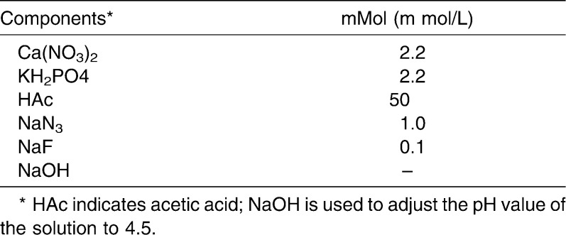
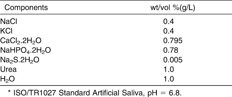
Eighty freshly extracted bovine incisors were obtained from a slaughterhouse. All teeth were stored in physiological saline after extraction and examined under a stereomicroscope to remove cracks and erosion. The teeth were soaked in demineralization buffer (Table 1) for 2 weeks to produce an artificially demineralized area (Figure 1). Two weeks later, the anti-oil varnish was washed away, and the teeth were stored in artificial saliva (Table 2) at 37°C.
Table 1.
Composition of the Demineralization Buffer
Figure 1.
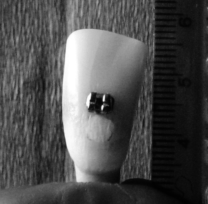
Standard edgewise brackets with a slot size of 0.018 × 0.025 inches were bound on the labial surface of the bovine incisor, and a 4 × 4 mm demineralized area was formed in the demineralization buffer for 2 weeks.
Table 2.
Composition of Artificial Saliva*
Remineralization of Enamel
Eighty teeth were randomly divided into four groups (n = 20) as follows:
—Group A: The demineralized area was coated with a thin film of CPP-ACP tooth mousse (GC Corp, Tokyo, Japan) every day for 5 minutes in artificial saliva.
—Group B: The demineralized area was coated with a thin film of fluoride toothpaste (Colgate Palmolive Corp, New York, NY) every day for 5 minutes in artificial saliva.
—Group C: The demineralized area was first coated with a thin film of fluoride toothpaste for 5 minutes, then with CPP-ACP tooth mousse for 5 minutes in artificial saliva every day.
—Group D: The teeth were stored in artificial saliva of 37°C and not coated with any agent. The artificial saliva was refreshed every 3 days.
The remineralization was arranged at 8:00 AM, 12:00 AM, and 5:00 PM. Afterward, the samples were soaked in artificial saliva at 37°C after the CPP-ACP tooth mousse was washed off with distilled water. The duration of remineralization was 12 weeks, and at the end of the 3rd, 6th, 9th, and 12th weeks, circularly polarized digital images were taken.
Production of Circularly Polarized Digital Images
Standardized images of the teeth were taken using a Canon Power Shot A720 IS digital camera (Canon Co, Japan) with a 5.8–34.8 mm, 1:2.8–4.8, 6 × IS Canon Zoom Lens. The image size was set at 2048 × 1536 pixels, image quality fine, and ISO sensitivity 400. Images were stored as a JPEG in the computer.
A CPL (HOYA Filter-Filtre, Tokia Co, Tokyo, Japan) was used to produce the polarized images (Figure 2). To standardize the photo-taking procedure, images were taken at 9:00 AM every time, and before images were taken, each tooth was dried with a triple spray for 15 seconds.
Figure 2.
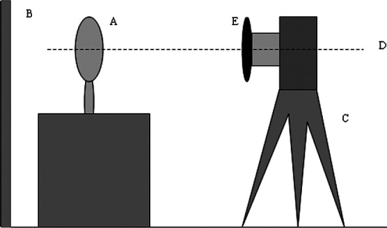
When the polarized images of each tooth (A) were taken, pink paper (B) was used as background to imitate the condition of the oral environment, and a tripod rest (C) helped to make sure the lens was perpendicular to the surface of the teeth (D). The CPL (E) was fixed in front of the lens via a piece of camera accessory to produce polarized light.
Computer-assisted Image Analysis of SDA and MGL
Computer-assisted image analysis of 360 tooth images was carried out using Adobe Photoshop CS2 Version 9.0 (Adobe Systems Inc, San Jose, Calif). A specific area of known size on a ruler beside the teeth was used for comparison for the SDA, and an area of normal enamel was used as reference for the MGL. The SDA and MGL were calculated as follows: SDA = Measured size of demineralized area on image/Measured size of comparison area on image × Actual size of comparison area on the ruler; MGL = (Measured MGL of demineralized area on image/Measured MGL of normal enamel − 1) × 100.
Statistical Analysis
Statistical analyses were performed using the SPSS 13.0 software (SPSS Inc, Chicago, Ill). Repeated measures variance analysis of three factors was used to determine whether significant differences exist between the SDA and MGL of the four groups at different examination times. Multiple factors variance analysis was used to determine whether there were differences in the SDA and MGL among the different groups at the same examination time. Pearson correlation coefficients were computed to evaluate the correlation between the SDA and MGL. A P value < .05 was considered statistically significant.
RESULTS
As Figure 3 shows, the SDA in the four groups all declined with time, and the rate of decline was relatively low in the first three weeks. The rates of decline in size in Group B and Group C were similar, and the rate for Group D was the lowest in the four groups. According to Table 3, at the end of the third week, the SDA of Group C was smaller (P = .039) than that of Group A. At the end of the 6th, 9th, and 12th weeks, the SDA of Groups A, B, and C were all smaller (P = .000) than that of Group D. The SDA of Group C was smaller than that of Group A at the end of the 6th week (P = .000), but it was not statistically different at the end of the 9th and 12th weeks. The SDA of Group C was smaller than that of Group B at the end of the 6th, 9th, and 12th weeks (P = .000, P = .005, P = .005, respectively). The SDA of Group A was smaller than that of Group B at the end of the 9th and 12th weeks (P = .013, P = .010, respectively).
Figure 3.
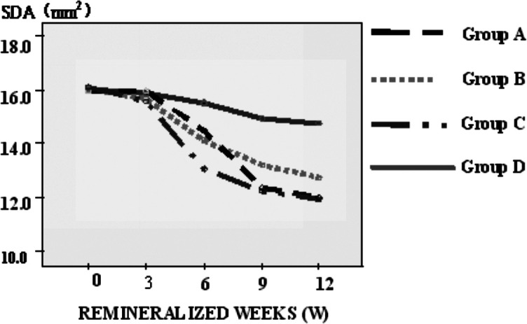
Comparison of the SDA at different times in the four groups: The SDAs in the four groups all declined with time, and the rates of decline were relatively low in the first 3 weeks. There was a rapid declining period in Group A from the 6th week to the 9th week.
Table 3.
P Values of the Comparison of SDA at the Same Test Times
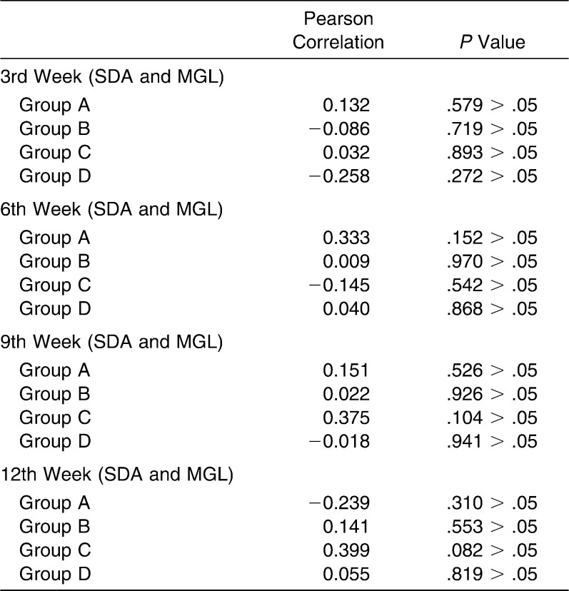
In Figure 4, the MGL in the all four groups decreased with time, and the rate of reduction was different. The rates of decline in MGL in Groups A, B, and C were similar, and Group D was the lowest in the four groups. According to Table 4, at the end of the third week, there was no statistical difference in the MGL of the four groups. At the end of the 6th, 9th, and 12th weeks, the MGL of Group D was the highest in the four groups. At the end of the sixth week, the MGL of Groups A and C were lower (P = .012 and P = .000, respectively) than that of Group B, while the MGL of Group C was lower than that of Group A (P = .037). At the end of the ninth week, the MGL of Group A and Group C were lower than that of Group B (P = .017 and P = .000, respectively). The MGL of Group C was lower than that of Groups A and B at the end of the 12th week (P = .004, P = .000, respectively). No statistically significant correlations were detected between SDA and MGL (Table 5).
Figure 4.
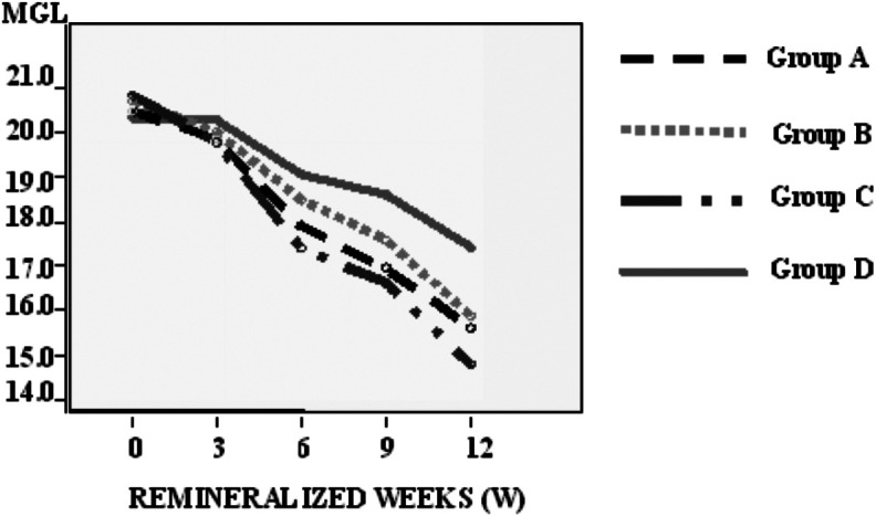
Comparison of the MGL at different times in the four groups: The MGL in the four groups all decreased with time, and the rates of reduction were different.
Table 4.
P Values of the Comparison of MGL at the Same Test Time
Table 5.
Correlations Between SDA and MGL
DISCUSSION
In this in vitro study, we examined the effect of CPP-ACP tooth mousse on bovine enamel remineralization through the analysis of circularly polarized digital images. Other studies have shown that bovine teeth are similar to human teeth in physical characteristics and chemical composition and are suitable for artificial caries research.16 In addition, bovine teeth can be easily collected from a slaughterhouse, and bovine incisors, which are wide and smooth-surfaced are convenient for taking digital images.
The demineralization of enamel adjacent to orthodontic brackets is a widely studied phenomenon, and the research about CPP-ACP is also a hot topic. The anticaries role of CPP-ACP is related to its containing calcium and phosphate, and the negative relationship between caries and the presence of calcium and phosphate has been generally accepted.17–20 At the same time, the fluoride ion has been shown to reduce the speed of demineralization and enhance the reproduction of enamel crystals.21–24 When CPP-ACP is combined with fluoride toothpaste, the fluoride ions react with CPP-ACP to form casein phosphopeptide–amorphous calcium fluoride phosphate. Reynolds25,26 established that the nanocomplex, casein phosphopeptide–amorphous calcium fluoride phosphate, provided calcium, phosphate, and fluoride ions to the surface of the teeth and therefore had a tremendous effect on enamel remineralization. The results of our experiment are consistent with these studies.
Image analysis of clinical digital photographs is a promising method for detecting and measuring demineralized areas and has attracted wide attention.27 However, because of light reflection from the environment and the flash light from the camera, which produces a false white lesion on the surface of enamel, the prevalence of enamel demineralization is usually overestimated when evaluated by digital photographs. A polarized filter removes unwanted reflections from nonmetallic surfaces and elevates the precision of the evaluation. Robertson and Toumba28 found that 100% of polarized digital images were suitable for evaluating enamel demineralization, whereas only 10% of nonpolarized digital images were valid. The studies of Benson et al.29,30 demonstrated that linearly polarized digital images had high repeatability in evaluating the size of demineralized areas. However, LPL may mislead the optic element of the camera to focus light, and it is usually used in primitive manual cameras.31 CPL emerges after LPL, and it includes a thin membrane of one-fourth wavelength that helps to produce circularly polarized light.32,33 This kind of design makes CPL more suitable for up-to-date automatic focusing and automatic exposure digital cameras.34,35 Recently, cameras with an AF lens mostly use CPL as polarizer. Therefore, we adopted CPL to obtain clear images of the demineralized area.
In this study, we chose the SDA and MGL of the demineralized area as parameters according to the protocol of both Benson et al.15 and Livas et al.13 The SDA represented the extent of the demineralized area, and the MGL provided optical properties, such as luminance of the demineralized area. According to Benson and colleagues,15 the measurement of SDA is more consistent and has a smaller random error when using cross-polarization, while the measurement of MGL shows a higher random error. Therefore, the sensitivity of SDA in measuring remineralization is higher than that of MGL. This explains why the sizes of the white spots but not the grey levels were different at week six among the groups. Other reports have shown that enamel remineralization is evident in the first 3–6 weeks, and it then progresses slowly and steadily after 12 weeks. This may well explain why the differences among the groups were evident in the early period and then almost disappeared after 12 weeks. In our experiment, no correlation was detected between SDA and MGL, and the relationship between these two parameters calls for further study.
The results of this study indicate that the SDA and MGL of demineralized enamel are positively reduced when the enamel is treated with CPP-ACP tooth mousse, and it is even more effective with the combined application of fluoride toothpaste and CPP-ACP tooth mousse.
This study provides information on the application of a CPL to digital cameras in a clinical setting. Circularly polarized images in this study are adequate for evaluating enamel remineralization for orthodontic purposes.
CONCLUSIONS
All of the null hypotheses have to be rejected.
CPP-ACP application can promote remineralization of bovine enamel by decreasing the SDA and MGL. The combination of fluoride toothpaste and CPP-ACP tooth mousse improves the remineralization effect.
Computer-assisted image analysis of circularly polarized images is an effective method of detecting enamel demineralization. Further research on the application of this technique is warranted.
Acknowledgments
We thank Dr. Jian Wang, PhD, State Key Laboratory of Oral Diseases, Department of Prosthodontics, West China Stomatology Hospital, Sichuan University, for reviewing the grammar and statistical evaluation of the data; and we appreciate the GC Company (Tokyo, Japan) for providing the oral hygiene amenities.
REFERENCES
- 1.Zachrisson B. U. A post-treatment evaluation of direct bonding in orthodontics. Am J Orthod. 1977;71:173–189. doi: 10.1016/s0002-9416(77)90394-3. [DOI] [PubMed] [Google Scholar]
- 2.Mizrahi E. Enamel demineralization following orthodontic treatment. Am J Orthod Dentofacial Orthop. 1982;82:62–67. doi: 10.1016/0002-9416(82)90548-6. [DOI] [PubMed] [Google Scholar]
- 3.Mitchell L. Decalcification during orthodontic treatment with fixed appliances—an overview. Br J Orthod. 1992;19:199–205. doi: 10.1179/bjo.19.3.199. [DOI] [PubMed] [Google Scholar]
- 4.Millet D. T, Nunn J. H, Welbury R. R, Gordon P. H. Decalcification in relation to brackets bonded with glass ionomer cement or a resin adhesive. Angle Orthod. 1999;69:65–70. doi: 10.1043/0003-3219(1999)069<0065:DIRTBB>2.3.CO;2. [DOI] [PubMed] [Google Scholar]
- 5.Derks A, Katsaros C, Frencken J. E, van't Hof M. A, Kuijpers-Jagtman A. M. Caries-inhibiting effect of preventive measures during orthodontic treatment with fixed appliances. A systematic review. Caries Res. 2004;38:413–420. doi: 10.1159/000079621. [DOI] [PubMed] [Google Scholar]
- 6.Reynolds E. C. Anticariogenic complexes of amorphous calcium phosphate stabilized by casein phosphopeptide: a review. Spec Care Dent. 1998;18:8–16. doi: 10.1111/j.1754-4505.1998.tb01353.x. [DOI] [PubMed] [Google Scholar]
- 7.Bailey D. L, Adams G. G, Tsao C. E, Hyslop A, Escobar K, Manton D. J, Reynolds E. C, Morgan M. V. Regression of post-orthodontic lesions by a remineralizing cream. J Dent Res. 2009;88:1148–1153. doi: 10.1177/0022034509347168. [DOI] [PubMed] [Google Scholar]
- 8.Reynolds E. C. Casein phosphopeptide-amorphous calcium phosphate: the scientific evidence. Adv Dent Res. 2009;21(1):25–29. doi: 10.1177/0895937409335619. [DOI] [PubMed] [Google Scholar]
- 9.Walker G. D, Cai F, Shen P, Bailey D. L, Yuan Y, Cochrane N. J, Reynolds C, Reynolds E. C. Consumption of milk with added casein phosphopeptide-amorphous calcium phosphate remineralizes enamel subsurface lesions in situ. Aust Dent J. 2009;54:245–249. doi: 10.1111/j.1834-7819.2009.01127.x. [DOI] [PubMed] [Google Scholar]
- 10.Yengopal V, Mickenautsch S. Caries preventive effect of casein phosphopeptide-amorphous calcium phosphate (CPP-ACP): a meta-analysis. Acta Odontol Scand. 2009;21:1–12. doi: 10.1080/00016350903160563. [DOI] [PubMed] [Google Scholar]
- 11.Zero D. T. Recaldent—evidence for clinical activity. Adv Dent Res. 2009;21(1):30–34. doi: 10.1177/0895937409335620. [DOI] [PubMed] [Google Scholar]
- 12.Benson P. E. Evaluation of white spot lesions on teeth with orthodontic brackets. Semin Orthod. 2008;14:200–208. [Google Scholar]
- 13.Livas C, Kuijpers-Jagtman A. M, Bronkhorst E, Derks A, Katsaros C. Quantification of white spot lesions around orthodontic brackets with image analysis. Angle Orthod. 2008;78:585–590. doi: 10.2319/0003-3219(2008)078[0585:QOWSLA]2.0.CO;2. [DOI] [PubMed] [Google Scholar]
- 14.Benson P. E, Shah A. A, Wilmot D. R. Measurement of white lesions surrounding orthodontic brackets: captured slides vs. digital camera images. Angle Orthod. 2005;75:226–230. doi: 10.1043/0003-3219(2005)075<0222:MOWLSO>2.0.CO;2. [DOI] [PubMed] [Google Scholar]
- 15.Benson P. E, Shah A, Willmot D. Polarized versus nonpolarized digital images for the measurement of demineralization surrounding orthodontic brackets. Angle Orthod. 2008;78:288–293. doi: 10.2319/121306-511.1. [DOI] [PubMed] [Google Scholar]
- 16.Esser M, Tinschert J, Marx R. Material characteristics of the hard tissues of bovine versus human teeth. Dtsch Zahnarztl Z. 1998;53:713–717. [Google Scholar]
- 17.Rose R. K. Binding characteristics of streptococcus mutants for calcium and casein phosphopeptide. Caries Res. 2000;34:427–431. doi: 10.1159/000016618. [DOI] [PubMed] [Google Scholar]
- 18.Llena C, Forner L, Baca P. Anticariogenicity of casein phosphopeptide-amorphous calcium phosphate: a review of the literature. J Contemp Dent Pract. 2009;10(3):1–9. [PubMed] [Google Scholar]
- 19.Neuhaus K. W, Lussi A. Casein phosphopeptide–amorphous calcium phosphate (CPP-ACP) and its effect on dental hard tissues. Schweiz Monatsschr Zahnmed. 2009;119:110–116. [PubMed] [Google Scholar]
- 20.Pai D, Bhat S. S, Taranath A, Sargod S, Pai V. M. Use of laser fluorescence and scanning electron microscope to evaluate remineralization of incipient enamel lesions remineralized by topical application of casein phosphopeptide amorphous calcium phosphate (CPP-ACP) containing cream. J Clin Pediatr Dent. 2008;32:201–206. doi: 10.17796/jcpd.32.3.d083470201h58m13. [DOI] [PubMed] [Google Scholar]
- 21.Hicks M. J, Flaitz C. M. Enamel caries formation and lesion progression with a fluoride dentifrice and a calcium-phosphate containing fluoride dentifrice: a polarized light microscopic study. ASDC J Dent Child. 2000;67(1):21. [PubMed] [Google Scholar]
- 22.Chin M. Y, Sandham A, Rumachik E. N, Ruben J. L, Huysmans M. C. Fluoride release and cariostatic potential of orthodontic adhesives with and without daily fluoride rinsing. Am J Orthod Dentofacial Orthop. 2009;136:547–553. doi: 10.1016/j.ajodo.2007.10.053. [DOI] [PubMed] [Google Scholar]
- 23.Magalhães A. C, Wiegand A, Rios D, Honório H. M, Buzalaf M. A. Insights into preventive measures for dental erosion. J Appl Oral Sci. 2009;17(2):75–86. doi: 10.1590/S1678-77572009000200002. [DOI] [PMC free article] [PubMed] [Google Scholar]
- 24.Benson P. E, Shah A. A, Millett D. T, Dyer F, Parkin N, Vine R. S. Fluorides, orthodontics and demineralization: a systematic review. J Orthod. 2005;32(2):102–114. doi: 10.1179/146531205225021033. [DOI] [PubMed] [Google Scholar]
- 25.Reynolds E. C. (North Balwyn, University of Melbourne). Calcium phosphopeptide complexes. Sept. 17, 1998, United States Patent #7312193. International Application No. PCT/AU1998/000160 [Google Scholar]
- 26.Reynolds E. C, Cain C. J, Webber F. L, Johnson I. H, Perich J. W. Anticariogenicity of calcium phosphate complexes of tryptic casein phosphopeptide in the rat. J Dent Res. 1995;74:1272–1279. doi: 10.1177/00220345950740060601. [DOI] [PubMed] [Google Scholar]
- 27.Benson P. E, Pender N, Higham S. M. Enamel demineralization assessed by computerized image analysis of clinical photographs. J Dent. 2000;28:319–326. doi: 10.1016/s0300-5712(00)00002-6. [DOI] [PubMed] [Google Scholar]
- 28.Robertson A. J, Toumba K. J. Cross-polarized photography in the study of enamel defects in dental pediatrics. J Audiov Media Med. 1999;22:63–70. doi: 10.1080/014051199102179. [DOI] [PubMed] [Google Scholar]
- 29.Benson P. E, Pender N, Higham S. M. Quantifying enamel demineralization from teeth with orthodontic brackets—a comparison of two methods. Part 2: validity. Eur J Orthod. 2003;25:159–165. doi: 10.1093/ejo/25.2.159. [DOI] [PubMed] [Google Scholar]
- 30.Benson P. E, Pender N, Higham S. M. Quantifying enamel demineralization from teeth with orthodontics brackets—a comparison of two methods. Part 1: repeatability and agreement. Eur J Orthod. 2003;25:149–158. doi: 10.1093/ejo/25.2.149. [DOI] [PubMed] [Google Scholar]
- 31.Millard H. The polarizer. Modern Photography. 1985;48(7):43. [Google Scholar]
- 32.Li L, Dobrowolski J. A, Sullivan B. T, Pang Z. Novel thin film polarizing beam-splitter and its application in high efficiency projection displays. Proc SPIE. 1999;3634:52–62. [Google Scholar]
- 33.Li L, Dobrowolski J. A. Visible broadband, wide-angle, thin film multilayer polarizing beam splitter. Appl Opt. 1996;35:2221–2225. doi: 10.1364/AO.35.002221. [DOI] [PubMed] [Google Scholar]
- 34.Ray S. F. Applied Photographic Optics. Oxford, UK: Focal Press; 2002. pp. 15–30. [Google Scholar]
- 35.Chipman R. A. Polarimetry. Handbook Opt. 2004;22(22):1–37. [Google Scholar]


