Abstract
1.1. Background:
It was reported that Hemoglobin A1c (HbA1c) values of African-Americans (AAs) were on average higher than whites whereas AAs with Sickle-Cell-Trait (SCT) had lower HbA1c values compared to AAs without SCT despite controlling for average glycaemia. We evaluated the HbA1c-glucose relationship between AAs with and without SCT, and whites using data from two cohort studies.
1.2. Methods:
We pooled data from Coronary-Artery-Risk-Development-Study-in-Young-Adults (CARDIA, n= 5,115, 2005–2011) and the Jackson-Heart-Study (JHS, n=5,301, 2000–2013). Whole exome sequencing in JHS and TaqMan-SNP-Genotyping Assays in CARDIA determined the SCT status in AAs. HbA1c was measured by two NGSP-certified assays without reportedly clinically significant interference from hemoglobin S. Participants without data on SCT or with hemoglobin SS, CC or AC were excluded, resulting in 6,623 participants (n=3,575 from CARDIA and n=3,048 from JHS). Generalized-estimating-equations estimated the cross-sectional association between fasting glucose and HbA1c(outcome) amongst whites, AAs with SCT, and AAs without SCT controlling for clinical-demographic factors.
1.3. Results:
Our analyses included 2,003 whites, 4,253 AAs without SCT and 367 AAs with SCT. AAs with and without SCT had similar clinical-demographic characteristics, whereas whites have lower fasting- and 2-hour-glucose values than AAs. Despite higher fasting-glucose values in AAs with SCT versus whites, their HbA1c values were similar (p=0.39). In the subset with 2-hour-glucose values, HbA1c values in AAs with SCT were lower than whites (p=0.007) despite higher 2-hour-glucose values.
1.4. Conclusions:
AAs with SCT have at least similar, if not lower, levels of mean HbA1c values than whites despite higher levels of glycaemia. Future research is warranted to assess whether these findings translate to clinical outcomes.
2. Introduction
It has been previously shown that for any given level of glycaemia, African Americans (AAs) have higher average Hemoglobin A1c (HbA1c) values compared to whites [1]. However, it was recently reported that among AAs the presence of Sickle Cell Trait (SCT) may affect the HbA1c-glucose relationship. Specifically, for any given fasting or 2-hour-postprandial glucose level, HbA1c was approximately 0.3 percentage points lower among AAs with SCT compared to AAs without SCT [2]. An accompanying editorial interpreted these findings as suggesting that AAs with SCT have an HbA1c-glucose relationship that is similar to whites. We sought to directly evaluate this untested hypothesis using data from two longitudinal community-based cohort studies.
3. Methods
We pooled data from participants of the Coronary Artery Risk Development Study in Young Adults (CARDIA, n= 5,115, 2005–2011) and the Jackson Heart Study (JHS, n=5,301, 2000–2013) with data on HbA1c and either 8-hr fasting (both cohorts) or 2-hr post-prandial (oral glucose tolerance test, CARDIA only) glucose levels [3,4]. All participants included in the analyses provided written informed consent and institutional review board from each of the participating institutions in both CARDIA (University of Alabama at Birmingham, Northwestern University, University of Minnesota, and Johns Hopkins University School of Medicine) and JHS (University of Mississippi Medical Center) approved the study. Whole exome sequencing in JHS and TaqMan SNP Genotyping Assays (Life Technologies, Grand Island, NY) in CARDIA were used to determine SCT status in AAs. HbA1c was measured by two NGSP-certified assays (Tosoh 2.2 and Tosoh G7) reportedly without clinically significant interference from hemoglobin S [5]. Participants without data on SCT, with hemoglobin SS, with hemoglobin CC or hemoglobin AC were excluded, resulting in 6,623 participants in the analysis (n=3,575 from CARDIA and n=3,048 from JHS). We assumed SCT to be absent in whites given its very low prevalence in this population (0.3%) [6].
We compared participant characteristics amongst whites, AAs with SCT and AAs without SCT using chi2 tests and ANOVA for discrete and continuous variables, respectively. We used Generalized-Estimating-Equations (GEE) using random effects at the participant level and an exchangeable correlation matrix to estimate the cross-sectional association between glucose measures (fasting and 2-hour glucose) and HbA1c (outcome) amongst whites, AAs with SCT, and AAs without SCT while accounting for repeated measures within individuals over time.
4. Results
Our analyses included 2,003 whites, 4,253 AAs without SCT and 367 AAs with SCT with a total of 10,997 observations with fasting glucose and 4,043 with 2-hr glucose from two follow-up visits in CARDIA (years 20 and 25 from 2005–2011), and three visits in JHS (baseline and years 5 and 10 follow-ups from 2000–2013). Compared to AAs (with and without SCT), whites were more likely to be male and younger, to have lower BMI, ferritin, and fasting and 2-hour glucose values; and less likely to have diabetes or be on diabetes medications (Table 1).
Table 1:
Baseline Characteristics of the Study Population.
| Mean (SD) unless otherwise specified |
Whites (n=2003) |
AA - non- SCT (n=4253) |
p-value (Whites vs AA non- SCT) |
AA - SCT (n=367) |
p-value (Whites vs AA - SCT) |
|---|---|---|---|---|---|
| Male, n (%) | 942 (47.1) | 1634 (38.4) | <0.0001 | 15 (41.1) | 0.04 |
| Age in years | 46.44 (3.8) | 52.2 (11.8) | <0.0001 | 53.9 (12.2) | <0.0001 |
| BMI (kg/m2) | 28.0 (6.1) | 31.8 (7.5) | <0.0001 | 31.7 (7.5) | <0.0001 |
| Ferritin (ng/mL) | 115.4 (141.0) | 156.3 (171.8) | <0.0001 | 151.4 (143.7) | <0.0001 |
| eGFR (ml/min/1.73m2) | 93.6 (13.6) | 97.5 (21.2) | <0.0001 | 92.5 (23.3) | 0.2 |
| Fasting Plasma Glucose (mg/dL) | 97.7 (21.3) | 101.5 (33.2) | <0.0001 | 102.7 (34.2) | 0.0002 |
| Hemoglobin A1c (%) | 5.4 (0.7) | 5.9 (1.2) | <0.0001 | 5.7 (1.2) | <0.0001 |
| 2-hour plasma glucose (mg/dL, n = 2900) | 104.2 (36.3) | 113.8 (44.9) | <0.0001 | 116.1 (39.0) | 0.005 |
| Diabetes medications,a n (%) | 57 (2.9) | 523 (12.5) | <0.0001 | 54 (14.9) | <0.0001 |
| Diabetes diagnosis,b n (%) | 172 (8.6) | 622 (14.7) | <0.0001 | 63 (17.2) | <0.0001 |
| Physical activity,c n (%) | <0.0001 | <0.0001 | |||
| Poor | 20 (1.0) | 1445 (34.1) | 139 (38.1) | ||
| Intermediate | 419 (21.0) | 1368 (32.3) | 113 (31.0) | ||
| Ideal | 1559 (78.0) | 1427 (33.7) | 113 (31.0) | ||
| Diet,d n (%) | <0.0001 | <0.0001 | |||
| Poor | 616 (37.4) | 2292 (58.5) | 206 (59.4) | ||
| Intermediate | 961 (58.4) | 1577 (40.3) | 133 (38.3) | ||
| Ideal | 69 (4.2) | 46 (1.2) | 8 (2.3) | ||
| Smoking, n (%) | 0.001 | 0.33 | |||
| Poor | 301 (15.2) | 734 (17.5) | 57 (15.8) | ||
| Intermediate | 61 (3.1) | 77 (1.8) | 6 (1.7) | ||
| Ideal | 1621 (81.7) | 3385 (80.7) | 298 (82.6) |
Abbreviations: AA = African Americans, SCT = Sickle Cell Trait, BMI = Body Mass Index (calculated as weight in kilograms divided by height in meters squared); eGFR = Estimated Glomerular Filtration Rate (calculated using the CKD-EPI equation), A1C.
Diabetes medications is defined as use of a medication for the treatment of diabetes taken in the 2 weeks prior to the study exam.
Diabetes diagnosis is defined as self-reported use of diabetes medications or self-reported physician diagnosis since the goal was to control for any potential confounding that may arise as a result of treatment for diagnosed diabetes (pharmaceutical or lifestyle).
Physical Activity is defined as Poor, Intermediate or Ideal based on minutes/week of moderate or vigorous physical activity: Poor (0 minutes/week); Intermediate (>0 - <150 minutes/week); Ideal (≥150 minutes/week)
Nutrition is defined as Poor, Intermediate or Ideal based on the number of diet components achieved. Components (based on a 2000-calorie diet): ≥4.5 cups of fruits and vegetables/day; ≥7 ounces of fish/week; <1500 mg of sodium/day; <450 calories/week of sugar-sweetened beverages; ≥3 servings/day of whole grains. Poor Health (0-1 components); Intermediate Health (2-3 components); Ideal Health (4-5 components).
Smoking is defined as Poor, Intermediate or Ideal based on current and former smoking status. Poor (Current smoker); Intermediate (Quit < 12 months ago); Ideal (Never smoked or quit ≥12 months ago).
4.1. AAs with SCT versus Whites
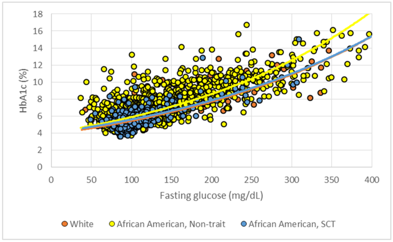
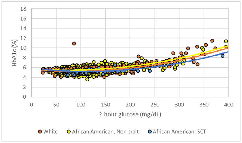
Despite significantly higher mean fasting glucose values in AAs with SCT (102.7±34.2 mg/dL) versus whites (97.7±21.3 mg/dL), p=0.0002, regression analyses revealed similar mean HbA1c values in AAs with SCT (5.83%, 95% CI: 5.75, 5.91) compared to whites (5.86%, 95% CI: 5.80%, 5.92%), p=0.39, after adjustment for age, gender, BMI, ferritin, estimated glomerular filtration rate, diabetes, current use of diabetes medications, physical activity, diet and smoking status (Figure 1). In the subset of CARDIA participants with glucose tolerance test results (n=2900), adjusted analyses (Figure 2) showed that mean HbA1c values in AAs with SCT (5.40%, 95% CI: 5.26%, 5.55%) were significantly lower than whites (5.54%, 95% CI: 5.41%, 5.66%), p=0.007, despite significantly higher mean 2-hour post-prandial glycemic values in AAs with SCT (116.1±39.0 mg/dL) versus whites (104.17±36.3 mg/dL), p=0.005.
Figure 1:

Scatterplot of hemoglobin A1C (%) versus fasting plasma glucose (mg/dL). Whites: Observed data points are represented by orange dots and the regression line is solid orange. AA Non-trait (African Americans without sickle cell trait): Observed data points are represented by yellow dots and the regression line is solid yellow. AA SCT (African Americans with sickle cell trait): Observed data points are represented by blue dots and the regression line is solid blue. Covariates in adjusted models are centered at the mean. Adjusted for age, BMI, ferritin, eGFR, male, diabetes medications, diabetes diagnosis, physical activity, diet and smoking.
Figure 2:

Scatterplot of hemoglobin A1C (%) versus 2-hour postprandial plasma glucose (mg/dL). Whites: Observed data points are represented by orange dots and the regression line is solid orange. AA Non-trait (African Americans without sickle cell trait): Observed data points are represented by yellow dots and the regression line is solid yellow. AA SCT (African Americans with sickle cell trait): Observed data points are represented by blue dots and the regression line is solid blue. Covariates in adjusted models are centered at the mean. Adjusted for age, BMI, ferritin, eGFR, male, diabetes medications, diabetes diagnosis, physical activity, diet and smoking.
4.2. AAs without SCT versus Whites
Adjusted analyses showed that after controlling for fasting glucose and same covariates listed above, mean HbA1c values in AAs without SCT (6.16%, 95% CI: 6.10 – 6.21) were still significantly higher than whites, p<0.0001. Results remained similar for the subset of participants with 2-hour glucose values, where AAs without SCT had significantly higher mean HbA1c values (5.76%, 95% CI: 5.64% - 5.89%) than whites, p<0.0001, even after adjustment for 2-hour glucose values and the same covariates listed above.
5. Discussion
In this analysis of 2 large, well-established cohorts, despite significantly higher mean fasting glucose values in AAs with SCT versus whites, mean HbA1c values in AAs with SCT were similar compared to whites. In the subset of participants with 2-hour postprandial glucose values, mean HbA1c values in AAs with SCT were significantly lower than whites despite significantly higher mean 2-hour post-prandial glycemic values in AAs with SCT. To our knowledge, this study is one of the first to report this relationship with important clinical implications given previous data showing AAs to have higher HbA1c values than whites [1]. These findings suggest that generalizations about AAs having higher levels of HbA1c than whites for similar levels of glycaemia are unnecessary simplifications.
Consistent with previous literature [1], this report also showed that, mean HbA1c values in AAs without SCT were significantly higher than whites, despite adjustment for fasting (overall study sample) or 2-hour (CARDIA only) glucose values. The potential mechanisms for the observed differences in Hba1c between the white and the AA population with or without SCT are likely multifactorial given the lack of a gold standard for HbA1c values and the inherent variation within assays being employed. Discrepancy in HbA1c values observed can be biological as supported by studies that utilized continuous plasma glucose monitoring (gold standard for which HbA1c is intended to measure), that suggest differences in red blood cell lifespan and hemoglobin glycation kinetics as potential sources of variation [7]. On the other hand, reports from Rohlfing, Hanson, Little and colleagues have consistently shown a statistically significant negative bias in the assays utilized for HbA1c in individuals with SCT [8–11], but the assay bias alone cannot explain the A1c differences between whites and AAs without SCT. As such, we hypothesize that the observed differences are likely a combination of biological and assay mechanisms. The mechanisms above will also likely result in some discrepancy in the relationship between HbA1c and either fasting blood glucose and 2-hour glucose values observed in our study. Irrespective of the mechanism, the tests being employed in this report are common in clinical practice and the results shown in this analysis are relevant to the day-to-day clinical practice.
6. Conclusions
AAs with SCT have at least similar, if not lower, levels of mean HbA1c values than whites despite higher levels of fasting and 2-hour postprandial glycaemia. Further research is warranted to assess whether these findings in HbA1c levels in relation to race and SCT status translate into distinct clinical outcomes in the setting of racial variations in metabolic risk profiles.
7. Acknowledgements
The Coronary Artery Risk Development in Young Adults Study (CARDIA) is conducted and supported by the National Heart, Lung, and Blood Institute (NHLBI) in collaboration with the University of Alabama at Birmingham (HHSN268201300025C & HHSN268201300026C), Northwestern University (HHSN268201300027C), University of Minnesota (HHSN268201300028C), Kaiser Foundation Research Institute (HHSN268201300029C), and Johns Hopkins University School of Medicine (HHSN268200900041C). CARDIA is also partially supported by the Intramural Research Program of the National Institute on Aging (NIA) and an intra-agency agreement between NIA and NHLBI (AG0005). This manuscript has been reviewed by CARDIA for scientific content.
The Jackson Heart Study is supported and conducted in collaboration with Jackson State University (HHSN268201300049C and HHSN268201300050C), Tougaloo College (HHSN268201300048C), and the University of Mississippi Medical Center (HHSN268201300046C and HHSN268201300047C) contracts from the National Heart, Lung, and Blood Institute (NHLBI) and the National Institute for Minority Health and Health Disparities (NIMHD). The authors thank the participants and data collection staff of the Jackson Heart Study.
Footnotes
Disclaimer
The views expressed in this manuscript are those of the authors and do not necessarily represent the views of the National Heart, Lung, and Blood Institute; the National Institutes of Health; or the U.S. Department of Health and Human Services or the Department of Veterans Affairs.
Conflicts of Interest
None of the authors have any conflicts of interest to disclose.
References
- 1.Bleyer AJ, Hire D, Russell GB, Xu J, Divers J, et al. (2009) Ethnic variation in the correlation between random serum glucose concentration and glycated hemoglobin. Diabet Med 26: 128–133. [DOI] [PubMed] [Google Scholar]
- 2.Lacy ME, Wellenius GA, Sumner AE, Correa A, Carnethon MR, Li, et al. (2017) Association of Sickle Cell Trait With Hemoglobin A1c in African Americans. JAMA 317: 507–515. [DOI] [PMC free article] [PubMed] [Google Scholar]
- 3.Friedman GD, Cutter GR, Donahue RP, Hughes GH, Hulley SB, et al. (1988) CARDIA: study design, recruitment, and some characteristics of the examined subjects. J Clin Epidemiol 41: 1105–1116. [DOI] [PubMed] [Google Scholar]
- 4.Taylor HA Jr, Wilson JG, Jones DW, Sarpong DF, Srinivasan A, et al. (2005) Toward resolution of cardiovascular health disparities in African Americans: design and methods of the Jackson Heart Study. Ethn Dis 15: S6–S4. [PubMed] [Google Scholar]
- 5.National Glycohemoglobin Standardization Program (NGSP). Factors that interfere with HbA1c test results. 2016; 2017. [Google Scholar]
- 6.Ojodu J, Hulihan MM, Pope SN, Grant AM (2014) Centers for Disease C and Prevention. Incidence of sickle cell trait--United States, 2010. MMWR Morb Mortal Wkly Rep 63: 1155–1158. [PMC free article] [PubMed] [Google Scholar]
- 7.Malka R, Nathan DM, Higgins JM (2016) Mechanistic modeling of hemoglobin glycation and red blood cell kinetics enables personalized diabetes monitoring. Sci Transl Med 8: 359ra130. [DOI] [PMC free article] [PubMed] [Google Scholar]
- 8.Rohlfing C, Hanson S, Little RR (2017) Measurement of Hemoglobin A1c in Patients With Sickle Cell Trait. JAMA 317: 2237. [DOI] [PubMed] [Google Scholar]
- 9.Lin CN, Emery TJ, Little RR, Hanson SE, Rohlfing CL, et al. (2012) Effects of hemoglobin C, D, E, and S traits on measurements of HbA1c by six methods. Clin Chim Acta 413: 819–821. [DOI] [PMC free article] [PubMed] [Google Scholar]
- 10.Mongia SK, Little RR, Rohlfing CL, Hanson S, Roberts RF, et al. (2008) Effects of hemoglobin C and S traits on the results of 14 commercial glycated hemoglobin assays. Am J Clin Pathol 130: 136–140. [DOI] [PubMed] [Google Scholar]
- 11.Roberts WL, Safar-Pour S, De BK, Rohlfing CL, Weykamp CW, et al. (2005) Effects of hemoglobin C and S traits on glycohemoglobin measurements by eleven methods. Clin Chem 51: 776–778. [DOI] [PubMed] [Google Scholar]


