Key Points
The level of baseline ctDNA correlated with PFS and OS in patients with R/R DLBCL receiving pola plus BR or BR alone.
Patients with a CR had a significantly greater median decrease in ctDNA levels at end of treatment than patients without a CR.
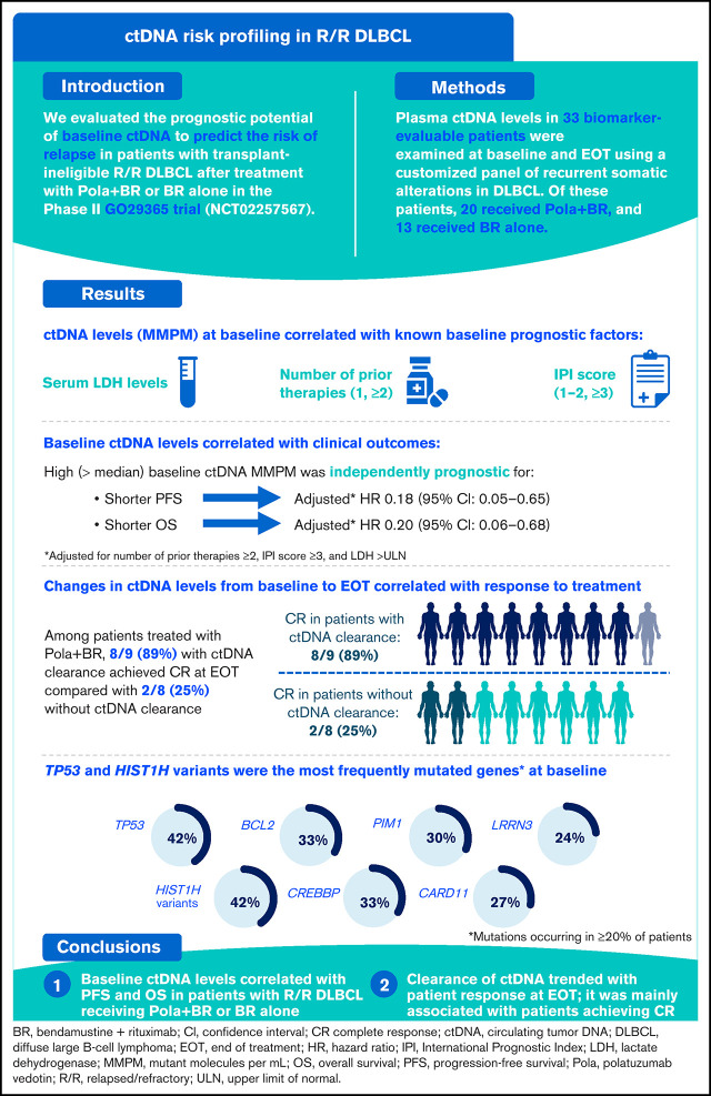
Visual Abstract
Abstract
Patients with relapsed/refractory (R/R) diffuse large B-cell lymphoma (DLBCL) have heterogeneous outcomes; durable remissions are infrequently observed with standard approaches. Circulating tumor DNA (ctDNA) assessment is a sensitive, potentially prognostic tool in this setting. We assessed baseline ctDNA to identify patients with R/R DLBCL at high risk of relapse after receiving polatuzumab vedotin and bendamustine plus rituximab (BR) or BR alone. Patients were transplant ineligible and had received ≥1 prior line of therapy. The ctDNA assay, based on a customized panel of recurrently mutated genes in DLBCL, measured mutant molecules per mL (MMPM) at baseline and end of treatment (EOT). Endpoints included progression-free survival (PFS) and overall survival (OS) in subgroups stratified by baseline ctDNA and log-fold change in ctDNA at EOT vs baseline. In biomarker-evaluable patients (n = 33), baseline ctDNA level correlated with serum lactate dehydrogenase (LDH) concentration, number of prior therapies, stage, and International Prognostic Index (IPI). After adjusting for number of prior therapies ≥2, IPI score ≥3, and LDH above the upper limit of normal, high (greater than median) baseline ctDNA MMPM was independently prognostic for shorter PFS (adjusted hazard ratio [HR], 0.18 [95% CI, 0.05-0.65]) and OS (adjusted HR, 0.20 [95% CI, 0.06-0.68]). In 23 patients with baseline and EOT samples, a significantly greater decrease in ctDNA MMPM was observed in patients with complete response (CR) (n = 13) than those without CR (n = 10); P = .0025. Baseline ctDNA assessment may identify patients at high risk of progression and should be further evaluated as a monitoring tool in R/R DLBCL. This trial was registered at www.clinicaltrials.gov as #NCT02257567.
Introduction
Approximately 30% to 40% of patients with diffuse large B-cell lymphoma (DLBCL) are either primary refractory or will relapse (R/R) following frontline treatment with rituximab, cyclophosphamide, doxorubicin, vincristine, and prednisolone (R-CHOP).1 Although novel treatment options have become available in recent years, including chimeric antigen receptor (CAR) T-cell therapy and antibody-drug conjugates,1 most patients will not achieve a durable response with standard therapeutic approaches and have poor outcomes.
Polatuzumab vedotin is an antibody-drug conjugate that comprises an anti-CD79b antibody conjugated to a potent cytotoxic microtubule inhibitor, monomethyl auristatin E.2,3 Regulatory approval of polatuzumab vedotin by the US Food and Drug Administration4 and the European Medicines Agency5 was based on the primary results of the pivotal phase 1b/2 GO29365 study. This study evaluated patients with R/R DLBCL receiving polatuzumab vedotin in combination with bendamustine (B) and rituximab (R) vs BR alone. Patients in the polatuzumab vedotin plus BR (pola plus BR) arm achieved a significantly higher complete response (CR) rate (40.0%) than those in the BR arm (17.5%); n = 40 for both arms (P = .026). In addition, progression-free survival (PFS) and overall survival (OS) were longer in patients who received pola plus BR compared with BR alone.6
Circulating tumor DNA (ctDNA) is a potential prognostic biomarker in patients with DLBCL. Baseline and on-treatment assessments of ctDNA have identified patients with previously untreated DLBCL who are at high risk of relapse.7-9 Baseline ctDNA levels correlate with disease burden and survival outcomes in patients with DLBCL7,8; furthermore, detectable interim on-treatment ctDNA is associated with early disease progression (PD).8 The magnitude of early on-treatment changes in ctDNA levels is also an independent prognostic factor of survival outcomes.7
To date, there are only preliminary data evaluating ctDNA in the R/R DLBCL setting. The analysis conducted by Kurtz et al7 included a proportion of patients with R/R DLBCL receiving various treatment regimens (n = 36, 25%) and showed an association between presalvage treatment ctDNA levels and event-free survival and OS. Additionally, Frank et al showed that 28 days after infusion with the CAR T-cell therapy axicabtagene ciloleucel, levels of ctDNA-based minimal residual disease (MRD) were significantly associated with PFS and OS in patients with R/R DLBCL.10
Thus, ctDNA-based assessments may represent a highly sensitive, noninvasive biomarker with potential for utility as a prognostic tool in DLBCL. However, further validation is needed to support the rational design of ctDNA-driven adaptive clinical trials to target patients who are most likely to benefit from a particular therapeutic approach; studies are particularly warranted in the setting of R/R DLBCL. Here, as part of the GO29365 study, we report the clinical utility of ctDNA measurements to identify patients with R/R DLBCL at high risk of PD following treatment with either pola plus BR or BR alone.
Materials and methods
Trial conduct
The GO29365 study protocol was approved by applicable ethics committees and institutional review boards in accordance with the International Conference on Harmonization guidelines for Good Clinical Practice and the Declaration of Helsinki. All patients gave informed consent before screening.
Patients
The GO29365 study included patients aged ≥18 years with R/R DLBCL confirmed by biopsy, with an Eastern Cooperative Oncology Group performance status (ECOG PS) of 0 to 2, grade ≤1 peripheral neuropathy, and who had received at least 1 prior line of therapy. Patients were eligible for the study if they were considered by the treating physician to be transplant ineligible or had experienced treatment failure with prior autologous stem cell transplantation. Patients with double- or triple-hit lymphomas were eligible for inclusion; patients with transformed lymphoma were excluded. Further details of inclusion/exclusion criteria have been previously published.6 The current analysis included patients for whom blood samples for ctDNA analysis were available at baseline.
Study design
The current analysis was based on the randomized phase 2 cohorts of the open-label, multicenter GO29365 study of pola plus BR vs BR, which has been described in full previously.6 Briefly, patients were stratified by duration of response to last therapy (≤12 months or >12 months) before being randomized 1:1 to receive pola plus BR or BR alone. Patients treated with polatuzumab vedotin received 1.8mg/kg intravenously on Day 2 of Cycle 1 and Day 1 of subsequent cycles. In addition, all patients received intravenous (IV) bendamustine 90mg/m2 on Days 2 and 3 of Cycle 1, and then on Days 1 and 2 of subsequent cycles, in combination with IV rituximab (375mg/m2 on Day 1 of each cycle). Patients were treated with up to 6 21-day cycles.
Data from patients receiving either pola plus BR, or BR alone, were combined for this analysis of the utility of ctDNA assessments.
Study assessments
The rate of CR was assessed by [18F] fluorodeoxyglucose positron emission tomography (PET)-computed tomography using modified Lugano response criteria at end of treatment (EOT; 6 to 8 weeks after Day 1 of Cycle 6 or last dose of study treatment).
Measurements of ctDNA were performed as an exploratory correlative analysis. At baseline, samples of whole blood were collected in 10 mL lavender top EDTA tubes and shipped at ambient temperature on the day of collection for the separation of peripheral blood mononuclear cells, which were used as a germline control. Plasma samples were collected at baseline and at EOT. Plasma was isolated from 6 mL of peripheral blood collected in EDTA tubes. Samples were centrifuged at 1500 to 2000 g for 15 minutes at 2 to 8°C at the study site within 60 minutes of collection; the plasma layer was transferred to polypropylene sample tubes and frozen and stored at or below −20°C before shipping to the central laboratory.
Next-generation sequencing library preparation was performed using an updated version of the AVENIO ctDNA analysis workflow.7,11 The AVENIO ctDNA assay is based on the Cancer Personalized Profiling by Deep Sequencing assay published by Scherer et al.12 Genomic regions with recurrent somatic alterations (single nucleotide variants [SNVs] and insertions/deletions from multiple whole-exome and whole-genome sequencing studies) in DLBCL are included in the panel.12 DNA was prepared for ligation and unique molecular identifier–containing adapters were ligated onto the DNA fragments; the library was then amplified with universal polymerase chain reaction primers targeting the unique molecular identifier adapters, which also contain unique dual-index sample indices. Half of the polymerase chain reaction product for each sample was captured with a ∼320 Kb panel designed to cover regions relevant for determination of the cell of origin and MRD in DLBCL7; each library was amplified to obtain a final sequencing library for each sample. The custom DLBCL panel was designed for high sensitivity to MRD by maximizing the number of expected variants for each patient with DLBCL. To accomplish this, the panel included additional nonexonic target regions that are recurrently mutated in DLBCL and are primarily activation-induced cytidine deaminase hotspot regions.7
Plasma-depleted whole blood from baseline samples was used as a source of germline DNA to filter out non–tumor-specific variants. The tumor burden of these samples was estimated by calculating the number of tumor genome copies per mL of plasma. This calculation, labeled mutant molecules per mL (MMPM), incorporates the allele fractions (AF) of the variant calls and the circulating free DNA mass of the sample, specifically:
SNVs were called from plasma sequencing data using updated versions of the AVENIO ctDNA analysis variant callers. These variant callers are based on previously described algorithms for ctDNA variant calling,7,11 and variants were annotated using SnpEff (version 4.2).13 Briefly, molecule deduplication and background error reduction were performed as described by Newman et al.11 For each sample, variant calling thresholds were adaptively set for each of the 12 SNV types (A>C, A>G, A>T, etc.) based on sample-specific background error models.
Variants were removed from consideration if any of the following were true: (1) variants present in the Single Nucleotide Polymorphism Database (dbSNP) common or with AF >0.1% in any of 1000 Genomes Project catalogue or Exome Aggregation Consortium populations unless reported by Catalogue of Somatic Mutations in Cancer or The Cancer Genome Atlas; (2) variants in low-complexity regions of the genome; (3) variants present in multiple samples in a set of 22 healthy donor samples previously sequenced with the workflow; (4) positions with <25% of the median depth within a sample; and (5) presence in the paired peripheral blood mononuclear cell sample at >0.25% AF.
Study endpoints
The study endpoints included PFS assessed by investigator in subgroups stratified by baseline ctDNA levels (above vs below the median MMPM at baseline, above vs below 25% of highest baseline quantitative values, above vs below 75% of highest baseline quantitative values), OS in subgroups stratified by ctDNA levels (above vs below median MMPM at baseline), and log-fold change in ctDNA at EOT vs baseline and correlation with EOT response.
Statistical analysis
Analyses were exploratory in nature; results are reported descriptively, without correction for multiple testing. Detectable ctDNA was determined using empirical P values (P < .05) through a bootstrap algorithm.12,14 ctDNA clearance at EOT was defined using 2 criteria: (1) empirical P value measuring the significance of the tumor-specific variant signal over the background <5% and (2) <5% baseline variants detectable at EOT. Satisfaction of either criterion was considered ctDNA clearance. To ensure that low levels of non–tumor-specific variants did not obscure ctDNA clearance, if >95% of variants from the pretreatment sample were absent in the posttreatment sample, patients were considered to have ctDNA clearance.
Associations between ctDNA levels and PFS/OS were performed using univariate and multivariate Cox regression. Variables included in the multivariate analysis were number of prior therapies ≥2, International Prognostic Index (IPI) ≥3, and lactate dehydrogenase (LDH) levels above the upper limit of normal as determined by the study site. Hazard ratios (HR) with 95% Wald confidence intervals (CIs) are presented. A Wilcoxon test was performed to determine the relationship between log-fold change in ctDNA MMPM (measured at baseline and EOT) and EOT response (defined as patients achieving CR at EOT or not).
Results
Patient disposition and baseline characteristics
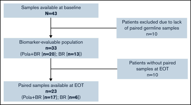
In the primary GO29365 study,6 80 patients were included in the intention-to-treat (ITT) population. No patients in the study had double- or triple-hit lymphomas. Forty-three patients in the ITT population had samples available for ctDNA analysis at baseline (Figure 1). Thirty-three patients had available germline data and provided separate consent for analysis (n = 20 [pola plus BR]; n = 13 [BR]). These patients comprised the biomarker-evaluable population (BEP) for the correlative analyses; of these patients, 23/33 (n = 17 [pola plus BR]; n = 6 [BR]) had paired samples available at EOT.
Figure 1.
Flow diagram of patient disposition in the ITT population.
Patient baseline characteristics in the BEP and ITT populations are shown in Table 1. The median follow-up period was 29.5 months in both the ITT population and BEP. Numerical differences were seen in the proportions of patients with refractory disease (67.5% in ITT and 42.4% in BEP), ECOG PS 1 (44.9% in ITT and 63.6% in BEP), and ECOG PS 2 (18.0% in ITT and 6.1% in BEP). Investigator-assessed PFS was similar between the BEP and the ITT population (supplemental Figure 1).
Table 1.
Baseline patient demographics in the ITT population and BEP
| Population | ITT (N = 80) | BEP (n = 33) |
|---|---|---|
| Median age, y | 68.5 | 72 |
| Sex, % | ||
| F | 33.8 | 33.3 |
| M | 66.2 | 66.7 |
| ECOG PS, % | ||
| 0 | 37.2 | 30.3 |
| 1 | 44.9 | 63.6 |
| 2 | 18.0 | 6.1 |
| Baseline IPI, % | ||
| High (≥3) | 63.8 | 57.6 |
| Low (0-2) | 36.3 | 42.4 |
| Cell of origin, % | ||
| ABC | 45.0 | 51.5 |
| GCB | 42.5 | 39.4 |
| NOS | 8.8 | 6.1 |
| Unclassified | 3.8 | 3.0 |
| Refractory, % * | ||
| No | 32.5 | 57.6 |
| Yes | 67.5 | 42.4 |
| Median follow-up period, mo | 29.5† | 29.5 |
ABC, activated B-cell–like; GCB, germinal center B-cell; NOS, not otherwise specified.
No response or progression or relapse within 6 months of last antilymphoma therapy.
Two patients did not complete their first treatment cycle and were not included.
Correlation between baseline characteristics and ctDNA levels
Baseline ctDNA was detected in all available samples (n = 43). In the BEP (n = 33), the median number of variants detected by the assay at baseline was 133 (range, 3-405). The distribution of ctDNA variants detected at baseline in the 33 patients is shown in supplemental Figure 2. As expected, given the panel design, protein-coding variants accounted for only a small proportion of the variants detected.
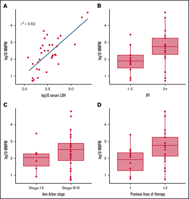
In the BEP, baseline ctDNA MMPM correlated with known prognostic factors including serum LDH levels (r2 = 0.52), number of prior therapies (1 and ≥2), and IPI score (1-2, ≥3) (Figure 2). Baseline ctDNA MMPM also trended weakly with Ann Arbor stages (I-II and III-IV).
Figure 2.
Correlation between baseline ctDNA MMPM and known prognostic factors. (A) serum LDH, (B) IPI score, (C) Ann Arbor stage, and (D) number of prior therapies.
Correlation between baseline ctDNA levels and clinical outcomes
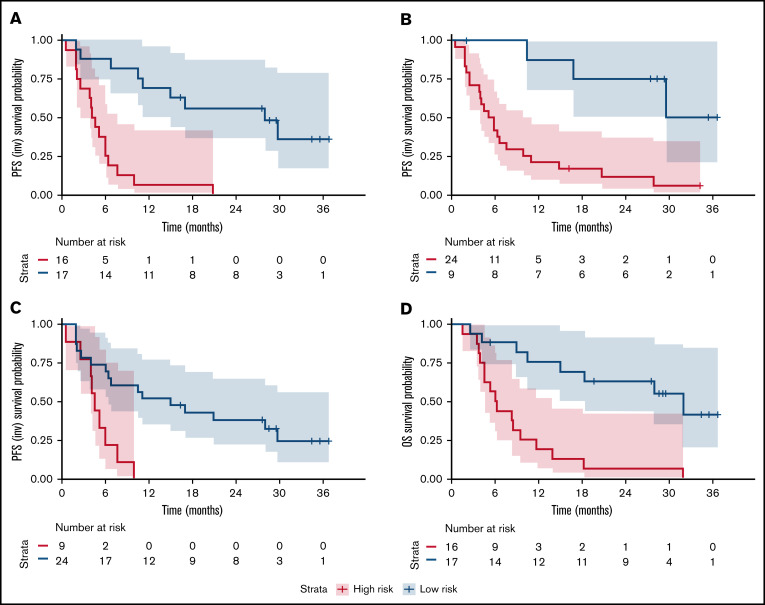
High ctDNA (above the median) at baseline was associated with a shorter PFS (Figure 3A). When stratified by baseline ctDNA, the unadjusted HR was 0.14 (95% CI, 0.05-0.37). After adjusting for number of prior therapies ≥2, IPI score ≥3, and LDH above the upper limit of normal, the HR was 0.18 (95% CI, 0.05-0.65), suggesting that baseline ctDNA was an independent predictor of PFS. The prognostic value of ctDNA for PFS was consistent for all baseline quartile stratifications (Figure 3B-C).
Figure 3.
Survival outcomes according to baseline ctDNA levels. (A) Progression-free survival in patients stratified by ctDNA MMPM at baseline (above and below median), (B) patients with baseline ctDNA MMPM above and below the lower quartile of quantitative values, and (C) patients with baseline ctDNA MMPM above and below the upper quartile of quantitative values. (D) OS in patients stratified by ctDNA MMPM at baseline (above and below median). INV, investigator-assessed.
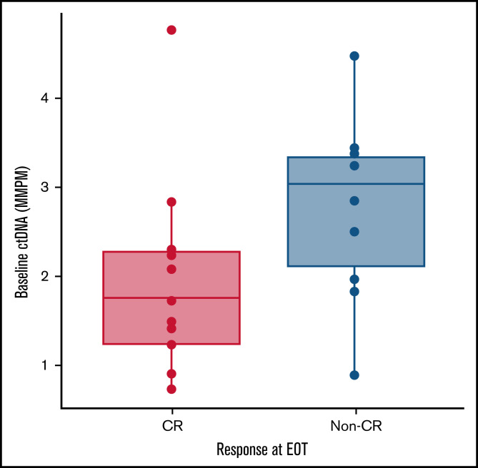
High ctDNA at baseline was also associated with shorter OS (Figure 3D). When patients were stratified by median baseline ctDNA levels, the unadjusted HR was 0.19 (95% CI, 0.08-0.47). After adjusting for number of prior therapies ≥2, IPI score ≥3, and serum LDH concentration above the upper limit of normal, the HR was 0.20 (95% CI, 0.06-0.68). Patients achieving a CR at EOT (n = 13) had significantly lower median ctDNA MMPM at baseline than patients who did not achieve CR (n = 10); P = .049 (Figure 4).
Figure 4.
ctDNA MMPM at baseline in patients with or without CR at EOT; P = .049 for difference between patients with CR and no CR by Wilcoxon test.
Correlation between EOT ctDNA levels and clinical outcomes
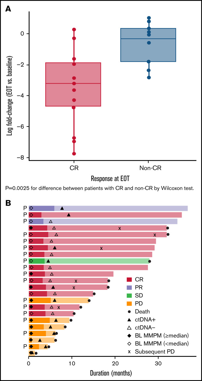
On-treatment log-fold changes in ctDNA MMPM correlated with patient response at EOT. Patients with a CR (n = 13) had a significantly greater median decrease in ctDNA MMPM across timepoints than patients who did not achieve CR (n = 10); P = .0025 (Figure 5A).
Figure 5.
Correlation between post-treatment ctDNA levels and patient response. (A) Log-fold change in ctDNA at EOT vs baseline in patients with or without a CR at EOT. (B) Swimlane plot of individual response to treatment showing patients with baseline ctDNA MMPM above and below the median and ctDNA clearance at EOT; P indicates patients who received polatuzumab vedotin. BL, baseline; PR, partial response; SD, stable disease.
ctDNA clearance at EOT
At EOT, 4 patients had undetectable levels of ctDNA (ie, the signal from tumor-specific variants determined from the baseline sample was indistinguishable from background), and 7 patients had few detectable variants (<5% of total variants identified at baseline); these 11 patients were considered cleared of ctDNA at EOT (supplemental Figure 3). The remaining 12 patients had a considerable number of baseline variants that were still detected at EOT (supplemental Figure 4).
Of the 17 patients in the pola plus BR cohort, 9 (53%) had ctDNA clearance compared with 2 (33%) of the 6 patients in the BR cohort (Table 2). Clinical responses in the 11 patients considered cleared of ctDNA at EOT and in 12 patients in whom tumor-specific variants were detected are shown in Table 2. Among the 9 patients with ctDNA clearance treated with pola plus BR, 8 (89%) achieved a CR at EOT, whereas among the 8 patients without ctDNA clearance treated with pola plus BR, 2 (25%) achieved a CR at EOT.
Table 2.
Summary of responses in patients with and without ctDNA clearance at EOT in the pola plus BR and BR cohorts
| Response | ctDNA cleared at EOT (n = 11) | ctDNA not cleared at EOT (n = 12) | ||
|---|---|---|---|---|
| Pola + BR (n = 9) | BR (n = 2) | Pola + BR (n = 8) | BR (n = 4) | |
| CR | 8 | 2 | 2 | 1 |
| PR | 1 | 0 | 1 | 0 |
| SD | 0 | 0 | 1 | 0 |
| PD | 0 | 0 | 4 | 3 |
Clearance of ctDNA trended with patient response at EOT: it was mainly associated with patients achieving CR, whereas patients with detectable ctDNA at EOT more commonly experienced PD. A swimlane plot of individual response to treatment in patients with baseline ctDNA MMPM above and below the median and those with detectable/nondetectable ctDNA at EOT is shown (Figure 5B).
In patients with ctDNA clearance, no clear trend was observed between the log-fold reduction of ctDNA at EOT and investigator-assessed PFS or OS.
Mutational analysis
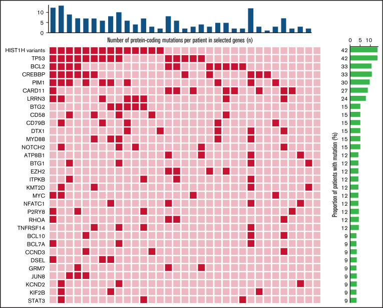
Protein-coding mutations were identified in all but 1 patient in the BEP. The most frequently mutated genes (mutated in at least 3 patients) are shown in the mutational heatmap in Figure 6. Almost half of the patients had mutations in the tumor suppressor gene TP53 at baseline (14/33, 42%). Other frequently mutated genes include histone H1 variants (14/33, 42%), BCL2 (11/33, 33%), CREBBP (11/33, 33%), PIM1 (10/33, 30%), CARD11 (9/33, 27%), LRRN3 (8/33, 24%), and BTG2 (5/33, 15%). CD79b mutation was observed in baseline ctDNA samples in 5 patients, with allelic frequencies ranging from 0.23% to 22.4%. One patient with a CD79b mutation in their baseline ctDNA sample received pola plus BR and achieved a CR at EOT, and 4 patients with a CD79b mutation received BR treatment and had PD at EOT. The distribution of the most frequently mutated genes, in patients with ctDNA clearance at EOT vs those without ctDNA clearance at EOT, is summarized in supplemental Table 1.
Figure 6.
Heatmap showing most frequently mutated genes (mutated in at least 3 patients) in the BEP at EOT. Dark red squares indicate presence of mutation.
Discussion
Outcomes for patients with R/R DLBCL are heterogeneous, and existing prognostic tools fail to consistently predict treatment failure. Despite recent treatment advances, including approvals for pola plus BR and CAR T-cell therapy, most patients with R/R DLBCL will not attain a durable response with any individual approach. Previous studies have used baseline IPI scores and interim PET scans to select subgroups of patients who may benefit from intensified therapy; however, no clear improvement in survival outcomes in these subgroups was demonstrated.15-19 Identifying patients with R/R DLBCL who are at the highest risk of treatment failure is critical to allow the move toward more personalized therapeutic approaches.
ctDNA profiling using targeted panels could facilitate the identification of patients with R/R DLBCL who are at risk of adverse outcomes. Use of a sequencing panel including customized genomic regions known to be enriched in patients with DLBCL, such as SNVs, insertions/deletions, breakpoints of fusions, and IgVH/IgJH, maximizes the number of patients and mutations detected per panel while minimizing the panel size and sequencing cost.12 In the current study, the comprehensive design of our customized panel allowed identification of a median of 133 mutations per patient for tumor monitoring and allowed detection of ctDNA in all patients with DLBCL included in the study.
The current study provides evidence that ctDNA can improve the identification of patients with R/R DLBCL who are at high risk of PD or relapse following treatment with pola plus BR or BR alone. Similar to results reported previously by Roschewski et al,8 in the current study baseline ctDNA levels correlated with known clinical risk factors and demonstrated independent prognostic value for response, PFS, and OS in patients with R/R DLBCL. Notably, baseline ctDNA level was still independently prognostic for PFS and OS after adjustment for LDH above the upper limit of normal. Changes in ctDNA levels from baseline to EOT also correlated with clinical response at EOT.
The utility of ctDNA as a biomarker for the prognosis of survival outcomes has been demonstrated in patients with previously untreated DLBCL, with few assessments of its utility in the R/R DLBCL setting. Pretreatment ctDNA levels were significantly associated with event-free survival in patients with DLBCL receiving either front-line therapy (HR, 2.6 [95% CI, 1.3-5.2]; P = .007) or salvage therapy (HR, 2.9 [95% CI, 1.3-6.4]; P = .01).7 High ctDNA levels were also predictive of significantly worse OS in the salvage setting (HR, 3.3 [95% CI, 1.4-7.5]; P = .0053). In addition, early and large decreases in ctDNA, after mainly first-line treatments for DLBCL, were associated with superior clinical outcomes.7 In patients receiving first-line R-CHOP, ctDNA sequencing enabled the detection of an additional 50% of relapsing cases within 2 years compared with interim PET scans.20 Furthermore, in previously untreated patients with DLBCL, significantly fewer patients with detectable ctDNA at interim assessment were disease free at 5 years compared with patients with undetectable ctDNA.8 In a recent large study, Alig et al evaluated the prognostic value of pretreatment ctDNA levels in a large cohort of patients with treatment-naïve DLBCL. Patients with a shorter diagnosis-to-treatment interval (DTI) had higher pretreatment ctDNA levels (P < .001) than patients with a longer DTI. Both ctDNA levels and DTI were associated with disease burden. Moreover, pretreatment ctDNA levels were prognostic of event-free survival independent of DTI.9 PD after CAR T-cell therapy was also predicted by ctDNA-based MRD in patients with R/R DLBCL.10 Measured 28 days after axicabtagene ciloleucel infusion, median PFS was 93 days in patients who were positive for MRD and was not reached in patients who were negative for MRD (log-rank test P = .001). Median OS was 281 days for patients with detectable MRD and was not reached for those without detectable MRD (log-rank test P = .0399).
The current analysis supports the findings of these previous studies in that ctDNA has the potential to be an important prognostic marker of survival for patients with DLBCL and is the first prospective clinical trial to report on ctDNA measurement of patients in the R/R DLBCL setting.
The mutation profile of ctDNA in patients with R/R DLBCL is not fully understood. In the current study, almost half of the biomarker-evaluable patients had a TP53 mutation, which is high compared with that seen in newly diagnosed DLBCL. In patients with newly diagnosed DLBCL, the frequency of TP53 mutations in DNA extracted from tumor tissue usually ranges from 12.5% to 22.1%,21-23 and TP53 mutations are an independent marker of poor survival outcomes in patients with DLBCL.21 Similar to our findings, in an analysis of ctDNA by Rushton et al, of patients with R/R DLBCL participating in clinical trials, 51% of patients had TP53 mutations.24 Rushton et al concluded that TP53 may have a role in DLBCL primary treatment resistance; however, the number of patients in the current study is limited, and our findings would need to be verified in a larger population before any conclusions can be drawn. In addition, we observed that for some patients with detectable ctDNA at EOT, almost all of the variants exhibited a similar change in MMPM by EOT; for the rest of the patients with detectable ctDNA at EOT, a subset of the variants were not detectable at EOT (those on the x-axis of supplemental Figure 4), whereas the remaining variants were detected. One possible explanation for this discordant pattern is that in the latter group of patients, a proportion of the disease clones were cleared by the treatment, whereas the rest did not respond to the treatment. Interestingly, in our study, among the 23 patients who had paired samples available at EOT, 9 had TP53 mutations at baseline, 2 of whom had ctDNA clearance at EOT. TP53 mutations were detected in 6 of the 7 patients who did not have ctDNA clearance at EOT. Future studies with a larger sample size are needed to validate any differential clearance based on mutation type.
A potential limitation of the methods used in this analysis is the current lack of consensus on threshold concentrations of ctDNA that might be used to predict patient response to therapy.25 In the multivariate Cox regression, there was multicollinearity in the model due to overlap of several prognostic factors. Furthermore, this analysis was based on a relatively small sample size, which limited the identification of an optimal cutoff for risk stratification. Ideally, a traditional method, such as receiving operator characteristic curves, would have been used to determine the optimal ctDNA threshold. However, adopting such an approach would have led to over-fitting of the data; therefore, the median MMPM was used as a prespecified cutoff in our analysis. The relatively small dataset also prevented the evaluation of ctDNA as a predictive biomarker of response to treatment with BR with or without polatuzumab vedotin. However, ctDNA samples were collected prospectively as part of a clinical trial with relative uniformity of treatment, which is an advantage of the design of this study. In addition, well-characterized patients participating in the GO29365 clinical trial were studied; this enabled multivariate analyses and correlations between ctDNA and other known prognostic factors to be performed. Despite this, our findings will require validation in future studies.
Although not performed in the current study, analysis of ctDNA methylation may also have prognostic value in DLBCL. In one study, aberrant DAPK1 methylation in plasma samples was an independent prognostic marker for OS (HR, 8.9 [95% CI, 2.7-29.3]; P < .0007) in patients with DLBCL.26 In addition, in a multivariate analysis, global hypomethylation in ctDNA samples was an independent risk factor for poor OS (HR, 11.87 [95% CI, 2.8-50.2]; P = .001) in patients with DLBCL.27 These data warrant further evaluation of the prognostic value of ctDNA methylation in future studies.
In conclusion, assessment of ctDNA may have value as a prognostic biomarker in DLBCL. It is a highly sensitive, noninvasive method that has the potential to enhance prognostication and prediction of treatment outcome in patients with DLBCL. ctDNA measurements may also enable the enhancement of tailored treatment approaches for DLBCL. As the utility of ctDNA in R/R DLBCL is further validated and ctDNA analyses become more refined with faster turnaround and standardized thresholds, it may be possible to use real-time, ctDNA-based interventions to predict responses during treatment with potential to switch from an unsuccessful therapeutic strategy at an earlier timepoint. Overall, the findings of the current study support the further evaluation of the use of ctDNA as a monitoring tool in the R/R DLBCL setting.
Supplementary Material
The full-text version of this article contains a data supplement.
Acknowledgments
The authors thank the participating patients and their families and the research nurses, study coordinators, and operations staff. Bioinformatics analysis was performed by Hai Lin and Ehsan Tabari of Roche Sequencing Solutions. Plasma samples were processed and next-generation sequencing (NGS) libraries were prepared and sequenced by Josh Lefkowitz of Roche Sequencing Solutions. Third-party medical writing assistance, under the direction of the authors, was provided by Angela Rogers and Carla Smith of Ashfield MedComms, an Ashfield Health company, and was funded by F. Hoffmann-La Roche Ltd.
This study was sponsored by Genentech, Inc. and F. Hoffmann-La Roche Ltd. A.F.H. is supported by the Emmet and Toni Stephenson Leukemia and Lymphoma Society Scholar Award and the Lymphoma Research Foundation Larry and Denise Mason Clinical Investigator Career Development Award.
Authorship
Contribution: A.F.H. contributed to the study concept and clinical study design; S.T. and J.N.P. performed the statistical analysis; and all authors contributed to the collection and assembly of study data and to the data analysis and interpretation, reviewed the data, vouch for the completeness and accuracy of the results and the trial’s fidelity to the protocol, reviewed the manuscript, and agreed on its submission for publication.
Conflict-of-interest disclosure: A.F.H. reports research funding from Bristol Myers Squibb, Merck, Genentech, Inc., F. Hoffmann-La Roche Ltd, Gilead Sciences, Seattle Genetics, AstraZeneca, and ADC Therapeutics; and consultancy for Bristol Myers Squibb, Merck, Genentech, Inc., F. Hoffmann-La Roche Ltd, Kite Pharma/Gilead, Seattle Genetics, Karyopharm, Takeda, Tubulis, AstraZeneca, and ADC Therapeutics. S.O. reports research funding from AbbVie, AstraZeneca, Beigene, Gilead, Epizyme, Janssen, Merck, F. Hoffmann-La Roche Ltd, and Takeda; and consultancy for and honoraria from AbbVie, AstraZeneca, Celgene, Gilead, Janssen, Merck, F. Hoffmann-La Roche Ltd, Sandoz, and Takeda. L.H.S. reports research funding from Teva; and consultancy for and honoraria from AbbVie, Acerta, Amgen, Apobiologix, AstraZeneca, Celgene, Gilead, Incyte, Janssen, Kite, Karyopharm, Lundbeck, Merck, Morphosys, F. Hoffmann-La Roche Ltd, Genentech, Inc., Sandoz, Seattle Genetics, Teva, Takeda, TG Therapeutics, and Verastem Oncology. A.F.L. was formerly employed by Genentech, Inc., holds stock in F. Hoffmann-La Roche Ltd, is employed by Freenome, and holds stock options in Freenome. B.C. was formerly employed by Genentech, Inc. and holds stock in F. Hoffmann-La Roche Ltd. S.T., J.R., L.M., J.N.P., and Y.J. are employed by Genentech, Inc. and hold stock in F. Hoffmann-La Roche Ltd.
Correspondence: Alex F. Herrera, City of Hope, 1500 E. Duarte Rd, Duarte, CA 91010; e-mail: aherrera@coh.org.
References
- 1.Sehn LH, Salles G. Diffuse large B-cell lymphoma. N Engl J Med. 2021;384(9):842-858. [DOI] [PMC free article] [PubMed] [Google Scholar]
- 2.Polson AG, Calemine-Fenaux J, Chan P, et al. Antibody-drug conjugates for the treatment of non-Hodgkin’s lymphoma: target and linker-drug selection [published correction appears in Cancer Res. 2010;70(3):1275]. Cancer Res. 2009;69(6):2358-2364. [DOI] [PubMed] [Google Scholar]
- 3.Okazaki M, Luo Y, Han T, Yoshida M, Seon BK. Three new monoclonal antibodies that define a unique antigen associated with prolymphocytic leukemia/non-Hodgkin’s lymphoma and are effectively internalized after binding to the cell surface antigen. Blood. 1993;81(1):84-94. [PubMed] [Google Scholar]
- 4.U.S. Food and Drug Administration. POLIVY U.S. Prescribing Information. https://wwwaccessdatafdagov/drugsatfda_docs/label/2019/761121s000lbl.pdf. Accessed 5 March 2021.
- 5.EMA. POLIVY: product information. https://www.ema.europa.eu/en/documents/product-information/polivy-epar-product-information_en.pdf. Accessed 5 March 2021.
- 6.Sehn LH, Herrera AF, Flowers CR, et al. Polatuzumab vedotin in relapsed or refractory diffuse large B-cell lymphoma. J Clin Oncol. 2020;38(2):155-165. [DOI] [PMC free article] [PubMed] [Google Scholar]
- 7.Kurtz DM, Scherer F, Jin MC, et al. Circulating tumor DNA measurements as early outcome predictors in diffuse large B-Cell lymphoma. J Clin Oncol. 2018;36(28):2845-2853. [DOI] [PMC free article] [PubMed] [Google Scholar]
- 8.Roschewski M, Dunleavy K, Pittaluga S, et al. Circulating tumour DNA and CT monitoring in patients with untreated diffuse large B-cell lymphoma: a correlative biomarker study. Lancet Oncol. 2015;16(5):541-549. [DOI] [PMC free article] [PubMed] [Google Scholar]
- 9.Alig S, Macaulay CW, Kurtz DM, et al. Short diagnosis-to-treatment interval is associated with higher circulating tumor DNA levels in diffuse large B-cell lymphoma. J Clin Oncol. 2021;39(23):2605-2616. [DOI] [PMC free article] [PubMed] [Google Scholar]
- 10.Frank MJ, Hossain NM, Bukhari AA, et al. Monitoring ctDNA in r/r DLBCL patients following the CAR T-cell therapy axicabtagene ciloleucel: day 28 landmark analysis. J Clin Oncol. 2019;37(15 _suppl):7552-7552. [Google Scholar]
- 11.Newman AM, Lovejoy AF, Klass DM, et al. Integrated digital error suppression for improved detection of circulating tumor DNA. Nat Biotechnol. 2016;34(5):547-555. [DOI] [PMC free article] [PubMed] [Google Scholar]
- 12.Scherer F, Kurtz DM, Newman AM, et al. Distinct biological subtypes and patterns of genome evolution in lymphoma revealed by circulating tumor DNA. Sci Transl Med. 2016;8(364):364ra155. [DOI] [PMC free article] [PubMed] [Google Scholar]
- 13.Cingolani P, Platts A, Wang le L, et al. A program for annotating and predicting the effects of single nucleotide polymorphisms, SnpEff: SNPs in the genome of Drosophila melanogaster strain w1118; iso-2; iso-3. Fly (Austin). 2012;6(2):80-92. [DOI] [PMC free article] [PubMed] [Google Scholar]
- 14.Pati A, Lovejoy A, Shi PW, et al. Detection of minimal residual disease from less than one cell equivalent in liquid biopsy samples using the AVENIO ctDNA Surveillance Kit. Cancer Res. 2018;78(13 suppl):4574. [Google Scholar]
- 15.Casasnovas RO, Ysebaert L, Thieblemont C, et al. FDG-PET-driven consolidation strategy in diffuse large B-cell lymphoma: final results of a randomized phase 2 study. Blood. 2017;130(11):1315-1326. [DOI] [PubMed] [Google Scholar]
- 16.Stiff PJ, Unger JM, Cook JR, et al. Autologous transplantation as consolidation for aggressive non-Hodgkin’s lymphoma. N Engl J Med. 2013; 369(18):1681-1690. [DOI] [PMC free article] [PubMed] [Google Scholar]
- 17.Swinnen LJ, Li H, Quon A, et al. Response-adapted therapy for aggressive non-Hodgkin’s lymphomas based on early [18F] FDG-PET scanning: ECOG-ACRIN Cancer Research Group study (E3404). Br J Haematol. 2015;170(1):56-65. [DOI] [PMC free article] [PubMed] [Google Scholar]
- 18.Hertzberg M, Gandhi MK, Trotman J, et al. ; Australasian Leukaemia Lymphoma Group (ALLG) . Early treatment intensification with R-ICE and 90Y-ibritumomab tiuxetan (Zevalin)-BEAM stem cell transplantation in patients with high-risk diffuse large B-cell lymphoma patients and positive interim PET after 4 cycles of R-CHOP-14. Haematologica. 2017;102(2):356-363. [DOI] [PMC free article] [PubMed] [Google Scholar]
- 19.Chiappella A, Martelli M, Angelucci E, et al. Rituximab-dose-dense chemotherapy with or without high-dose chemotherapy plus autologous stem-cell transplantation in high-risk diffuse large B-cell lymphoma (DLCL04): final results of a multicentre, open-label, randomised, controlled, phase 3 study. Lancet Oncol. 2017;18(8):1076-1088. [DOI] [PubMed] [Google Scholar]
- 20.Macaulay C, Alig S, Kurtz DM, et al. Interim circulating tumor DNA as a prognostic biomarker in the setting of interim PET-based adaptive therapy for DLBCL. Am J Hematol. 2019;134(suppl 1):1600. [Google Scholar]
- 21.Xu-Monette ZY, Wu L, Visco C, et al. Mutational profile and prognostic significance of TP53 in diffuse large B-cell lymphoma patients treated with R-CHOP: report from an International DLBCL Rituximab-CHOP Consortium Program Study. Blood. 2012;120(19):3986-3996. [DOI] [PMC free article] [PubMed] [Google Scholar]
- 22.Peroja P, Pedersen M, Mantere T, et al. Mutation of TP53, translocation analysis and immunohistochemical expression of MYC, BCL-2 and BCL-6 in patients with DLBCL treated with R-CHOP. Sci Rep. 2018;8(1):14814. [DOI] [PMC free article] [PubMed] [Google Scholar]
- 23.Young KH, Weisenburger DD, Dave BJ, et al. Mutations in the DNA-binding codons of TP53, which are associated with decreased expression of TRAILreceptor-2, predict for poor survival in diffuse large B-cell lymphoma. Blood. 2007;110(13):4396-4405. [DOI] [PMC free article] [PubMed] [Google Scholar]
- 24.Rushton CK, Arthur SE, Alcaide M, et al. Genetic and evolutionary patterns of treatment resistance in relapsed B-cell lymphoma. Blood Adv. 2020;4(13):2886-2898. [DOI] [PMC free article] [PubMed] [Google Scholar]
- 25.Pessoa LS, Heringer M, Ferrer VP. ctDNA as a cancer biomarker: a broad overview. Crit Rev Oncol Hematol. 2020;155:103109. [DOI] [PubMed] [Google Scholar]
- 26.Kristensen LS, Hansen JW, Kristensen SS, et al. Aberrant methylation of cell-free circulating DNA in plasma predicts poor outcome in diffuse large B cell lymphoma. Clin Epigenetics. 2016;8(1):95. [DOI] [PMC free article] [PubMed] [Google Scholar]
- 27.Wedge E, Hansen JW, Garde C, et al. Global hypomethylation is an independent prognostic factor in diffuse large B cell lymphoma. Am J Hematol. 2017;92(7):689-694. [DOI] [PubMed] [Google Scholar]
Associated Data
This section collects any data citations, data availability statements, or supplementary materials included in this article.


