For the study relating to this case report see A. I. Guaricci, G. Chiarello, E. Gherbesi, L. Fusini, N. Soldato, P. Siena, et al. Coronary-specific quantification of myocardial deformation by strain echocardiography may disclose the culprit vessel in patients with non–ST-segment elevation acute coronary syndrome. European Heart Journal Open 2022; doi: 10.1093/ehjopen/oeac010. https://doi.org/10.1093/ehjopen/oeac010.
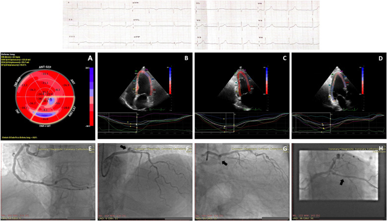
A 64-year-old patient was admitted with chest pain hypothetically attributed to gastro-oesophageal reflux. The 12-lead-electrocardiogram showed sinus rhythm at 61 b.p.m. and non-specific left ventricle (LV) repolarization changes. The diagnosis of non-ST elevation myocardial infarction (NSTEMI) was ascertained with the evidence of a high level of hs-TnI of 23 930.4 pg/mL (99th percentiles of hs-TnI in the normal subject was 74.9 pg/mL). The echocardiographic exam revealed the absence of LV wall motion abnormalities (wall motion score index =1) with preserved left ventricular ejection fraction = 50% (Videos 1 and 2). Territorial longitudinal strain-left anterior descending (TLS-LAD), -right coronary artery (TLS-RCA), and -left circumflex artery (TLS-CX) were −18, −16, and −11.7, respectively (Figure 1). The Global Longitudinal Strain (GLS) was −14.8.
Figure 1.
Upper panel shows 12-lead-electrocardiogram obtained at admission. Middle panels show echocardiographic images: (A) Bull’s eye presenting strain values corresponding to each myocardial segment and the GLS value. Segments delineated by the yellow mark represent the territorial longitudinal strain relative to the myocardial portion vascularized by circumflex artery. (B–D) Myocardial deformation images in apical four-, two-, and three-chamber views, respectively. Lower panels show invasive coronary angiograms: (E) Patent right coronary artery; (F, G) patent left anterior descending and occluded circumflex artery (arrows); (H) patent circumflex artery after percutaneous transluminal coronary angioplasty.
At invasive coronary angiography, right coronary was mildly diseased, the left main presented with a short non-significant stenosis of 25–30% at bifurcation, left anterior descending showed a brief stenosis less than 50% at the level of the middle tract and CX was occluded immediately after the origin. The patient underwent percutaneous coronary angioplasty and stent implantation (PCI+DES) on Cx-OM2 with thrombolysis in myocardial infarction Grade III flow at the end of the procedure. Left circumflex artery was demonstrated to be a non-dominant vessel.
In the setting of NSTE-ACS patients, the conventional echocardiographic assessment may reveal regional myocardial wall motion abnormality of the LV as well as the absence of kinetic alteration (25–76% of cases).1 Speckle tracking echocardiography (STE) is a validated and accurate technique able to detect subtle changes in the regional systolic LV function.2 In this case, coronary-specific quantification of myocardial deformation by STE revealed the LCX as the culprit vessel irrespective of visual wall motion evaluation.
Strain imaging would be of additional benefit in clinical practice since patients affected by myocardial infarction often present with multiple unstable lesions and sometimes more than one culprit lesion.3 Current guidelines recommend invasive coronary angiography within 24 h of hospitalization of NSTEMI, but increased demands on hospital systems mean that additional clinical factors need to be considered to prioritize patients. Territorial longitudinal strain may provide an additional tool that is very fast, non-invasive, and easily accessible to help prioritize the timing of coronary angiography. However, the use of TLS in this setting has not been validated, and further studies are needed.
Consent: The authors confirm that written consent for submission and publication of this case report including images and associated text has been obtained from the patient in line with COPE guidance.
Conflict of interest: None declared.
Funding: None declared.
References
- 1.Collet JP, Thiele H, Barbato E, Barthélémy O, Bauersachs J, Bhatt DL, et al. ESC Scientific Document Group. 2020 ESC Guidelines for the management of acute coronary syndromes in patients presenting without persistent ST-segment elevation. Eur Heart J 2021;42:1289–1367. [DOI] [PubMed] [Google Scholar]
- 2.Atici A, Barman HA, Durmaz E, Demir K, Cakmak R, Tugrul S. et al. Predictive value of global and territorial longitudinal strain imaging in detecting significant coronary artery disease in patients with myocardial infarction without persistent ST-segment elevation. Echocardiography 2019;36:512–520. [DOI] [PubMed] [Google Scholar]
- 3.Goldstein JA, Demetriou D, Grines CL, Pica M, Shoukfeh M, O'Neill W.. Multiple complex coronary plaques in patients with acute myocardial infarction. N Engl J Med 2000;343:915–922. [DOI] [PubMed] [Google Scholar]


