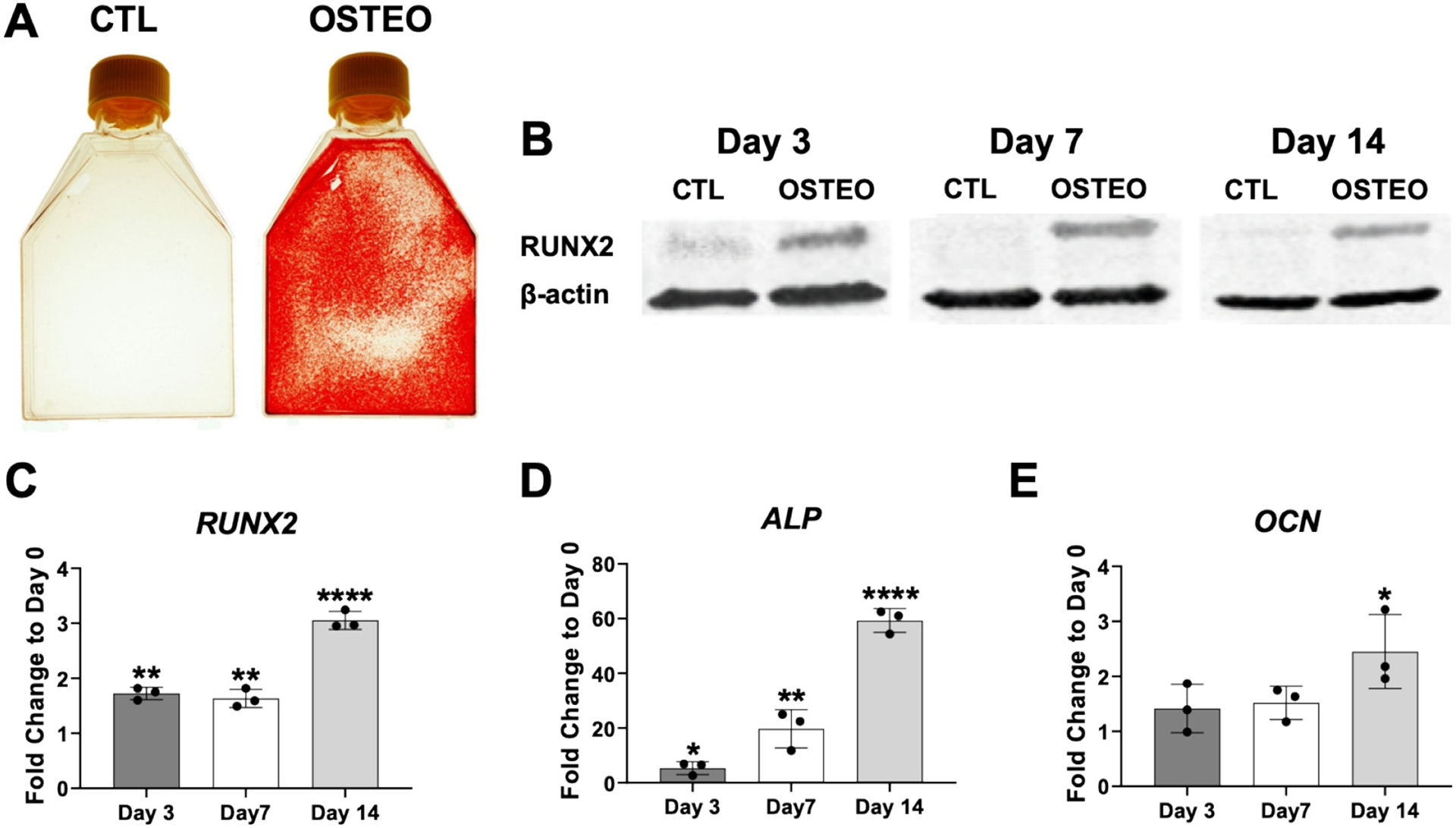
Fig. 1.

Confirmation of osteogenic differentiation. Panel (A) shows positive Alizarin Red staining following osteogenic differentiation (OSTEO) of human bone marrow-derived MSCs at 3 weeks. No staining was detected in control (CTL) MSC cultures grown at the same time in regular growth medium. Panel (B) shows increased levels of RUNX2 in protein lysates harvested from MSCs at days 3, 7 and 14 of osteogenic induction when compared to control, non-induced cultures. Panels C-E show expression patterns for RUNX2, alkaline phosphatase (ALP) and osteocalcin (OCN) at different time points during osteogenesis (fold change expression relative to day 0). Data in A, B is from MSC-Sample 1 (MSC-S1) cell line that was used in the microarray study. Similar results were shown for MSC-S2 and MSC-S3 samples (results not shown). Data in C-E is from the three MSC samples used for the microarray (data expressed +/− SD, n = 3; *p < 0.05; ** p < 0.01; **** p < 0.0001).
