Abstract
Purpose
To measure visual fields using two-color dark-adapted chromatic perimetry in a subset of participants in the Rate of Progression of USH2A-related Retinal Degeneration (RUSH2A), a study of USH2A-mediated syndromic (USH2) and autosomal recessive nonsyndromic retinitis pigmentosa, determine percentage retaining rod function, and explore relationships between dark-adapted visual fields (DAVF) and rod function from ERG and full-field stimulus thresholds (FST).
Methods
Full-field rod mean sensitivity, number of rod loci, maximum sensitivity, DAVF full-field hill of vision (DAVF VTOT), and 30° hill of vision (DAVF V30) were measured in one eye for DAVF ancillary study participants (n = 49). Loci where cyan relative to red sensitivity was more than 5 dB on dark-adapted chromatic perimetry were considered rod mediated. Correlation coefficients between the DAVF measures and standard clinical measures were estimated, as were kappa statistics (κ) for agreement between DAVF and other measures of rod function.
Results
Of 49 participants tested with DAVF, 38 (78%) had evidence of rod function, whereas 15 (31%) had measurable rod ERGs. DAVF maximum sensitivity was highly correlated with FST white thresholds (r = −0.80; P < .001). Although not statistically significant, the number of rod loci and DAVF VTOT were lower in eyes with longer disease duration by 0.82 (95% confidence interval, −1.76, 0.12) loci/year and 0.59 (95% confidence interval, −1.82, 0.64) dB-steradians/year, respectively.
Conclusions
Rod-mediated function on FST and DAVF is present in many patients with symptomatic USH2A-related retinal degeneration, including some without measurable rod ERGs. RUSH2A longitudinal data will determine how these measures change with disease progression and whether they are useful for longitudinal studies in inherited retinal degenerations.
Keywords: visual fields, dark adaptation, autosomal recessive retinitis pigmentosa, Usher syndrome type 2, full-field stimulus test
The Rate of Progression of USH2A-related Retinal Degeneration (RUSH2A) 4-year natural history study of Usher syndrome type 2 (USH2) and nonsyndromic autosomal recessive retinitis pigmentosa (ARRP) was initiated in 2017 to estimate annual change in state-of-the-art outcome measures in patients with USH2A-related retinal degeneration.1 Measures such as best-corrected visual acuity (BCVA), static perimetry, and contrast sensitivity measure primarily cone-mediated retinal function. However, loss of rod function is known to cause night blindness in patients with USH2A disease, and this is often the earliest symptom reported by patients. The RUSH2A study incorporates rod ERGs at baseline and full-field sensitivity testing (FST) annually to gain some insight into the progression of rod loss in these patients. However, full-field rod ERGs were not measurable in most participants at baseline.2 FST provides a measure at the most sensitive location but does not provide any topographical information about rod loss.3,4 To provide more extensive monitoring of rod function in RUSH2A, dark-adapted visual field (DAVF) testing at selected sites was added beginning at the 12-month RUSH2A visit, hereafter referred to as the DAVF initial visit.
Here we identify and measure rod-mediated visual field thresholds using two-color dark-adapted chromatic (DAC) perimetry5 in a subset of participants in the RUSH2A study. One aim of this DAVF ancillary study is to estimate the proportion of participants with evidence of rod function at DAVF initial visit testing. Additional aims are to test for the association of rod function with age and disease duration, and for correlation with measures of cone function such as BCVA and photopic static perimetry. We also explore the relationship between DAVF from DAC perimetry and parameters of rod function from ERG and FST. Since ERG measures a pan-retinal response, and FST measures only the most sensitive region, there could be large changes in localized regions that go undetected using these modalities. The present study will address a major knowledge gap in outcome measures for rod–cone degenerations by providing localized information about rod function in patients with USH2A-related retinal degeneration.
Methods
Study Design
The RUSH2A multicenter, international, longitudinal natural history study has enrolled participants at 16 clinical sites in North America, South America, and Europe.1 The protocol and informed consent process adhered to the tenets of the Declaration of Helsinki and were approved by the ethics boards of all participating sites. Written informed consent was obtained from all study participants before enrollment.
Eligibility and Enrollment
Participants in the RUSH2A study had USH2 or nonsyndromic ARRP with at least two homozygous or heterozygous disease-causing variants in USH2A, based on a genetic report from a CLIA-certified laboratory or the equivalent in non-US countries.1 Participants with USH2 who had at least two disease-causing mutations and participants with homozygous disease-causing mutations did not undergo segregation analysis, but segregation studies were performed to confirm inheritance of all compound heterozygous pathogenic variants in participants with ARRP. A patient-reported history of hearing loss and review of the baseline audiology examination by an independent audiologist determined the diagnosis of USH2 (vs. ARRP). Additional criteria for the RUSH2A study were baseline ETDRS visual acuity letter score of 54 or more (approximate Snellen equivalent of 20/80 or better), stable fixation, and clinically determined kinetic visual field III4e diameter of 10° or more in every meridian of the central field of the study eye (defined as the eye with better BCVA and determined at investigator discretion if both eyes had the same BCVA) using the Octopus 900 Pro (Haag-Streit, Mason, OH) as described previously.1
DAVFs
Six RUSH2A sites that had access to a Medmont DAC perimeter (Medmont, Nunawading, Australia) participated in the DAVF Ancillary study. The equipment was purchased by the sites independent of and before the initiation of the RUSH2A study. Adult participants in the RUSH2A primary cohort who consented to DAVF testing were tested for DAVF rod function (only in the study eye) at the 12-month RUSH2A study visit. Phenylephrine 2.5% and tropicamide were used for dilation and cycloplegia. The study eye was dark adapted for 45 minutes before testing. A 1.72° stimulus (equivalent to the Goldman size V) was presented for 200 ms. The response time was set to 400 ms and the interval between stimuli was fixed at 1.1 seconds. The maximum luminance of the cyan stimulus (dominant wavelength = 505 nm; half bandwidth = 28 nm) was 12.58 cd/m2 and the dynamic range was approximately 75 dB. The maximum luminance of the red stimulus (dominant wavelength = 625 nm, half bandwidth = 25 nm) was 4.64 cd/m2.
Trial lenses were used in the central field as needed for correction of refractive errors and age-related accommodation loss. The test eye was aligned in the infrared viewing window and the position of the pupil was monitored throughout the examination. Patient-controlled pauses were encouraged as often as needed to prevent fatigue. Test points (n = 75) in the grid were separated by 12° and extended 132° across the temporal to nasal field and 78° across the superior to inferior field. Far eccentric loci were tested after an automated relocation of the fixation target. The examination paused while the location of the fixation target changed. The participant's eye was realigned in the viewing window before the test continued. Care was taken to instruct and confirm proper head alignment (the head remains stationary, and the eyes move to a new fixation location). A two-down, one-up staircase algorithm used a bracketing strategy to determine the threshold for stimulus detection at each point. Each field took approximately 15 minutes to complete.
The quality of each participant's examination was assessed by the percentage of false-positive responses. Exams with more than 15% false positives were excluded; based on this criterion, no participants were excluded from analysis.
Defining Rod-Mediated Stimulus Detection
Two-color perimetry was used to determine whether rods or cones were mediating detection at each location.6,7 Under dark-adapted conditions, mean normal sensitivity values are 20 to 25 dB higher to cyan (505 nm) than to red (625 nm).5 This finding is consistent with the scotopic luminosity curve, where the rods are approximately 2.5 log units more sensitive to 505 nm than to 625 nm.8 As the rods degenerate, sensitivity to cyan decreases, whereas sensitivity to red decreases slightly, but is then mediated by cones. Sensitivity to cyan continues to decrease as rods degenerate until it, too, is mediated by cones. The cyan stimulus is 4.4 dB brighter than the red stimulus in cd/m2, which is consistent with the mean normal sensitivity to cyan being approximately 4 dB greater than to the red under light-adapted conditions.5 Thus, the spectral sensitivity difference (cyan – red) at each point stipulated whether rods were mediating the detection of the stimulus. Participants were considered to have evidence of rod function if the cyan–red difference was more than 5 dB for at least one cluster of three or more locations within the field. This criterion of three or more points in a cluster was adopted to avoid a spurious high cyan value, leading to a participant being considered to have rod function. Topographic analysis of cyan values for all rod-mediated locations was provided by visual field modeling and analysis, which produces three-dimensional surface models of the hill of vision (HOV).9 The total volume (VTOT) in decibels-steradians beneath the surface of the thin plate spline representation of the HOV and within the external boundary of the grid was quantified, as was the volume within the central 30° of the field (V30). Participants with evidence of rod function at the DAVF initial visit (RUSH2A 12-month visit) will complete additional DAVF testing at 12, 24, and 36 months (RUSH2A 24-, 36-, and 48-month visits) subsequent to the initial visit.
Statistical Analysis
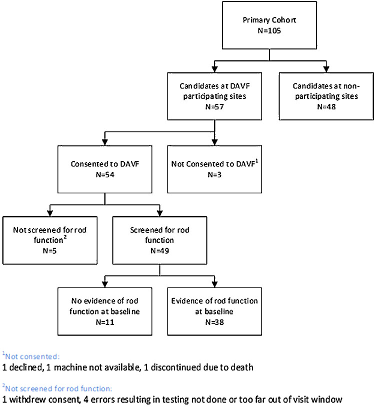
Forty-nine participants were tested for DAVF rod function (Fig. 1). Logistic regression was used to test for differences in the proportion with rod function on DAVF among the USH2 and ARRP groups, adjusted for disease duration (the difference between participant age and the self-reported age of symptom onset). Differences in the proportion with rod function on DAVF among age and disease duration groups were tested using Fisher's exact test. Linear regression was carried out to estimate the rate of decline and 95% confidence interval (CI) of the DAVF continuous measures as a function of disease duration. Spearman correlation coefficients were estimated between DAVF measures (VTOT, V30, maximum/mean sensitivity, and full-field rod mean sensitivity treated as continuous variables for analysis) and standard summary measures of visual field obtained using Octopus 900 Pro.1 Spearman correlation coefficients were also estimated between DAVF initial visit measures (at RUSH2A 12-month visit) and clinical measures obtained at the same visit from the FST white stimulus and FST blue stimulus. ERG data (the amplitude of the b-wave from the dark adapted dim-flash 0.01 cd.s/m2 ERG response) obtained at RUSH2A baseline was used to obtain correlations with DAVF measures, because ERG testing was not performed at the initial DAVF visit. Bias-corrected accelerated bootstrap method was used to get 95% CIs for the correlation coefficients.10,11 ANOVA was used to evaluate the relationship between maximum DAVF sensitivity (highest sensitivity value within the testing field) and FST threshold, and residuals were regressed on FST threshold to determine whether the variance of maximum sensitivity increased with FST thresholds. Evidence for rod function by FST was defined as a threshold of less than −30 dB.2 ERG rod function was defined as a b-wave amplitude of greater than 0.2 Kappa statistics (κ) were calculated to measure agreement between evidence of rod function by DAVF versus other modalities. Owing to the small sample size, the bias-corrected percentile bootstrap method was used to get 95% CIs for kappa estimates.12,13 Analyses were performed using SAS version 9.4 (SAS Institute, Cary, NC).
Figure 1.
DAVF ancillary study enrollment flow chart.
Results
The enrollment flow chart for the study is shown in Figure 1. Forty-nine of 105 participants in the primary RUSH2A cohort were tested for DAVF rod function. The baseline characteristics of this subset of participants at the DAVF initial visit are shown in Table 1. Of the screened participants, 29 (59%) were female, 44 (90%) identified themselves as White, and 31 (63%) were enrolled from the United States or Canada. The median age of participants was 37 years (interquartile range [IQR], 30–48), the median age at onset was 19 years (IQR, 14–27), and the median disease duration was 14 years (IQR, 7–21].
Table 1.
Characteristics of Study Participants at the DAVF Initial Visit
| Evidence of | ||
|---|---|---|
| Rod Function at | ||
| DAVF Initial Visit | ||
| DAVF Initial | ||
| Visit Characteristics | No Evidence | Evidence |
| (N = 11) | (N = 38) | |
| Gender | ||
| Female | 8 (28%) | 21 (72%) |
| Male | 3 (15%) | 17 (85%) |
| Clinical diagnosis | ||
| USH2 | 9 (32%) | 19 (68%) |
| ARRP | 2 (10%) | 19 (90%) |
| Race/ethnicity | ||
| White | 8 (18%) | 36 (82%) |
| Hispanic | 2 (67%) | 1 (33%) |
| Asian | 1 (50%) | 1 (50%) |
| Enrollment area | ||
| United States/Canada | 5 (16%) | 26 (84%) |
| Europe/UK | 6 (33%) | 12 (67%) |
| Age at visit, years | ||
| Median (IQR) | 40.5 (31.0, 45.8) | 37.2 (28.0, 48.8) |
| [Min, Max] | [26.4, 64.1] | [17.8, 69.7] |
| <40 | 5 (18%) | 23 (82%) |
| ≥40 | 6 (29%) | 15 (71%) |
| Age at onset, years | ||
| Median (IQR) | 19.0 (13.0, 24.0) | 19.5 (14.0, 37.0) |
| [Min, Max] | [12.0, 27.0] | [7.0, 56.0] |
| <16 | 3 (19%) | 13 (81%) |
| [16, 25] | 6 (35%) | 11 (65%) |
| ≥25 | 2 (13%) | 14 (88%) |
| Duration of disease, years | ||
| Median (IQR) | 17.7 (12.8, 27.9) | 13.1 (7.1, 20.3) |
| [Min, Max] | [7.0, 45.1] | [2.0, 37.4] |
| <10 | 2 (13%) | 14 (88%) |
| [10, 20] | 5 (28%) | 13 (72%) |
| ≥20 | 4 (27%) | 11 (73%) |
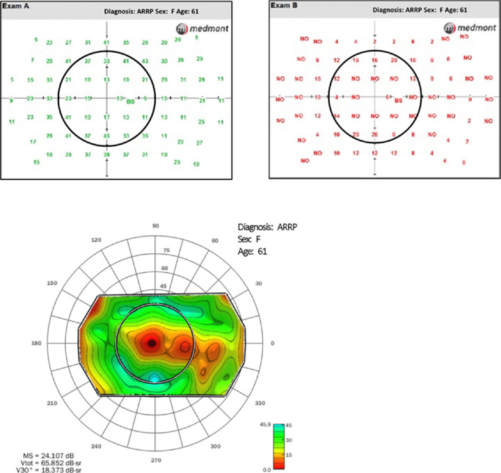
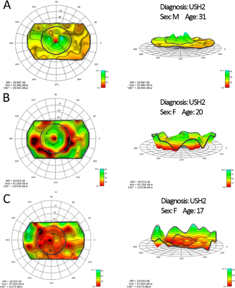
DAVF results from an eye retaining evidence of rod function are shown in Figure 2. Sensitivity at most field locations is higher for cyan than for red (Fig. 2a). The HOV model for rod-mediated sensitivity is shown in Figure 2b. This participant eye showed minimal rod-mediated function in the central retina, but retained high rod sensitivity in the periphery. Other patterns of rod visual field are shown in Figure 3, with top-down views shown in the left column and side views shown in the right column. Row A shows a participant with preserved rod function throughout the periphery and peaks in the central 30°, row B shows a participant with a deep mid peripheral scotoma and row C shows a participant with residual rod function only in the far periphery.
Figure 2.
Derivation of rod visual fields in a participant with ARRP. Fields were obtained twice, once with a cyan test and once with a red test (a). Not seen points are indicated with NO. Locations where the cyan-red difference was greater than 5 dB were considered rod mediated. Topographic analysis of cyan values for all rod-mediated locations was provided by visual field modeling and analysis (b).
Figure 3.
Representative rod visual fields, with top-down views shown in the left column and side views shown in the right column. Row A shows a participant with preserved rod function in the central 30°. Row B shows a participant with a deep mid peripheral scotoma, and row C shows a participant with only residual rod function in the far periphery.
Thirty-eight of the 49 screened participants (78%) had evidence of rod function. As shown in Table 1, the age of the reported onset of disease and the age at DAVF initial visit was comparable in those with and without DAVF evidence of rod function. Median disease duration for participants with rod function was 13 years (IQR, 7–20 years) vs. 18 years (IQR, 13–28 years) for participants with no evidence of DAVF rod function.
The clinical diagnosis was USH2 in 28 participants and ARRP in 21 participants; evidence of rod function was found in 19 (68%) of USH2-diagnosed participants and 19 (90%) of ARRP-diagnosed participants (P = 0.09, adjusted for disease duration) (Fig. 4). For USH2-diagnosed participants in the less than 40 year and 40-year and older age groups, evidence of rod function was seen in 15 (75%) patients and 4 (50%) patients, respectively (P = 0.37); for ARRP-diagnosed participants, evidence was seen in 8 (100%) and 11 (85%) participants, respectively (P = 0.50) (Table 2). For USH2-diagnosed participants in the less than 15 year and 15-year and more disease duration groups, evidence of rod function was seen in 10 (77%) and 9 (60%) participants, respectively (P = 0.43); for ARRP-diagnosed participants evidence was seen in 12 (100%) and 7 (78%) participants, respectively (P = 0.17) (Table 3).
Figure 4.
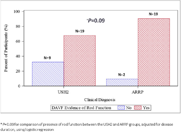
DAVF evidence of rod function by clinical diagnosis.
Table 2.
Proportion with DAVF Rod Function by Clinical Diagnosis and Age Group
| Clinical Diagnosis | ||||||||
|---|---|---|---|---|---|---|---|---|
| USH2 | ARRP | |||||||
| Age Group (Years) | Age Group (Years) | |||||||
| Evidence of Rod Function | <40 N (%) | ≥40 N (%) | All N (%) | P | <40 N (%) | ≥40 N (%) | All N (%) | P |
| DAVF | 0.37 | 0.50 | ||||||
| No | 5 (25%) | 4 (50%) | 9 (32%) | 0 (0%) | 2 (15%) | 2 (10%) | ||
| Yes | 15 (75%) | 4 (50%) | 19 (68%) | 8 (100%) | 11 (85%) | 19 (90%) | ||
| All | 20 (100%) | 8 (100%) | 28 (100%) | 8 (100%) | 13 (100%) | 21 (100%) | ||
Table 3.
Proportion With DAVF Rod Function by Clinical Diagnosis and Disease Duration
| Clinical Diagnosis | ||||||||
|---|---|---|---|---|---|---|---|---|
| USH2 | ARRP | |||||||
| Disease Duration (Years) | Disease Duration (Years) | |||||||
| Evidence of Rod Function | <15 N (%) | ≥15 N (%) | All N (%) | P | <15 N (%) | ≥15 N (%) | All N (%) | P |
| DAVF | 0.43 | 0.17 | ||||||
| No | 3 (23%) | 6 (40%) | 9 (32%) | 0 (0%) | 2 (22%) | 2 (10%) | ||
| Yes | 10 (77%) | 9 (60%) | 19 (68%) | 12 (100%) | 7 (78%) | 19 (90%) | ||
| All | 13 (100%) | 15 (100%) | 28 (100%) | 12 (100%) | 9 (100%) | 21 (100%) | ||
Of the 49 participants, 15 (31%) had measurable rod ERGs. Of the 15 participants with measurable rod ERGs, 14 (93%) showed evidence of DAVF rod function. Of the 34 with unmeasurable rod ERGs, 24 (71%) showed evidence of rod function on DAVF. Agreement as measured by the kappa statistic regarding evidence of rod function by DAVF versus FST white, FST blue, and rod ERG was 0.38 (95% CI, 0.18 to 0.58), 0.40 (95% CI, 0.08 to 0.72), and 0.16 (95% CI, 0.005 to 0.31), respectively. In USH2 participants alone, agreement regarding evidence of rod function by DAVF versus FST white, FST blue, and rod ERG was 0.25 (95% CI, 0.04 to 0.46), 0.40 (95% CI, 0.03–0.77), and 0.07 (95% CI, −0.13 to 0.27), respectively. In ARRP participants, agreement regarding evidence of rod function by DAVF versus FST white, FST blue, and rod ERG was 0.34 (95% CI, −0.17 to 0.86), −0.06 (95% CI, −0.13 to 0.02), and 0.17 (95% CI, −0.06 to 0.40), respectively. In the less than 40-year age group, agreement regarding evidence of rod function by DAVF for FST white, FST blue, and rod ERG was 0.31 (95% CI, 0.06 to 0.55), 0.26 (95% CI, −0.18 to 0.71), and 0.03 (95% CI, −0.15 to 0.20), respectively. For the 40-year and older age group, agreement regarding evidence of rod function with DAVF for FST white, FST blue and rod ERG was 0.49 (95% CI, 0.15–0.82), 0.56 (95% CI, 0.13 to 1.00), and 0.40 (95% CI, 0.10 to 0.69), respectively (Table 4). FST white and FST blue showed fair agreement (kappa = 0.21–0.40) with DAVF regarding evidence of rod function, whereas rod ERG showed slight agreement (kappa = 0.01–0.20) with DAVF regarding evidence of rod function.13 There was some evidence agreement varied with age and diagnosis groups. However, the sample sizes were too small to make a definitive conclusion.
Table 4.
Agreement of Evidence of Rod Function by DAVF Versus Other Modalities (Stratified by Clinical Diagnosis and Age Group); All (N = 49)
| Evidence of Rod Function Other Modalities | |||||||||
|---|---|---|---|---|---|---|---|---|---|
| FST White Stimulus† | FST Blue Stimulus‡ | ERG Rod B-Wave§ | |||||||
| DAVF* | No N (%) | Yes N (%) | κ (95% CI) || | No N (%) | Yes N (%) | κ (95% CI) || | No N (%) | Yes N (%) | κ (95% CI)|| |
| All | |||||||||
| No | 10 (22%) | 0 (0%) | 0.38 | 5 (11%) | 5 (11%) | 0.40 | 10 (20%) | 1 (2%) | 0.16 |
| Yes | 15 (33%) | 21 (46%) | (0.18, 0.58) | 4 (9%) | 32 (70%) | (0.08, 0.72) | 24 (49%) | 14 (29%) | (0.005, 0.31) |
| <40 years | |||||||||
| No | 5 (19%) | 0 (0%) | 0.31 | 2 (7%) | 3 (11%) | 0.26 | 4 (14%) | 1 (4%) | 0.03 |
| Yes | 10 (37%) | 12 (44%) | (0.06, 0.55) | 3 (11%) | 19 (70%) | (−0.18, 0.71) | 17 (61%) | 6 (21%) | (−0.15, 0.20) |
| ≥40 years | |||||||||
| No | 5 (26%) | 0 (0%) | 0.49 | 3 (16%) | 2 (11%) | 0.56 | 6 (29%) | 0 (0%) | 0.40 |
| Yes | 5 (26%) | 9 (47%) | (0.15, 0.82) | 1 (5%) | 13 (68%) | (0.13, 1.00) | 7 (33%) | 8 (38%) | (0.10, 0.69) |
| USH2 | |||||||||
| No | 9 (33%) | 0 (0%) | 0.25 | 5 (19%) | 4 (15%) | 0.40 | 8 (29%) | 1 (4%) | 0.07 |
| Yes | 12 (44%) | 6 (22%) | (0.04, 0.46) | 3 (11%) | 15 (56%) | (0.03, 0.77) | 15 (54%) | 4 (14%) | (−0.13, 0.27) |
| ARRP | |||||||||
| No | 1 (5%) | 0 (0%) | 0.34 | 0 (0%) | 1 (5%) | −0.06 | 2 (10%) | 0 (0%) | 0.17 |
| Yes | 3 (16%) | 15 (79%) | (−0.17, 0.86) | 1 (5%) | 17 (89%) | (−0.13, 0.02) | 9 (43%) | 10 (48%) | (−0.06, 0.40) |
Defined as at least one cluster of rod-mediated points in visual field where cyan relative to red sensitivity is >5 dB.
Defined as a white threshold of less than −30. Three participants missing data for FST White
Defined as a blue threshold of less than −25. Three participants missing data for FST Blue.
Defined as ERG rod function b-wave amplitude of >0.
Bias-corrected percentile bootstrap method used to get 95% CIs for Kappa estimate.
Along with mean sensitivity and maximum sensitivity, an HOV analysis of the DAVF results provided volumetric measures of total field (VTOT) and the volume of the central 30° (V30). The correlations among DAVF measures of rod visual field and light adapted static HOV parameters were low, ranging from 0.31 to 0.40 (Table 5). However, DAVF parameters were highly correlated with FST measures of rod function in these participants. DAVF maximum sensitivity decreased with higher FST white stimulus intensity (Fig. 5) (r = −0.80; P < .001; 95% CI, −0.92 to −0.59); the variance in the maximum sensitivity increased as the FST stimulus intensity increased (P = 0.05), especially above the −30 dB level, where FST thresholds may be influenced by cones (area to the right of the vertical red line). DAVF mean sensitivity, DAVF VTOT, and DAVF V30 were moderately correlated with FST white thresholds (r = −0.71 [95% CI, −0.86 to −0.50] r = −0.72 [95% CI, −0.87 to −0.51], and r = −0.61 [95% CI, −0.81 to −0.31]), respectively; all P < .001). Correlations with FST blue thresholds were similar to those with FST white thresholds (Table 5). Among participants with evidence of rod function, the number of rod loci and the DAVF VTOT were lower in participants with longer disease duration by −0.82 loci per year (95% CI, −1.76 to 0.12) and −0.59 decibels-steradians per year (95% CI, −1.82 to 0.64) of disease duration, respectively (Figs. 6 & 7). The number of rod loci and DAVF VTOT also were lower in USH2 than ARRP participants (mean difference, −16; 95% CI, −33 to −0.3; P = 0.046; and mean difference, −20; 95% CI, −41 to 0.7; P = 0.058, respectively).
Table 5.
Correlation of DAVF Measures With Standard Measures
| DAVF Measures | ||||||||||
|---|---|---|---|---|---|---|---|---|---|---|
| Mean | Maximum | Full Field Rod | ||||||||
| Sensitivity | VTOT | V30 | Sensitivity | Mean Sensitivity | ||||||
| Standard (non-DAVF) | Correlation | Correlation | Correlation | Correlation | Correlation | |||||
| Measures | Coefficient | 95% CI* | Coefficient | 95% CI* | Coefficient | 95% CI* | Coefficient | 95% CI* | Coefficient | 95% CI* |
| Mean sensitivity | 0.37 | 0.04 to 0.66 | 0.38 | 0.05 to 0.67 | 0.38 | 0.07 to 0.66 | 0.19 | −0.17 to 0.49 | 0.33 | −0.07 to 0.64 |
| Octopus-VTOT | 0.40 | 0.05 to 0.72 | 0.40 | 0.04 to 0.71 | 0.33 | −0.04 to 0.65 | 0.24 | −0.16 to 0.56 | 0.37 | −0.04 to 0.70 |
| Octopus-V30 | 0.31 | −0.04 to 0.61 | 0.33 | −0.02 to 0.63 | 0.37 | 0.04 to 0.64 | 0.07 | −0.30 to 0.38 | 0.26 | −0.13 to 0.57 |
| FST White | −0.71 | −0.86 to −0.50 | −0.72 | −0.87 to 0.51 | −0.61 | −0.81 to −0.31 | −0.80 | −0.92 to −0.59 | −0.50 | −0.73 to −0.18 |
| FST Blue | −0.78 | −0.89 to −0.59 | −0.78 | −0.89 to −0.60 | −0.64 | −0.83 to −0.38 | −0.85 | −0.94 to −0.67 | −0.56 | −0.78 to −0.26 |
| ERG | 0.43 | 0.01 to 0.69 | 0.43 | 0.02 to 0.68 | 0.35 | −0.04 to 0.61 | 0.54 | 0.15 to 0.78 | 0.41 | 0.06 to 0.67 |
Bias-corrected accelerated bootstrap method used to get 95% CIs for spearman correlation coefficient.
Figure 5.
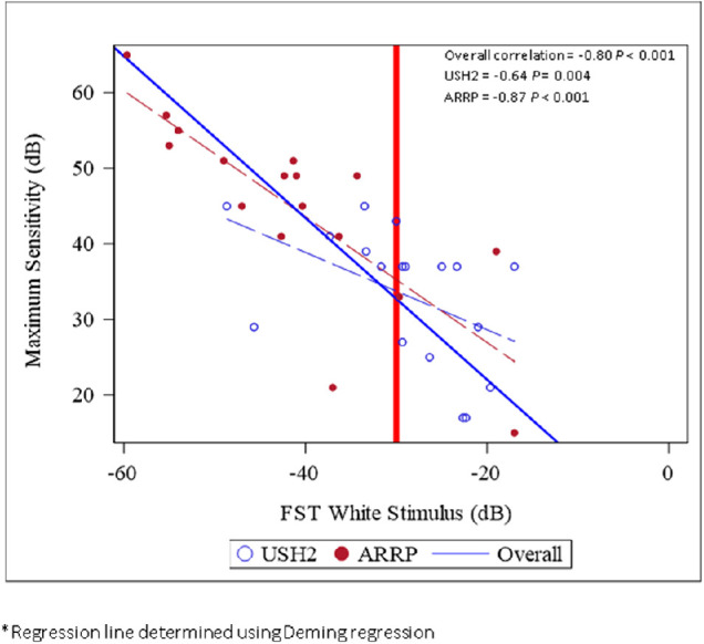
Maximum rod field sensitivity was inversely correlated with FST white thresholds. FST thresholds are mediated by rods for points to the left of the red vertical line.
Figure 6.
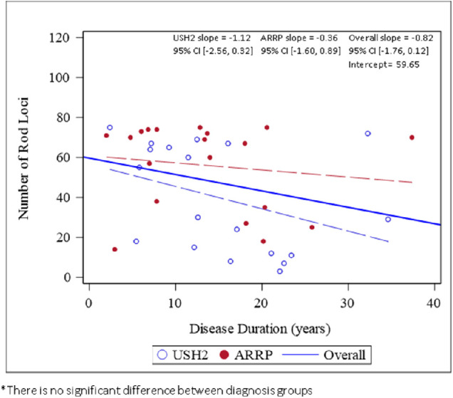
The number of rod-mediated loci with the DAVF decreased with increasing duration of disease.
Figure 7.

The volume of VTOT tended to decrease with increasing disease duration, but examples of substantial rod function were found at all durations.
Discussion
The inclusion criteria for the primary cohort of the RUSH2A trial included BCVA letter score of 54 or more (approximate Snellen equivalent of 20/80 or better) and a visual field diameter of 10° or more in every meridian of the central field, resulting in a population of syndromic (USH2) and nonsyndromic (ARRP) participants with intermediate stage disease. We showed that the rod ERG was not useful for following these participants, with approximately 50% in RUSH2A showing unmeasurable responses, whereas FST seemed promising as a reliable measure of rod function.2 However, because the FST only measures the most sensitive region, there could be large changes elsewhere that go undetected.
The ancillary study reported here uses two-color DAC perimetry to derive rod visual fields. Two-color perimetry, originally pioneered by Wald and Zeavin,14 has long been used to map rod and cone sensitivity. With this technique, the sensitivity difference (cyan–red) to chromatic test stimuli can be used to determine whether rods, cones or both photoreceptor systems mediate the threshold at a given location in the retina. Two-color perimetry has been performed using a variety of modified perimeters, including the Goldmann–Weekers, Tubingen, Octopus, and Humphrey instruments.6,7,15 Although they provide useful data for single-site studies, these custom devices are not readily adaptable to multicenter trials. The Medmont DAC perimeter is commercially available and in use at six of the sites participating in RUSH2A, allowing us to conduct an ancillary study in approximately half of the enrolled RUSH2A participants.
The majority of participants in the ancillary study showed evidence of rod-mediated detection in at least one cluster of points in the field. This included 14 of 15 patients (93%) retaining measurable rod ERGs, but more importantly 24 of 34 patients (71%) with unmeasurable rod ERGs. This finding is consistent with data in autosomal dominant RP, where the average number of rod-mediated loci was 31 ± 17 in patients with a measurable response to the 3.0 cd.s/m2 standard flash, but an unmeasurable response to the rod-only 0.01 cd.s/m2 flash.5 Although the RUSH2A study has shown that USH2 participants tend to be more severely affected than ARRP participants at a similar age,1 our sample size was too small to establish a difference based on diagnosis for DAVF. There was a tendency, however, for a higher percentage of participants with ARRP (90%) than with USH2 (68%) to retain measurable DAVF. The severity of mutations between diagnosis groups is the subject of analysis in a separate manuscript that has been submitted for peer review.16 There was also a trend for a greater number of rod loci and higher VTOT in participants having shorter disease duration with either USH2 or ARRP. A weakness in this analysis is that the reported disease duration is extremely subjective and USH2 patients may be more attentive to vision problems than patients with ARRP. But although not significant, these trends in the cross-sectional data suggest that longitudinal measures in patients may be sensitive to disease progression.
The correlations between DAVF parameters and light-adapted static perimetric parameters ranged from 0.31 to 0.4. This outcome suggests that DAVF is capturing a dimension of visual experience that is distinct from the standard visual field. It will be interesting in the future to compare the topography of rod and cone fields to determine possible relationships between regional rod and cone loss. The value at the most sensitive region within the rod visual field (maximum sensitivity) was highly (inversely) correlated with FST white or blue thresholds. This finding is reassuring, because it has been shown in previous studies that FST measures the response from the photoreceptors with the greatest sensitivity and best function.17 The correlation is greatest over the region where FST white threshold are clearly determined by rods, that is, up to −30 dB.2 At higher stimulus intensities, FST is likely mediated by a combination of rod and cone function. This factor results in a greater variance and a worse correlation with DAVF maximum sensitivity for FST values greater than −30 dB.
It is encouraging that rod-mediated DAVFs can be measured in the majority of participants tested in the RUSH2A study. The intertest and intratest variability in DAVF has been shown to be only slightly higher than for standard perimetry.18 Future analyses will determine annual rate of change in sensitivity in rod-mediated regions at 12, 24, and 36 months after the initial DAVF visit (which will occur at the RUSH2A 24-, 36-, and 48-month visits). Longitudinal measures of rod function in the RUSH2A study will provide insight into regional variations in rod degeneration and relationships to patterns of cone loss.
Acknowledgments
Katarina Stingl and Isabelle Audo are members of the ERN-EYE (European Reference Network for Rare Eye Diseases). David Birch has support from NIH EY09076.
Funded by Foundation Fighting Blindness.
Disclosure: D.G. Birch, Biogen (C,F), Nacuity (C), ProQR (C,F), Editas (C), AGTC (C,F), Iveric (C), 4D Therapeutics (C,F), DTx Therapeutics (C); L. Samarakoon, None; M. Melia, None; J.L. Duncan, Acucela (F), Allergen/Abbvie (F), Biogen/Nightstarx Therapeutics (F), Neurotech USA (F),Second Sight Medical Products (F), 4D Therapeutics (C), AGTC (C), Astellas (C), DTx Therapeutics (C), Editas (C), Eloxx (C), Eyevensys (C); Gyroscope (C), Horama (C), ProQR Therapeutics (C), Spark Therapeutics (C), SparingVision (C), Vedere Bio (C), Nacuity (C), Partner-Rx Sight (I); A.R. Ayala, None; I. Audo, None; J.K. Cheetham, DTx Pharma (C); T.A. Durham, None; A. Iannaccone, None; M.E. Pennesi, None; K. Stingl, Drs. Pennesi, Birch and Duncan serve on advisory boards for Foundation Fighting Blindness, ViGeneron (C), Novartis (C), CRA (R), ProQR (C,F)
References
- 1. Duncan JL, Liang W, Maguire MG, et al.. Baseline visual field findings in the RUSH2A Study: associated factors and correlation with other measures of disease severity. Am J Ophthalmol . 2020; 219: 87–100. [DOI] [PMC free article] [PubMed] [Google Scholar]
- 2. Birch DG, Cheng P, Duncan JL, et al.. The RUSH2A study: best-corrected visual acuity, full-field electroretinography amplitudes, and full-field stimulus thresholds at baseline. Transl Vis Sci Technol . 2020; 9(11): 9. [DOI] [PMC free article] [PubMed] [Google Scholar]
- 3. Roman AJ, Cideciyan AV, Aleman TS, Jacobson SG.. Full-field stimulus testing (FST) to quantify visual perception in severely blind candidates for treatment trials. Physiol Meas . 2007; 28(8): N51–N56. [DOI] [PubMed] [Google Scholar]
- 4. Klein M, Birch DG.. Psychophysical assessment of low visual function in patients with retinal degenerative diseases (RDDs) with the Diagnosys full-field stimulus threshold (D-FST). Doc Ophthalmol . 2009; 119(3): 217–224. [DOI] [PMC free article] [PubMed] [Google Scholar]
- 5. Bennett LD, Klein M, Locke KG, Kiser K, Birch DG.. Dark-adapted chromatic perimetry for measuring rod visual fields in patients with retinitis pigmentosa. Transl Vis Sci Technol . 2017; 6(4): 15–15. [DOI] [PMC free article] [PubMed] [Google Scholar]
- 6. Birch DG, Herman WK, deFaller JM, Disbrow DT, Birch EE.. The relationship between rod perimetric thresholds and full-field rod ERGs in retinitis pigmentosa. Invest Ophthalmol Vis Sci . 1987; 28(6): 954–965. [PubMed] [Google Scholar]
- 7. Jacobson SG, Voigt WJ, Parel J-M, et al.. Automated light- and dark- adapted perimetry for evaluating retinitis pigmentosa. Ophthalmology . 1986; 93(12): 1604–1611. [DOI] [PubMed] [Google Scholar]
- 8. Wyszeck G, Stiles WS.. Color Science . New York: John Wiley & Sons; 1982. [Google Scholar]
- 9. Weleber RG, Smith TB, Peters D, et al.. VFMA: topographic analysis of sensitivity data from full-field static perimetry. Transl Vis Sci Technol . 2015; 4(2): 14. [DOI] [PMC free article] [PubMed] [Google Scholar]
- 10. Wicklin R. The bias-corrected and accelerated (BCa) bootstrap interval. 2017. Available at: https://blogs.sas.com/content/iml/2017/07/12/bootstrap-bca-interval.html. Accessed September 3, 2021, 2021.
- 11.SAS Institute Inc. Jackknife and bootstrap analyses. 2021. Available at: https://support.sas.com/kb/24/982.html. Accessed September 3, 2021.
- 12. Efron B. Better bootstrap confidence intervals. J Am Stat Assoc . 1987; 82(397): 171–185. [Google Scholar]
- 13. Vierkant R. Macro for calculating bootstrapped confidence intervals about a kappa coefficient. 1997. Available at: https://support.sas.com/resources/papers/proceedings/proceed-ings/sugi22/STATS/PAPER295.PDF. Accessed March 9, 2021.
- 14. Wald G, Zeavin BH.. Rod and cone vision in retinitis pigmentosa. Am J Ophthalmol . 1956; 42(4 Part 2): 253–269. [DOI] [PubMed] [Google Scholar]
- 15. Gunkel RD. Retinal profiles. A psychophysical test of rod and cone sensitivity. Arch Ophthalmol . 1967; 77(1): 22–25. [DOI] [PubMed] [Google Scholar]
- 16. Hufnagel RB, Guan B, Ullah E, et al.. RUSH2A: systematic cohort variant modeling reveals phenotypic correlates. Invest Ophthalmol Vis Sci . 2020; 61(7): 836–836. [Google Scholar]
- 17. Roman AJ, Schwartz SB, Aleman TS, et al.. Quantifying rod photoreceptor-mediated vision in retinal degenerations: dark-adapted thresholds as outcome measures. Exp Eye Res . 2005; 80(2): 259–272. [DOI] [PubMed] [Google Scholar]
- 18. Bennett LD, Metz G, Klein M, Locke KG, Khwaja A, Birch DG.. Regional variations and intra-/intersession repeatability for scotopic sensitivity in normal controls and patients with inherited retinal degenerations. Invest Ophthalmol Vis Sci . 2019; 60(4): 1122–1131. [DOI] [PMC free article] [PubMed] [Google Scholar]


