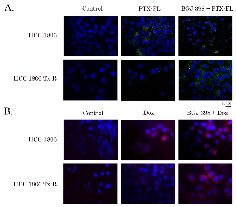
Figure 2.
Fluorescence microscopy analysis of the intracellular accumulation of Flutax-2 (PTX-FL) (A) and doxorubicin (DOX) (B) in HCC 1806 parental (upper panels) and Tx-R (lower panels) cells. The cells were first incubated with BGJ 398 (20 µM) (right) or DMSO as a control (middle) for 60min and then incubated with 3 µM PTX-FL (A) or 40 µM DOX (B) for an additional 60 min. After the wash-out with a pre-warmed FBS-free culture medium, the BGJ 398-treated cells were additionally incubated with BGJ 398 for 60 min. The non-fixed slides were counterstained with Hoechst 33342 (final concentration 3 µg/mL) for 5 min to outline the nuclei and processed for fluorescence microscopy to obtain the merged images.

