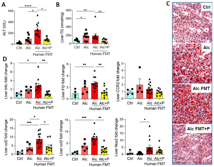
Figure 1.
Liver injury in control and alcohol-fed mice and pectin-treated alcohol-fed mice. Control (Ctrl) and Alcohol-fed (Alc) mice received a Lieber DeCarli diet with isocaloric maltodextrin or alcohol, respectively. Pectin-treated mice (Alc + P) received 6.5% pectin in addition to 5% ethanol. Human fecal microbiota transfer (FMT) groups received microbiota from a patient with alcoholic hepatitis. (A) Plasma alanine transaminase (ALT) levels. (B) Liver triglyceride (TG) levels. (C) Representative histological images of Oil-Red-O staining of the liver (scale bar: Oil-Red-O = 100 µm). (D) Liver mRNA levels of pro-inflammatory cytokines and chemokines (tumor necrosis factor α, tnfα; interleukin 1 β, il1β; C-C chemokine receptor type 2, CCR2; chemokine (C-C motif) ligand 2, ccl2; chemokine (C-C motif) ligand 3, ccl3; Nitric oxide synthase 2, Nos2). * p < 0.05, ** p < 0.01, *** p < 0.001, **** p < 0.0001.

