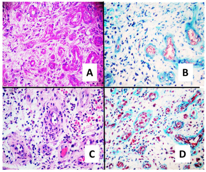
Figure 2.
Histopathological analysis of wound healing at day 21 of treatment. Representative microscope photographs of ulcers in each treatment group are shown. Haematoxylin-eosin staining (A,C) and Masson’s trichrome staining (B,D) shows that SuDe + Lp treatment (C,D) induced an abundant synthesis of ECM, and its accumulation in healed ulcers is evident when compared to SuDe (A,B). In addition, the ordered deposition of collagens is clear. Photos were taken with 40× objective.

