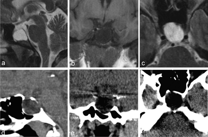
Figure 1:
Non-contrast MRI (panels a–c) of the pituitary gland with sagittal (a) and axial (c) T2-weighted as well as coronal T1-weighted dedicated 3-mm thin-sections from case 1, demonstrating thin rim of pituitary tissue along the anterior and inferior aspects of the enlarged CSF-filled sella turcica. Non-contrast multiplanar reformatted CT images (panels d–f) in the corresponding sagittal (d), coronal (e), and axial (f) planes demonstrating smooth remodelling of the osseous boundaries of the sella turcica with no evidence of bony erosion.

