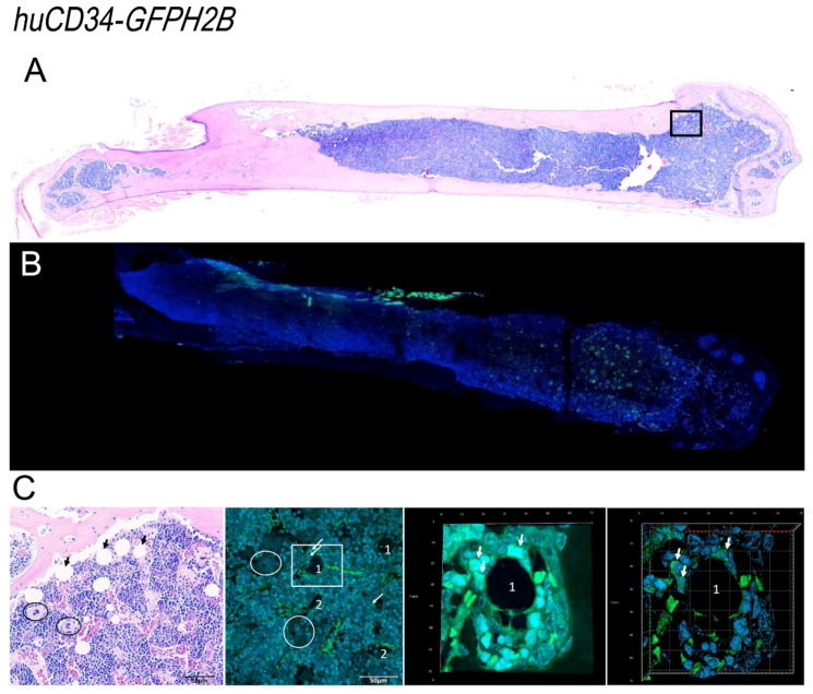
Figure 9.
Confocal microscopy analyses of the distribution of GFP-positive cells within the bone marrow architecture of huCD34-GFPH2B mice. (A) The H&E staining of a femur section from one representative huCD34-GFPH2B mouse reveals hypercellular bone marrow with scant residual adipocytes well visible in H&E figure in C. (B) Confocal microscopy analyses with GFP and DAPI of a femur from one representative huCD34-GFPH2B mouse revealing that GFP+ cells are mostly distributed in the epiphysis of the femur. (C) Representative panels showing, at larger magnification, the areas indicated in rectangles in A (H&E staining, left panel) and in B (confocal microscopy, second panel of the left). The rectangle on the second panel on the left is shown as stack image and as tridimensional reconstruction on the right. The tridimensional reconstruction is shown in detail in movie 3. Black arrows in A indicate adipocytes, circles indicate megakaryocyte clusters, and white arrows indicate GFP-positive cells. A and B are a photo-merge of 4× magnification pictures; in C, the magnifications are 20× for the first and second panel, and 40× for the third and fourth panel.

