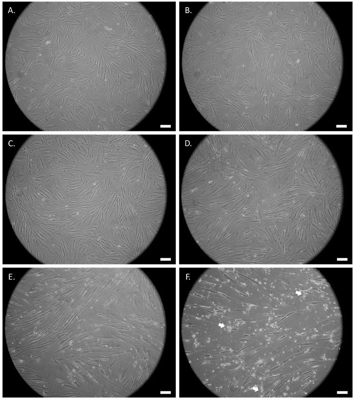
Figure 3.
Phase contrast microscopic images of SCAP treated with 1 mM of UDP-4 cultured for (A) 0 h (untreated), (B) 72 h, (C) 96 h, (D) 7 days, (E) 14 days, and (F) 21 days showing the progressive development of elongated neural-like morphology. White arrows denote cellular stress. Scale bars correspond to 50 µm.

