

Abstract
Background
Cannulation techniques have been recognized as being important in causing post‐endoscopic retrograde cholangiopancreatography (ERCP) pancreatitis (PEP). However, considerable controversy exists about the usefulness of the guidewire‐assisted cannulation technique for the prevention of PEP.
Objectives
To assess the effectiveness and safety of the guidewire‐assisted cannulation technique compared to the conventional contrast‐assisted cannulation technique for the prevention of PEP in people undergoing diagnostic or therapeutic ERCP for biliary or pancreatic diseases.
Search methods
For the previous version of this review, we searched CENTRAL (the Cochrane Library), MEDLINE, Embase, CINAHL and major conference proceedings, up to February 2012, with no language restrictions. An updated search was performed on 26 February 2021 for the current version of this review. Two clinical trial registries, clinicaltrials.gov and WHO ICTRP, were also searched in this update.
Selection criteria
Randomized controlled trials (RCTs) comparing the guidewire‐assisted cannulation technique versus the contrast‐assisted cannulation technique in people undergoing ERCP.
Data collection and analysis
Two review authors conducted study selection, data extraction, and methodological quality assessment independently. Using intention‐to‐treat analysis with random‐effects models, we combined dichotomous data to obtain risk ratios (RR) with 95% confidence intervals (CI). We assessed heterogeneity using the Chi² test (P < 0.10) and I² statistic (> 50%). To explore sources of heterogeneity, we conducted a priori subgroup analyses according to trial design, publication type, risk of bias, use of precut sphincterotomy, inadvertent guidewire insertion or contrast injection of the pancreatic duct (PD), use of a PD stent, cannulation device, and trainee involvement in cannulation. To assess the robustness of our results, we carried out sensitivity analyses using different summary statistics (RR versus odds ratio (OR)) and meta‐analytic models (fixed‐effect versus random‐effects) and per‐protocol analysis.
Main results
15 RCTs comprising 4426 participants were included. There was moderate heterogeneity among trials for the outcome of PEP (P = 0.08, I² = 36%). Meta‐analyses suggest that the guidewire‐assisted cannulation technique probably reduces the risk of PEP compared to the contrast‐assisted cannulation technique (RR 0.51, 95% CI 0.36 to 0.72, 15 studies, moderate‐certainty evidence). In addition, the guidewire‐assisted cannulation technique may result in an increase in primary cannulation success (RR 1.06, 95% CI 1.01 to 1.12, 13 studies, low‐certainty evidence), and probably reduces the need for precut sphincterotomy (RR 0.79, 95% CI 0.64 to 0.96, 10 studies, moderate‐certainty evidence). Compared to the contrast‐assisted cannulation technique, the guidewire‐assisted cannulation technique may result in little to no difference in the risk of post‐sphincterotomy bleeding (RR 0.87, 95% CI 0.49 to 1.54, 7 studies, low‐certainty evidence) and perforation (RR 0.93, 95% CI 0.11 to 8.23, 8 studies, very low‐certainty evidence). Procedure‐related mortality was reported by eight studies, and there were no cases of deaths in both arms (moderate‐certainty evidence). Subgroup analyses suggest that the heterogeneity for the outcome of PEP could be explained by differences in trial design. The results were robust in sensitivity analyses.
Authors' conclusions
There is moderate‐certainty evidence that the guidewire‐assisted cannulation technique probably reduces the risk of PEP compared to the contrast‐assisted cannulation technique. There is low‐certainty evidence that the guidewire‐assisted cannulation technique may result in an increase in primary cannulation success. There is low‐ and very low‐certainty evidence that the guidewire‐assisted cannulation technique may result in little to no difference in the risk of bleeding and perforation. No procedure‐related deaths were reported. Therefore, the guidewire‐assisted cannulation technique appears to be superior to the contrast‐assisted cannulation technique considering the certainty of evidence and the balance of benefits and harms. However, the routine use of guidewires in biliary cannulation will be dependent on local expertise, availability, and cost. Future research should assess the effectiveness and safety of the guidewire‐assisted cannulation technique in the context of other pharmacologic or non‐pharmacologic interventions for the prevention of PEP.
Plain language summary
Guidewire or contrast: which works better for the prevention of post‐endoscopic retrograde cholangiopancreatography pancreatitis?
Key messages:
• Endoscopic retrograde cholangiopancreatography (ERCP) combines endoscopy and x‐ray to diagnose and treat problems of the bile and pancreatic ducts. Compared to the traditional technique involving injection of contrast dye into the ducts with a catheter, using a guidewire technique to gain access to the bile duct probably reduces the risk of post‐ERCP pancreatitis (PEP) and may also increase the success rate of gaining access to the bile duct.
• Future research in this area should focus on the effects of the guidewire technique in addition to other options for reducing the risk of PEP (for example, rectally administered anti‐inflammatory drugs, a plastic tube inserted into the pancreatic duct).
What is post‐ERCP pancreatitis (PEP)?
ERCP combines endoscopy (examination inside the body using a medical instrument called an endoscope) and x‐ray to diagnose and treat problems of the bile and pancreatic ducts (structures that support the process of digestion). With the patient under sedation, an endoscope is passed down the oesophagus (windpipe) and into the small bowel, where the opening of the bile and pancreatic ducts (papilla) is located. A catheter is inserted through the endoscope and papilla into the bile duct. Contrast dye is injected into the bile duct, and x‐rays are taken to look for gallstones or blockage. However, the major risk of ERCP is the development of pancreatitis (inflammation of the pancreas) due to irritation of the pancreatic duct by the contrast material or catheter, which can occur in 5% to 10% of all procedures. This may be self‐limited and mild, but can also be severe and require hospitalisation. Rarely, it may be life‐threatening. There are also small risks of bleeding or making a hole in the bowel wall.
What did we want to find out?
There are two techniques for gaining access to the bile duct during ERCP. The traditional technique (contrast) involves inserting a catheter into the papilla and injecting contrast dye to confirm access to the bile duct. However, contrast dye may be unintentionally injected into the pancreatic duct. A second technique (guidewire) involves using a guidewire to probe the papilla to gain access to the bile duct. Once an x‐ray confirms the guidewire is in the bile duct, contrast dye is injected into the bile duct.
We wanted to find out:
• which technique for gaining access to the bile duct during ERCP works best to reduce the risk of PEP;
• which technique achieves better success in gaining access to the bile duct; and
• which technique causes fewer unwanted effects (for example, the need to use advanced techniques involving blind incision into the papilla to gain access to the bile duct, inadvertent entry of the pancreatic duct, bleeding, hole in the bowel wall, and death).
What did we do?
We searched for studies that compared the guidewire to the contrast technique in people undergoing ERCP for biliary or pancreatic diseases. We compared and summarized their results and rated our confidence in the evidence based on factors such as study methods and sizes.
What did we find?
We found 15 studies that involved 4426 people undergoing ERCP. The studies were conducted in various countries around the world. The biggest study was in 513 people, and the smallest study was in 88 people, with ages ranging from 18 to 96 years and roughly equal numbers of men and women. Nine studies declared no funding sources and conflicts of interest, while the other six studies did not report this information.
What are the main results of the review?
Compared to the contrast technique, using the guidewire technique probably reduces the risk of PEP and may increase the success rate of gaining access to the bile duct, and probably reduces the need to use advanced techniques to gain access to the bile duct. The guidewire technique may result in little to no difference in the risks of bleeding and hole in the bowel wall. There were no cases of procedure‐related death.
What are the limitations of the evidence?
We are moderately confident that the guidewire technique reduces the risk of PEP and reduces the need to use advanced techniques to gain access to the bile duct, but it is possible that physicians who performed the ERCP and assessed the outcomes may be biased, as they were aware of which technique(s) they used during the procedures. We are less confident in the results for the success rate of gaining access to the bile duct, and the results of further research could differ from ours. We are also less confident in our results for the risks of bleeding and hole in the bowel wall because of the low number of reported events. We are moderately confident in our results for mortality due to no events reported in a large number of people.
How up‐to‐date is this evidence?
This review updates our previous review published in 2012. The evidence is up‐to‐date to February 2021.
Summary of findings
Summary of findings 1. Guidewire‐assisted cannulation compared to contrast‐assisted cannulation for the prevention of post‐ERCP pancreatitis (PEP).
| Guidewire‐assisted cannulation compared to contrast‐assisted cannulation for the prevention of post‐ERCP pancreatitis (PEP) | ||||||
| Patient or population: Patients undergoing diagnostic or therapeutic ERCP Setting: Inpatient and outpatient Intervention: Guidewire‐assisted cannulation Comparison: Contrast‐assisted cannulation | ||||||
| Outcomes | Anticipated absolute effects* (95% CI) | Relative effect (95% CI) | № of participants (studies) | Certainty of the evidence (GRADE) | Comments | |
| Risk with contrast‐assisted cannulation | Risk with guidewire‐assisted cannulation | |||||
| Post‐ERCP pancreatitis (PEP) Follow‐up: range 1 days to 30 days | Study population | RR 0.51 (0.36 to 0.72) | 4426 (15 RCTs) | ⊕⊕⊕⊝ MODERATE 1 2 | ||
| 77 per 1000 | 39 per 1000 (28 to 55) | |||||
| Primary cannulation success | Study population | RR 1.06 (1.01 to 1.12) | 3962 (13 RCTs) | ⊕⊕⊝⊝ LOW 1 3 | ||
| 784 per 1000 | 831 per 1000 (792 to 878) | |||||
| Overall cannulation success | Study population | RR 1.01 (1.00 to 1.03) | 4426 (15 RCTs) | ⊕⊕⊕⊝ MODERATE 1 | ||
| 908 per 1000 | 917 per 1000 (908 to 935) | |||||
| Need for precut sphincterotomy | Study population | RR 0.79 (0.64 to 0.96) | 2849 (10 RCTs) | ⊕⊕⊕⊝ MODERATE 1 | ||
| 130 per 1000 | 102 per 1000 (83 to 124) | |||||
| Post‐sphincterotomy bleeding Follow‐up: range 1 days to 30 days | Study population | RR 0.87 (0.49 to 1.54) | 2122 (7 RCTs) | ⊕⊕⊝⊝ LOW 1 4 | ||
| 24 per 1000 | 21 per 1000 (12 to 37) | |||||
| Perforation | Study population | RR 0.93 (0.11 to 8.23) | 2522 (8 RCTs) | ⊕⊝⊝⊝ VERY LOW 1 4 5 | ||
| 3 per 1000 | 3 per 1000 (0 to 27) | |||||
| Mortality | Study population | Not estimable | 2276 (6 RCTs) | ⊕⊕⊕⊝ MODERATE 1 6 | ||
| 0 per 1000 | 0 per 1000 (0 to 0) | |||||
| *The risk in the intervention group (and its 95% confidence interval) is based on the assumed risk in the comparison group and the relative effect of the intervention (and its 95% CI). CI: Confidence interval; RR: Risk ratio | ||||||
| GRADE Working Group grades of evidence High certainty: We are very confident that the true effect lies close to that of the estimate of the effect. Moderate certainty: We are moderately confident in the effect estimate: The true effect is likely to be close to the estimate of the effect, but there is a possibility that it is substantially different. Low certainty: Our confidence in the effect estimate is limited: The true effect may be substantially different from the estimate of the effect. Very low certainty: We have very little confidence in the effect estimate: The true effect is likely to be substantially different from the estimate of effect. | ||||||
1 The certainty of evidence was rated down one level due to serious study limitations. All studies were judged to be at high risk of bias for blinding of participants and personnel. Lack of blinding of the endoscopist may have an impact on PEP, cannulation success, the use of precut sphincterotomy, or complications (post‐sphincterotomy bleeding, mortality) depending on the experience, expertise, and preference of the endoscopist performing the procedure.
2 The certainty of evidence for PEP was not rated down for inconsistency as the moderate heterogeneity (I² = 36%) for this outcome could be explained by differences in trial design. Subgroup analysis according to trial design indicated evidence of a difference between 'non‐cross‐over' and 'cross‐over' studies for the outcome of PEP, with the guidewire‐assisted cannulation technique favoured in 'non‐cross‐over' studies (P = 0.004). There was no important heterogeneity among each subgroup (I² = 0% for 'non‐cross‐over' studies and I² = 9% for 'cross‐over' studies).
3 The certainty of evidence for primary cannulation success was rated down for inconsistency due to unexplained substantial heterogeneity (I² = 83%).
4 The certainty of evidence for post‐sphincterotomy bleeding and perforation was rated down for imprecision due to few events and wide confidence intervals which included the possibility of no effect and important benefit or harm associated with guidewire‐assisted cannulation.
5 The certainty of evidence for perforation was rated down for inconsistency due to unexplained moderate heterogeneity (I² = 46%).
6 The certainty of evidence for mortality was not rated down for imprecision. If there are no events and the number of participants is large, judgment about the certainty of evidence (particularly judgments about imprecision) may be based on the absolute effect (as per the guidance of the Cochrane Handbook). Here, the certainty rating may be considered moderate (downgraded due to serious risk of bias but not for imprecision) as the outcome was appropriately assessed and the event, in fact, did not occur in 2276 studied participants.
Background
Description of the condition
Endoscopic retrograde cholangiopancreatography (ERCP) is a commonly performed endoscopic procedure that has both diagnostic and therapeutic roles in various hepatobiliary and pancreatic disorders. Despite its potential benefits, ERCP is not without risks. Acute pancreatitis is one of the most common serious complications of ERCP (Cotton 1991). The incidence of post‐ERCP pancreatitis (PEP) varies between 5% and 10%, although it may exceed 25% in certain high‐risk patient populations (Freeman 2004a). While most PEP manifests as a minor illness with two to three days of additional hospitalisation and an expected full recovery, severe pancreatitis is a devastating illness with significant morbidities, such as pancreatic necrosis, multi‐organ failure, and mortality. Severe pancreatitis has been reported to occur in 0.1% to 0.5% of ERCPs in prospective series (Freeman 2004a).
The pathophysiologic mechanisms of PEP are likely to be multifactorial and are incompletely understood (Freeman 2004a; Pezzilli 2002). These may include:
Mechanical injury to the papilla and pancreatic duct (PD) due to instrumental manipulation, resulting in obstruction or impairment of pancreatic flow;
Chemical injury due to contrast injection into the PD;
Hydrostatic injury due to contrast injection into the PD;
Thermal injury due to the electrosurgical current used for biliary or pancreatic sphincterotomy;
Enzymatic injury from the introduction of activated proteolytic enzymes into the PD;
Microbiological injury due to contamination or instillation of intestinal flora or bacteria into the PD.
Considerable efforts have been made to identify risk factors for PEP. Multivariate analyses of prospective studies have found a number of patient‐related risk factors for PEP, including young age, female gender, sphincter of Oddi dysfunction (SOD), recurrent pancreatitis, and history of PEP (Cheng 2006; Freeman 2001). Procedure‐related risk factors include difficult cannulation, multiple injections of the PD, precut sphincterotomy, pancreatic sphincterotomy, and biliary sphincter balloon dilation (Cheng 2006; Freeman 2001). Operator‐related risk factors such as the endoscopist's expertise, case volume, and trainee involvement in the procedure have been considered to be potential factors that can influence the outcome of ERCP. Indeed, low case volumes have been found to be associated with higher ERCP failure and complication rates (Freeman 1996; Loperfido 1998). However, large prospective studies have provided conflicting evidence whether any of these operator‐related risk factors increase the risk of PEP (Cheng 2006; Colton 2009; Freeman 1996; Freeman 2001; Loperfido 1998; Testoni 2010; Vandervoort 2002; Wang 2009; Williams 2007b). This is likely to be due to the fact that any difference in the rates of PEP between low‐ and high‐volume centres or endoscopists is often blunted by a disparity in case mix. In contrast, trainee participation has been shown to be a significant risk factor for the development of PEP (Cheng 2006). This increased risk is possibly due to multiple cannulation attempts by trainees.
In clinical practice, as recommended by current guidelines (Banks 2013), acute pancreatitis is diagnosed by the presence of two of the following three features:
Abdominal pain typical of acute pancreatitis;
Greater than or equal to three‐fold elevation in amylase or lipase;
Radiographic evidence of pancreatitis on cross‐sectional imaging.
However, much controversy remains about the definition of PEP. There are currently two definitions of PEP: the consensus definition (Cotton 1991) and the revised Atlanta Classification (Banks 2013). The European Society of Gastrointestinal Endoscopy (ESGE) guideline stated that both definitions of PEP may be used, but neither of these is ideal in the setting of PEP (Dumonceau 2014; Dumonceau 2020). The consensus definition was developed in 1991 based on data collected from more than 15,000 procedures (Cotton 1991). PEP was defined as a rise in serum amylase levels to greater than or equal to three‐fold above the upper limit of normal, 24 hours after ERCP, accompanied by abdominal pain characteristic of pancreatitis and requiring an unplanned hospital stay or an extension of a planned hospital stay by at least two days (Cotton 1991). The severity of PEP (mild, moderate, severe) was graded according to the length of stay and local or systemic complications related to pancreatitis. However, this consensus definition (Cotton 1991) has not been adopted widely, and varying definitions of PEP have been used in clinical trials. This likely reflects the ongoing controversy in defining PEP in the context of post‐ERCP complications. The revised Atlanta Classification from 2012 (Banks 2013) was an update of the 1992 Atlanta criteria for defining and diagnosing acute pancreatitis, regardless of aetiology. The revised Atlanta Classification requires two of three features for the diagnosis of PEP: 1) abdominal pain consistent with acute pancreatitis, 2) serum amylase or lipase greater than three times the upper limit of normal, and 3) characteristic findings of acute pancreatitis on abdominal imaging. This classification defines severity based on the presence or absence of organ failure and of local or systemic complications (Banks 2013). This definition, however, was not developed specifically for PEP, but for all‐cause acute pancreatitis (Banks 2013). Neither the consensus definition (Cotton 1991) nor the revised Atlanta Classification has been shown to reliably diagnose PEP. This is due to the fact that asymptomatic transient elevations in amylase or lipase levels, or both, are often seen post‐ERCP (up to 70%) (Conn 1991; Skude 1976; Testoni 1999). Asymptomatic hyperamylasaemia with levels more than five times the upper limit of normal and lasting for 24 hours after ERCP has been reported in about 27% of cases (Testoni 1999). Moreover, serum lipase is now considered to be more sensitive and specific than serum amylase in the diagnosis of acute pancreatitis (Yadav 2002). In addition, abdominal pain post‐procedure could be due to a multitude of factors other than PEP (for example air insufflation). The duration of pain is, therefore, essential for defining PEP because pain that subsides within 24 hours is unlikely to indicate pancreatitis. Moreover, mild pain disappearing within 24 to 48 hours and not requiring analgesics or prolonged hospital stay still does not fulfil the criteria for clinical pancreatitis. Taken together, these two common findings post‐ERCP (pain and elevation in amylase) may lead to over‐diagnosis of PEP. Because of the lack of specificity of pain and hyperamylasaemia after ERCP, computed tomography (CT) has been proposed as the most appropriate method to confirm the diagnosis of PEP (Badalov 2009; Kiriyama 2010). To add to the controversy, the need for diagnostic criteria for PEP distinct from those used for pancreatitis has been challenged by a recent study suggesting that the consensus definition (Cotton 1991) may under‐diagnose PEP (Artifon 2010). On the other hand, it is uncertain whether the revised Atlanta Classification may over‐diagnose PEP without having any significant impact on clinical management or patient outcomes.
Description of the intervention
Endoscopic retrograde cholangiopancreatography (ERCP) involves the passage of a side‐viewing endoscope into the duodenum and cannulation of the common bile duct (CBD) with a device (sphincterotome or catheter). Contrast can then be injected in a retrograde manner into the CBD. Selective deep cannulation of the CBD is a prerequisite to successful diagnostic and therapeutic ERCP.
Contrast‐assisted cannulation
Conventional contrast‐assisted cannulation of the CBD is the direct injection of contrast through a catheter or a sphincterotome into the papilla under fluoroscopy (Freeman 2005). With this technique, a catheter or a sphincterotome is first aligned with the CBD and advanced into the papilla. Contrast is then injected to determine if the CBD has been entered. Upon visualization of the CBD, more contrast can be injected for optimal opacification and the catheter or the sphincterotome is then advanced further into the CBD for deep cannulation. If contrast is noted to fill the pancreatic duct (PD), the catheter or sphincterotome is then withdrawn and reoriented to the direction of the CBD, and the above steps are repeated until the CBD is accessed. However, inadvertent contrast injection of the PD or the papilla itself (submucosal injection), as well as repeated cannulation attempts, may increase the risk of post‐ERCP pancreatitis (PEP) (Cheng 2006; Freeman 2001).
Guidewire‐assisted cannulation
Guidewires were initially designed and utilized to maintain access to the CBD during therapeutic manoeuvres such as stent placement and stone extraction. Increasingly, guidewires are used to facilitate selective deep cannulation of the CBD. With the guidewire‐assisted cannulation technique, a guidewire is used to confirm selective cannulation of the CBD before contrast injection. If the guidewire inadvertently enters the PD, the guidewire is withdrawn into the catheter or the sphincterotome and attempts repeated to enter the CBD. Once the guidewire is noted to enter the CBD, the catheter or the sphincterotome can be advanced deeper into the CBD, and contrast is injected for optimal opacification. It has been postulated that the guidewire‐assisted cannulation technique may improve biliary cannulation success and prevent PEP by avoiding papillary trauma and inadvertent contrast injection of the PD or the papilla itself. In general, there are two variations of the guidewire‐assisted cannulation technique (Freeman 2005):
A guidewire is extended slightly beyond the catheter or the sphincterotome and is advanced in small increments under fluoroscopy to probe and gain access to the CBD;
The tip of the catheter or the sphincterotome is first inserted into the papilla and oriented to the direction of the CBD followed by advancement of the guidewire to probe and gain access to the CBD.
Achieving deep cannulation of the CBD can be difficult. Success depends primarily on the skill and experience of the endoscopist but also on anatomical variations and underlying conditions. Even among experienced endoscopists, failure of biliary cannulation may occur in up to 10% to 20% of cases (Varadarajulu 2006; Williams 2007a). When access by conventional methods fails, a precut sphincterotomy, by means of an incision into or just above the papilla, is often employed as a last resort to achieve CBD cannulation (Freeman 2005; Siegel 1989). Use of precut sphincterotomy has been reported to be associated with an increased risk of complications including PEP, bleeding, and perforation (Cennamo 2010; Freeman 2001; Masci 2003). However, it remains controversial whether the increased risk is due to the precut itself or to the prolonged attempts at cannulation. In high‐risk patients, the placement of a prophylactic PD stent after ERCP has been shown to reduce the risk of PEP (Choudhary 2011; Mazaki 2010). However, PD stents can be technically difficult to place even for the most experienced endoscopists, with reported failure in up to 10% of cases (Freeman 2007). In high‐risk patients, PD manipulation followed by failure to place a PD stent may be associated with a higher risk of PEP than no attempt at all (Freeman 2004b). There is also a potential for inducing pancreatic ductal injury (Kozarek 1990).
How the intervention might work
Cannulation techniques have long been recognized to be important in causing post‐endoscopic retrograde cholangiopancreatography (ERCP) pancreatitis (PEP) (Freeman 2001; Freeman 2004a). Mechanical injury to the papilla and pancreatic duct (PD) from repeated cannulation attempts may lead to oedema and obstruction of pancreatic ductal flow. In addition, inadvertent injection of contrast into the PD may lead to both chemical and hydrostatic injuries of the pancreas. Contrast injection into the PD itself is independently associated with risk of PEP, and the risk increases with number of injections (Freeman 2001; Wang 2009). These factors are thought to play an important role in the development of PEP with conventional contrast‐assisted cannulation of the common bile duct (CBD) using a catheter or a sphincterotome. It has been postulated that the guidewire‐assisted cannulation technique may improve biliary cannulation success and prevent PEP by avoiding papillary trauma and inadvertent contrast injection of the PD or the papilla itself (submucosal injection). The rationale for more successful CBD cannulation with the guidewire‐assisted technique is that a small‐diameter guidewire with a hydrophilic tip can pass more easily through the small opening of the bile duct than a larger‐diameter catheter or sphincterotome. There are, however, potential concerns with the guidewire‐assisted cannulation technique including false passage, intramural dissection, perforation, and PD injury (Freeman 2005).
Why it is important to do this review
Prevention of post‐endoscopic retrograde cholangiopancreatography (ERCP) pancreatitis (PEP) has been the 'Holy Grail' of ERCP. Investigators have long searched for a pharmacologic agent that will prevent PEP, but nearly all agents evaluated (with the exception of rectal non‐steroidal anti‐inflammatory drugs) have failed to demonstrate efficacy in randomized controlled trials or logistic feasibility in real‐life settings (Akshintala 2021; Elmunzer 2012; Serrano 2019; Testoni 2006; Yang 2017). Aggressive intravenous hydration with Ringer's lactate in the peri‐procedural period (defined as 3 mL/kg/hour during ERCP and 20 mL/kg bolus after ERCP, and 3 mL/kg/hour for eight hours after ERCP) has been shown to reduce the risk of PEP (Akshintala 2021; Radadiya 2019), although the feasibility of such practice is limited and the benefits are uncertain in people with moderate to high risk of developing PEP who routinely receive prophylactic rectal non‐steroidal anti‐inflammatory drugs (Mok 2016; Weiland 2021). Similarly, numerous endoscopic interventions have been studied for the prevention of PEP (Freeman 2004a). The findings of these studies have often provided conflicting results due to different study designs, definitions of outcomes, patient populations, and interventions used. Nevertheless, pancreatic duct (PD) stenting has been shown to reduce the risk of PEP in high‐risk patients, but failed PD stenting carries an increased risk of PEP of up to 35% (Akshintala 2021; Choksi 2015; Njei 2020). As well, the role of PD stenting is unclear with the prophylactic use of rectal non‐steroidal anti‐inflammatory drugs (Bekkali 2017). Furthermore, considerable controversy remains about the usefulness of the guidewire‐assisted cannulation technique compared to the conventional contrast‐assisted cannulation technique for the prevention of PEP. A comprehensive meta‐analysis of the efficacy and safety of the guidewire‐assisted cannulation technique will allow us to make recommendations for clinical practice and research. This systematic review is part of a series of reviews examining endoscopic interventions for the prevention of PEP.
PEP is the most common serious complication of ERCP and carries significant morbidity and mortality. The cannulation technique is believed to be pivotal in the pathogenesis of PEP. This is an update of a systematic review previously published in 2012 (Tse 2012), which aims to evaluate the relative merits of the two different cannulation techniques for the prevention of PEP. Given the ongoing controversy about the relative benefits and risks of the guidewire‐associated cannulation techniques compared to the contrast‐assisted cannulation techniques, we aimed to update the literature search to identify any new studies that could potentially change or strengthen the conclusions of this review. The findings of this review are relevant to patients, physicians, and healthcare systems.
Objectives
We aimed to assess the clinical effectiveness of the guidewire‐assisted cannulation technique compared to the conventional contrast‐assisted cannulation technique for cannulation of the common bile duct (CBD) in the prevention of post‐endoscopic retrograde cholangiopancreatography (ERCP) pancreatitis (PEP) by systematic review and meta‐analysis of randomized controlled trials (RCTs).
The objectives of this review were two‐fold:
To assess the effects of the guidewire‐assisted cannulation technique for the prevention of PEP and other ERCP‐related complications (post‐sphincterotomy bleeding, cholangitis, perforation, mortality) compared to the contrast‐assisted cannulation technique in people undergoing diagnostic or therapeutic ERCP for biliary or pancreatic diseases.
To assess the technical success of selective CBD cannulation (cannulation success) of the guidewire‐assisted cannulation technique compared to the contrast‐assisted cannulation technique in people undergoing diagnostic or therapeutic ERCP for biliary or pancreatic diseases.
Methods
Criteria for considering studies for this review
Types of studies
Randomized controlled trials (RCTs) comparing the guidewire‐assisted cannulation technique versus the contrast‐assisted cannulation technique in people undergoing diagnostic or therapeutic endoscopic retrograde cholangiopancreatography (ERCP) for biliary or pancreatic diseases. Trials that permitted other concomitant therapies were eligible as long as the therapies were administered to both the intervention and the control arms. We considered published and unpublished studies, full articles, and abstracts for inclusion in this review.
We did not include trials that employed non‐random methods of allocation, such as judgment of the clinician or preference of the participant, results of a laboratory test or series of tests, or availability of the intervention, as the allocation was not truly random.
We included trials that permitted the technique 'cross‐over', in which participants were allowed to receive the alternative endoscopic technique only if the randomized technique failed. These trials are not considered conventional cross‐over trials in which all participants are randomized to a sequence of interventions rather than to an intervention. Conventional cross‐over trials can only be conducted in chronic diseases. It is not possible to conduct conventional cross‐over trials in an acute condition or short‐term illness like post‐ERCP pancreatitis (PEP) because of the carry‐over effect from the previous intervention on to the effect of the next intervention thereby altering the results. It is also not possible to have a wash‐out period (the time required for an intervention to be fully washed out during a procedure like ERCP). Therefore, it was not anticipated that we would find any conventional cross‐over trials for this review. We also did not anticipate any cluster‐RCTs, but study data would only be used if the authors had used appropriate statistical methods in taking the clustering effect into account.
Types of participants
Trials were eligible for inclusion in the review if they recruited men and women aged at least 18 years who were scheduled to undergo diagnostic or therapeutic endoscopic retrograde cholangiopancreatography (ERCP) for biliary or pancreatic diseases.
Types of interventions
Guidewire‐assisted cannulation technique compared with contrast‐assisted cannulation technique for cannulation of the common bile duct (CBD) using a catheter or a sphincterotome.
Types of outcome measures
We considered only dichotomous outcomes for inclusion.
Primary outcomes
The primary outcome measure was post‐endoscopic retrograde cholangiopancreatography (ERCP) pancreatitis (PEP), as defined by the primary studies. If different definitions of PEP were provided by the same study, the consensus definition (Cotton 1991) or the revised Atlanta Classification (Banks 2013) was used for the assessment of this outcome.
Secondary outcomes
The secondary outcome measures were as follows.
Severity of post‐endoscopic retrograde cholangiopancreatography (ERCP) pancreatitis (PEP), as defined by the primary studies. If different definitions of the severity of PEP were provided by the same study, the consensus criteria (Cotton 1991) or the revised Atlanta Classification (Banks 2013) were used for the assessment of this outcome.
Primary common bile duct (CBD) cannulation success with the randomized technique.
Secondary CBD cannulation success after technique 'cross‐over', as defined by cannulation success with the 'cross‐over' technique (in trials that allowed technique 'cross‐over' after failed attempts with the randomized technique).
Overall CBD cannulation success.
Precut sphincterotomy.
Inadvertent guidewire cannulation or contrast injection of the pancreatic duct (PD) (inadvertent PD manipulation).
Post‐sphincterotomy bleeding.
Post‐ERCP cholangitis.
Perforation.
Mortality.
Search methods for identification of studies
The search strategies were constructed by using a combination of subject headings and text words relating to endoscopic retrograde cholangiopancreatography (ERCP) and acute pancreatitis. We applied the standard Cochrane search strategy filter for identifying randomized controlled trials (RCTs) to all searches (Lefebvre 2019).
Electronic searches
We conducted a comprehensive literature search to identify all published and unpublished randomized controlled trials (RCTs), with no language restriction. We searched the following electronic databases to identify potential studies:
The search date was on Feb 2012 for the previous publication. We searched on February 26, 2021 for this updated version:
The Cochrane Central Register of Controlled Trials (CENTRAL, via Ovid Evidence‐Based Medicine Reviews Database (EBMR), from inception to February 26, 2021) (Appendix 1);
MEDLINE (via Ovid, 1946 to February 26, 2021) (Appendix 2);
Embase (via Ovid, 1974 to February 26, 2021) (Appendix 3);
CINAHL (Cumulative Index to Nursing and Allied HealthLiterature, via EBSCO, 1982 to February 26, 2021) (Appendix 4);
ClinicalTrials.gov (www.clinicaltrials.gov) (Appendix 5); and
World Health Organization International Clinical Trials RegistryPlatform (ICTRP; https://trialsearch.who.int/) (Appendix 6).
Searching other resources
Two review authors (YY, FT) hand‐searched the published abstracts from the conference proceedings in Digestive Disease Week (published in Gastroenterology and Gastrointestinal Endoscopy) and United European Gastroenterology Week (published in Gut) from 2004 to 2021. We hand‐searched references cited in studies found by the above search to identify further relevant trials.
Data collection and analysis
Selection of studies
Two review authors (YY, JL) independently screened titles and trial abstracts that were identified by the search strategy for potential inclusion in the review using predefined inclusion and exclusion criteria. We resolved differences by discussion and consensus. The same two review authors (YY, JL) retrieved and reviewed the complete reports of all selected articles. We contacted authors of trial reports if they were published only as abstracts or if additional data were required for analyses. In the case of duplicate publications, we retained only the most comprehensive report. A third review author (FT) independently reviewed and confirmed the inclusion and exclusion of studies for this review.
Data extraction and management
Two independent review authors (YY, JL) recorded the following study and participant characteristics with review and confirmation by a third review author (FT):
Setting (single or multicenter);
Country of origin;
Enrolment period;
Year of publication, format (abstract or full publication);
Study design;
Inclusion and exclusion criteria used;
Indications for endoscopic retrograde cholangiopancreatography (ERCP);
Types of ERCP performed (diagnostic or therapeutic ERCP);
Diagnostic criteria for and severity of post‐endoscopic retrograde cholangiopancreatography (ERCP) pancreatitis (PEP);
Endoscopists (number, trainee involvement);
Number of participants assigned per intervention;
Participant demographics and characteristics including gender, mean age, comorbidities, sphincter of Oddi (SOD), previous history of PEP or recurrent pancreatitis, difficult cannulation with definitions, or prior endoscopic sphincterotomy;
Endoscopic interventions evaluated;
Specific endoscopic interventions (types of guidewire, sphincterotome, catheter; electrosurgical generator and current used for sphincterotomy; use of pancreatic duct (PD) stent; use of precut sphincterotomy; therapeutic interventions including stone extraction, stent placement, balloon dilatation of sphincter, SOD manometry);
Pharmacological prophylaxis for PEP;
Outcomes (PEP, severity of PEP, primary common bile duct (CBD) cannulation success with the randomized technique, secondary CBD cannulation success after technique 'cross‐over', overall CBD cannulation success, precut, inadvertent guidewire cannulation or contrast injection of the PD, and other ERCP‐related complications including bleeding, cholangitis, perforation, and mortality);
Dropouts or loss to follow‐up; and
Study quality (generation of allocation sequence, allocation concealment, blinding, incomplete outcome data, selective reporting, other bias).
Studies were summarized and, if appropriate, meta‐analysis was undertaken.
Assessment of risk of bias in included studies
Two review authors (YY, JL) independently assessed the methodological quality of the included studies using Cochrane's Risk of Bias tool based on the Cochrane Handbook for Systematic Reviews of Interventions (Higgins 2011). The assessment was reviewed and confirmed by a third review author (FT). We assessed each included study regarding sequence generation, allocation sequence concealment, blinding, incomplete outcome data, selective outcome reporting, and other potential sources of bias. We resolved disagreements by discussion and consensus.
Random sequence generation
Low risk, if the allocation sequence was generated by a computer or a random number table.
Unclear, if the trial was described as randomized, but the method used for the generation of the allocation sequence was not described.
High risk, if a system involving dates, names, or hospital record numbers was used for the allocation of participants.
Allocation concealment
Low risk, if the allocation of participants involved central allocation or sequentially numbered, opaque, sealed envelopes.
Unclear, if there was insufficient information to permit judgment of 'low risk' or 'high risk'.
High risk, if the allocation was based on using an open random allocation schedule (e.g. a list of random numbers); assignment envelopes without appropriate safeguards; alternation or rotation; date of birth; case record number; or any other explicitly unconcealed procedure.
Blinding of participants and personnel (post‐ERCP pancreatitis)
Low risk, blinding of participants and key study personnel ensured, and unlikely that the blinding could have been broken.
Unclear risk, insufficient information to permit judgment of 'low risk' or 'high risk'.
High risk, no blinding or incomplete blinding, and the outcome was likely to be influenced by lack of blinding; blinding of study participants and personnel attempted but likely that the blinding could have been broken, and the outcome was likely to be influenced by lack of blinding.
Blinding of outcome assessment (post‐ERCP pancreatitis)
Low risk, blinding of outcome assessment ensured, and unlikely that the blinding could have been broken.
Unclear risk, insufficient information to permit judgment of 'low risk' or 'high risk'.
High risk, no blinding of outcome assessment, and the outcome measurement was likely to be influenced by lack of blinding; blinding of outcome assessment, but likely that the blinding could have been broken, and the outcome measurement was likely to be influenced by lack of blinding.
Incomplete outcome data
Low risk, if no missing outcome data; reasons for missing outcome data unlikely to be related to true outcome; missing outcome data balanced in numbers across intervention groups, with similar reasons for missing data across groups; the proportion of missing outcomes compared with observed event risk not enough to have a clinically relevant impact on the intervention effect estimate; missing data have been imputed using appropriate methods.
Unclear, if insufficient reporting of attrition or exclusions to permit judgment of ‘low risk’ or ‘high risk’ (e.g. number randomized not stated, no reasons for missing data provided).
High risk, if reasons for missing outcome data were likely to be related to true outcome, with either imbalance in numbers or reasons for missing data across intervention groups; the proportion of missing outcomes compared with observed event risk was enough to introduce clinically relevant bias in intervention effect estimate; per‐protocol analysis done with substantial departure of the intervention received from that assigned at randomization; potentially inappropriate application of simple imputation.
Selective reporting
Low risk, if the published reports included all expected outcomes, including those that were prespecified.
Unclear, if insufficient information to permit judgment of 'low risk' or 'high risk'.
High risk, if not all of the study’s prespecified primary outcomes have been reported; if one or more primary outcomes was reported using measurements, analysis methods, or subsets of the data that were not prespecified; one or more of the reported primary outcomes were not prespecified; one or more outcomes of interest were reported incompletely, or the study report failed to include results for a key outcome that would be expected to have been reported for such a study.
Other potential sources of bias
baseline imbalance between groups of participants
differential diagnostic activity
study changes due to interim results
deviations from the study protocol
inappropriate administration of an intervention or having co‐intervention(s)
Measures of treatment effect
Primary outcome
The primary outcome was post‐endoscopic retrograde cholangiopancreatography (ERCP) pancreatitis (PEP). We expected dichotomous data for PEP, and we expressed this as risk ratio (RR) with 95% confidence interval (CI). We defined RR as the risk of PEP in the guidewire‐assisted cannulation technique compared to the contrast‐assisted cannulation technique.
Secondary outcomes
We expressed dichotomous outcomes for severity of PEP, cannulation success (primary, secondary, overall), precut sphincterotomy, inadvertent guidewire cannulation or contrast injection of the pancreatic duct (PD), post‐ERCP complications (bleeding, cholangitis, perforation, mortality) as RR with 95% CI.
Unit of analysis issues
Trials that permitted technique 'cross‐over', in which participants were allowed to receive the alternative endoscopic technique if the randomized technique failed, were included in this review. However, these 'cross‐over' trials are at risk for contamination due to carry‐over effects in the subgroup of participants who received the alternative technique after failing the assigned technique. Therefore, we also performed subgroup analysis according to trial design (permission of technique 'cross‐over' versus non‐permission of technique 'cross‐over').
Dealing with missing data
We contacted authors for any outcome data missing from the included studies. We performed analyses on an intention‐to‐treat (ITT) basis, with the inclusion of data from all participants randomized whenever possible. Otherwise, we adopted the 'available‐case' analysis. We assumed there should not be any missing data with respect to cannulation success as this outcome is assessed during the procedure and is not dependent on the follow‐up of participants. We assumed most participants with post‐endoscopic retrograde cholangiopancreatography (ERCP) pancreatitis (PEP) would require admission to the hospital for treatment. Therefore, any missing data with respect to PEP is unlikely to be related to the actual outcome itself ('missing at random'). We did not assume a 'worst‐case scenario' (PEP) for the participants who were lost to follow‐up because the event rates for PEP were low, and this assumption may be unrealistic.
We did not contact authors to obtain further information where risk of bias could not be adequately assessed using published reports due to the concern of potential response bias and the uncertain value and validity of such practice.
Assessment of heterogeneity
We assessed heterogeneity using the Chi²test (P < 0.10, significant heterogeneity) and I² statistic (> 50%, substantial heterogeneity) using a random‐effects model along with visual inspection of forest plots. Following the guidance of the Cochrane Handbook (Higgins 2021), we defined I² = 0‐30% as not important heterogeneity, 31‐50% as moderate heterogeneity, 51‐90% as substantial heterogeneity, and 91‐100% as considerable heterogeneity. When substantial or considerable heterogeneity was found, possible explanations were investigated by subgroup and sensitivity analyses to test the robustness of the overall results. The potential sources of heterogeneity, hypothesized a priori, were the following.
Trial design (permission for technique 'cross‐over' versus non‐permission of technique 'cross‐over').
Precut sphincterotomy (yes versus no versus unclear).
Use of pancreatic duct (PD) stent (yes versus no versus unclear).
Cannulation device (sphincterotome versus catheter).
Involvement of trainees in cannulation (yes versus no versus unclear).
Publication type (abstract versus full text).
Risk of bias (high versus low versus unclear).
Assessment of reporting biases
This review was designed to include published and unpublished studies, with no language restriction. We assessed publication bias visually by examining the relationship between the treatment effects and the standard error of the estimate using a funnel plot.
Data synthesis
We conducted a meta‐analysis for the comparison of the guidewire‐assisted cannulation technique and the contrast‐assisted cannulation technique for cannulation of the common bile duct (CBD). We performed meta‐analysis only if two or more trials with similar comparisons and outcome measures were found. Where appropriate, we combined data using a random‐effects model (the Mantel‐Haenszel method) to determine a summary estimate of the RR and 95% CI. We calculated the RR of the incidence of post‐endoscopic retrograde cholangiopancreatography (ERCP) pancreatitis (PEP) as the primary outcome. We calculated the RRs of other dichotomous secondary outcomes including severity of PEP, primary CBD cannulation success, secondary CBD cannulation success, overall CBD cannulation success, precut sphincterotomy, inadvertent guidewire cannulation or contrast injection of the pancreatic duct (PD) (inadvertent PD manipulation), post‐sphincterotomy bleeding, post‐ERCP cholangitis, perforation, and mortality. The number needed to treat (NNT) with CI were obtained from the 1/(assumed comparator risk (ACR) x (1‐RR)). ACR is the risk that the outcome of interest would occur with the comparator intervention using the un‐weighted proportion for each analysis. We used the Cochrane Review Manager 5.4 software (RevMan 2020) to carry out the analysis based on the ITT principle. We presented results in forest plots, using a random‐effects model.
Subgroup analysis and investigation of heterogeneity
We decided to perform the following subgroup analyses for the incidence of post‐endoscopic retrograde cholangiopancreatography (ERCP) pancreatitis (PEP) a priori.
Risk of bias (high or unclear versus low).
Publication type (abstract versus full text).
Trial design (permission for technique 'cross‐over' versus non‐permission of technique 'cross‐over'). In technique 'cross‐over' trials, participants were permitted to receive the alternative endoscopic technique if the randomized technique failed. These 'cross‐over' trials are at risk for contamination due to carry‐over effects in the subgroup of participants who received the alternative technique after failing the assigned technique.
Among all trials and within trials that did not permit technique 'cross‐over' ('non‐cross‐over' trials) but provided data for the following variables, further subgroup analyses for the incidence of PEP were performed:
Precut sphincterotomy (yes versus no versus unclear);
Inadvertent guidewire insertion or contrast injection into the pancreatic duct (PD) (inadvertent PD manipulation) (yes versus no);
Use of PD stent (yes versus no versus unclear);
Cannulation device (sphincterotome versus catheter);
Involvement of trainees in cannulation (yes versus no versus unclear).
Among all trials and within trials that did not permit technique 'cross‐over' ('non‐cross‐over' trials) but provided data for the following variables, further subgroup analyses for primary cannulation success were performed:
Cannulation device (sphincterotome versus catheter);
Involvement of trainees in cannulation (yes versus no versus unclear).
We performed tests for subgroup differences based on the fixed‐effect model inverse‐variance method (implemented in RevMan 5.4) for the above outcomes, with P < 0.05 considered evidence of a difference between the subgroups.
Sensitivity analysis
Sensitivity analyses were as follows:
Intention‐to‐treat (ITT) versus per‐protocol (PP) analysis;
Summary statistic (risk ratio versus odds ratio); and
Meta‐analysis modelling (fixed‐effect versus random‐effects).
Summary of findings and assessment of the certainty of the evidence
Two review authors (YY, FT) used the GRADE approach (Grading of Recommendations Assessment, Development and Evaluation) for assessing the certainty of evidence for each clinical outcome reported in this systematic review (Guyatt 2008). The GRADE approach specifies four levels of the certainty of a body of evidence for a given outcome: high, moderate, low, and very low.
GRADE assessments of certainty were determined through consideration of five domains:
Risk of bias,
Inconsistency,
Indirectness,
Imprecision, and
Publication bias.
For evidence from non‐randomized studies and rarely randomized studies, assessments can be upgraded through consideration of three further domains: a dose‐response gradient, a large effect, or opposing plausible residual bias and confounding. Using the GRADEpro software (GRADEpro GDT), we prepared a Summary of Findings (SoF) table to provide key information concerning both the absolute and relative measures of the effect of the interventions examined for each main outcome (up to a maximum of seven as per the guidance of the Cochrane Handbook), the amount of available evidence and the certainty of available evidence. We included the following seven outcomes in the GRADE assessment:
Post‐endoscopic retrograde cholangiopancreatography (ERCP) pancreatitis (PEP),
Primary common bile duct (CBD) cannulation success,
Overall CBD cannulation success,
Precut sphincterotomy,
Post‐sphincterotomy bleeding,
Perforation, and
Mortality.
The justifications for downgrading were included in explanatory notes (footnotes) to the SoF table Table 1.
Results
Description of studies
See: Characteristics of included studies and Characteristics of excluded studies.
Results of the search
Search results for the previous 2012 version of this review:
The search strategy used for CENTRAL, MEDLINE, Embase, and CINAHL identified 3413 records. A recursive search of the reference lists of these articles and the hand‐searching of conference proceedings from Digestive Disease Week (published in Gastroenterology and Gastrointestinal Endoscopy) and United European Gastroenterology Week (published in Gut) (from 2004 to 2011) identified 26 further articles. After reviewing the abstracts of the above records, we excluded 3045 records as they were clearly not relevant. We retrieved the full text for the remaining 42 records. Of these, 30 did not meet the eligibility criteria and were excluded for the following reasons: non‐randomized trial design (Bailey 2006b; Ito 2010; Kamata 2011; Lee 2004; Mariani 2012; Nakai 2011; Trifan 2011), inappropriate interventions (Angsuwatcharakon 2010; Angsuwatcharakon 2012; Balderas 2011; Cha 2011; Cote 2010; De Tejada 2007; De Tejada 2009; Ito 2008; Maeda 2003; Zheng 2010), meta‐analyses (Cennamo 2009; Cheung 2009; Choudhary 2009; Choudhary 2010a; Choudhary 2010b; Epstein 2009; Madhoun 2009; Shao 2009), and preliminary or duplicate data (Artifon 2005; Bailey 2006a; Bailey 2006c; Nambu 2009; Park 2008). In the previous 2012 version of this review, 12 randomized controlled trials (RCTs) (Apostolopoulos 2005; Artifon 2007; Bailey 2008; Gruchy 2007; Katsinelos 2008; Kawakami 2012; Kobayashi 2013; Lee 2009; Lella 2004; Mangiavillano 2007; Mangiavillano 2011; Nambu 2011) comprising 3450 participants were included.
Search results for the current version of this review:
We performed an updated search on February 26, 2021 for the current version of this review (Figure 1). According to Cochrane MECIR guidance, we also searched two clinical trials registries (Clinicaltrial.gov and WHO ICTRP). This search yielded 208 records after duplicates were removed. After reviewing the abstracts of the above articles, we excluded 195 records as they were clearly not relevant. We retrieved the full text for the remaining 13 records. Of these, six did not meet the eligibility criteria and were excluded for the following reasons: inappropriate interventions (Bassan 2018; Gon 2016; Pereira‐Lima 2021) and meta‐analyses (De Moura 2016; Inaganti 2013; Ma 2016). Two new studies published in 2012 (Savadkoohi 2012) and in 2015 (Masci 2015) were identified. A study (Zhang 2007) published in Chinese in a non‐indexed journal was identified after reviewing the included studies of a Chinese systematic review published in 2016 (Ma 2016). A conference abstract in 2010 (Kobayashi 2010) that was included in our previous version was published in full in 2013 (Kobayashi 2013) with updated data. Hence, we included three more RCTs (Masci 2015; Savadkoohi 2012; Zhang 2007) and updated data of one RCT (Kobayashi 2013) for the current review. In total, 15 RCTs (Apostolopoulos 2005; Artifon 2007; Bailey 2008; Gruchy 2007; Katsinelos 2008; Kawakami 2012; Kobayashi 2013; Lee 2009; Lella 2004; Mangiavillano 2007; Mangiavillano 2011; Masci 2015; Nambu 2011; Savadkoohi 2012; Zhang 2007) comprising 4426 participants were included in this updated version. The results of the updated search are shown in Figure 1.
1.

Study flow diagram
We also performed a search of ClinicalTrials.gov and the WHO International Clinical Trials Registry Platform (ICTRP) portal, but we did not identify any additional relevant trials for inclusion in this review.
A detailed summary of all included and excluded studies can be found in Characteristics of included studies and Characteristics of excluded studies. No study was identified for inclusion in Studies awaiting classification or Characteristics of ongoing studies.
Included studies
Design
All 15 included studies were randomized controlled trials (RCTs). Of these, seven were 'non‐cross‐over' studies which did not report the use of the alternative technique when the randomized technique failed (Apostolopoulos 2005; Artifon 2007; Lee 2009; Lella 2004; Mangiavillano 2007; Savadkoohi 2012; Zhang 2007), two of these were in abstract format (Apostolopoulos 2005; Mangiavillano 2007). Eight were 'cross‐over' studies which allowed participants to receive the alternative endoscopic technique when the randomized technique failed due to difficult cannulation (Bailey 2008; Gruchy 2007; Katsinelos 2008; Kawakami 2012; Kobayashi 2013; Mangiavillano 2011; Masci 2015; Nambu 2011), two of which were in abstract format (Gruchy 2007; Mangiavillano 2011). One study did not report the permission of technique 'cross‐over' in the conference proceedings (Gruchy 2007). However, authors of the primary study (Gruchy 2007) were contacted and confirmed the use of technique 'cross‐over'. One 'cross‐over' study (Kawakami 2012) used a 2 x 2 factorial design and randomized participants to four intervention groups according to cannulation device (sphincterotome or catheter) and cannulation method (guidewire‐assisted or contrast‐assisted).
The criteria used to define difficult cannulation were highly variable among studies. Among the 'non‐cross‐over' studies, difficult cannulation was defined by a time limit of 20 minutes in one study (Apostolopoulos 2005) or greater than 10 unsuccessful cannulation attempts in two studies (Artifon 2007; Zhang 2007) prior to the use of precut sphincterotomy as a rescue technique. One 'non‐cross‐over' study defined difficult cannulation as after a time limit of 10 minutes or five unintentional pancreatic duct (PD) cannulation or two contrast injections into the PD (Lee 2009). Three 'non‐cross‐over' studies (Lella 2004; Mangiavillano 2007; Savadkoohi 2012) did not define difficult cannulation. Four 'cross‐over' studies defined difficult cannulation by a time limit of 10 minutes (Bailey 2008; Katsinelos 2008; Kawakami 2012; Nambu 2011). Two studies allowed 'cross‐over' after a time limit of five minutes or five unintentional PD cannulations or three contrast injections into the PD (Mangiavillano 2011; Masci 2015). One study allowed 'cross‐over' after three cannulation attempts (Gruchy 2007). In one 'cross‐over' study (Kawakami 2012), the subsequent cannulation techniques used to achieve selective biliary cannulation were left to the discretion of the endoscopists (including 'cross‐over' to the alternative technique and the use of precut sphincterotomy) after failure to achieve cannulation within 10 minutes. One 'cross‐over' study (Kobayashi 2013) defined difficult cannulation as failure to achieve cannulation within 20 minutes and a second endoscopist would take over for a further 10 minutes If biliary cannulation was not achieved within 30 minutes, it was defined as failure of primary cannulation with the assigned technique, and an alternative technique was applied.
Trainees were allowed to start cannulation in five studies (Bailey 2008; Gruchy 2007; Kawakami 2012; Kobayashi 2013; Nambu 2011). If cannulation was unsuccessful after a predefined cannulation time limit (five minutes in Bailey 2008, Kawakami 2012 and Nambu 2011; unclear in Kobayashi 2013 and Gruchy 2007), the experienced endoscopists took over the procedure. In other studies (Apostolopoulos 2005; Artifon 2007; Katsinelos 2008; Lee 2009; Lella 2004; Masci 2015), experienced endoscopists performed all procedures. Four studies (Mangiavillano 2007; Mangiavillano 2011; Savadkoohi 2012; Zhang 2007) did not provide information whether trainees were involved in cannulation. In one study (Apostolopoulos 2005), trainees manipulated the guidewire during cannulation.
Sample sizes
The number of participants per trial ranged from 88 (Mangiavillano 2011) to 513 (Zhang 2007). One study (Apostolopoulos 2005) excluded from the analysis any randomized participants who received precut sphincterotomy (N = 7). In one study (Bailey 2008), 17 participants were excluded after randomization because of the presence of unsuspected prior sphincterotomy or surgically altered anatomy. In one study (Nambu 2011), two cases of bilio‐duodenal fistula were excluded from the analysis after randomization. In one study (Gruchy 2007), participants who received precut sphincterotomy or a PD stent or were lost to follow‐up (N = 93) were excluded from the analysis after randomization.
According to the ITT principle, we included all randomized participants for the main analyses (N = 4426). We used per‐protocol sample sizes (N = 4267) in sensitivity analysis.
Setting
Nine studies were conducted in single centres (Apostolopoulos 2005; Bailey 2008; Gruchy 2007; Lee 2009; Lella 2004; Mangiavillano 2007; Nambu 2011; Savadkoohi 2012; Zhang 2007). Six were multicenter studies (Artifon 2007; Katsinelos 2008; Kawakami 2012; Kobayashi 2013; Mangiavillano 2011; Masci 2015). In seven studies, the procedures were performed by one or two experienced endoscopists (Apostolopoulos 2005; Artifon 2007; Bailey 2008; Katsinelos 2008; Lee 2009; Lella 2004; Zhang 2007). In five studies, the procedures were performed by multiple endoscopists at single (Nambu 2011) or multiple centres (Kawakami 2012; Kobayashi 2013; Mangiavillano 2011; Masci 2015). Three studies did not report on who performed the procedures (Gruchy 2007; Mangiavillano 2007; Savadkoohi 2012).
Participants
The 15 studies that were included in the main analyses comprised a total of 4426 participants (Apostolopoulos 2005; Artifon 2007; Bailey 2008; Gruchy 2007; Katsinelos 2008; Kawakami 2012; Kobayashi 2013; Lee 2009; Lella 2004; Mangiavillano 2007; Mangiavillano 2011; Masci 2015; Nambu 2011; Savadkoohi 2012; Zhang 2007). Of these, 2351 were randomized to the guidewire‐assisted cannulation technique and 2075 to the contrast‐assisted cannulation technique.
The included studies were heterogeneous in their patient selection criteria. The specific criteria for each study are outlined in the Characteristics of included studies section. In general, studies included participants with intact papilla who required endoscopic retrograde cholangiopancreatography (ERCP) for pancreaticobiliary diseases. One study (Masci 2015) included only participants with one or more risk factors for post‐endoscopic retrograde cholangiopancreatography (ERCP) pancreatitis (PEP) (common bile duct (CBD) diameter < 10 mm, previous episode of acute or recurrent acute pancreatitis, sphincter of Oddi dysfunction (SOD) type 1, female sex). Participants were excluded if they had previous sphincterotomy (Artifon 2007; Bailey 2008; Katsinelos 2008; Kawakami 2012; Kobayashi 2013; Lella 2004; Masci 2015; Nambu 2011; Savadkoohi 2012; Zhang 2007), surgically altered anatomy (Billroth II or Roux‐en‐Y anastomosis) (Artifon 2007; Bailey 2008; Katsinelos 2008; Kawakami 2012; Kobayashi 2013; Lee 2009; Lella 2004; Masci 2015; Nambu 2011), ampullary neoplasm (Bailey 2008; Katsinelos 2008; Kawakami 2012; Lee 2009; Masci 2015; Nambu 2011), pancreatic cancer (Bailey 2008; Masci 2015), balloon dilatation of sphincter (Kawakami 2012; Masci 2015; Nambu 2011), separate orifices of the CBD and PD (Katsinelos 2008; Kawakami 2012), acute pancreatitis (Artifon 2007; Kawakami 2012; Kobayashi 2013; Lee 2009), history of PEP (Kobayashi 2013), chronic pancreatitis (Kawakami 2012; Zhang 2007), impacted CBD stones (Kawakami 2012; Lee 2009), peri‐ampullary diverticulum (Katsinelos 2008; Masci 2015); indication for papillectomy (Kobayashi 2013), duodenal stenosis (Masci 2015), prior plastic or metal biliary stent placement (Kobayashi 2013; Masci 2015), oesophageal or gastroduodenal stenting (Masci 2015), and pancreaticobiliary malunion (long common channel) (Kawakami 2012; Lee 2009; Nambu 2011). One study excluded participants with "no successful cannulation" which we interpreted as prior failed cannulation (Savadkoohi 2012). Indications for the procedure were provided by all (Apostolopoulos 2005; Artifon 2007, Bailey 2008; Katsinelos 2008; Kawakami 2012; Kobayashi 2013; Lee 2009; Lella 2004; Mangiavillano 2011; Masci 2015; Nambu 2011; Zhang 2007) but three studies (Gruchy 2007; Mangiavillano 2007; Savadkoohi 2012): CBD stones (60.2%%), pancreaticobiliary malignancy (24.8%%), SOD dysfunction (3.0%%), idiopathic recurrent pancreatitis (4.2%%) and other indications (7.8%%).
The age range of participants was 18 to 96 years. The mean age of participants was reported by 11 studies: 53.4 years (Artifon 2007), 59.4 years (Bailey 2008), 69.0 years (Katsinelos 2008), 69.6 years (Kobayashi 2013), 63.2 years (Lee 2009), 61.2 years (Lella 2004), 65.8 years (Mangiavillano 2011),65.0 years (Masci 2015),70.5 years (Nambu 2011), 56.5 years (Savadkoohi 2012), and 64.5 years (Zhang 2007). One study (Kawakami 2012) reported a median age of 67.7 years. The gender of the participants was reported by 12 studies (Artifon 2007; Bailey 2008; Katsinelos 2008; Kawakami 2012; Kobayashi 2013; Lee 2009; Lella 2004; Mangiavillano 2011; Masci 2015; Nambu 2011; Savadkoohi 2012; Zhang 2007). Overall, there were roughly equal proportions of females and males: 100/200 (Artifon 2007), 251/162 (Bailey 2008), 193/139 (Katsinelos 2008), 147/253 (Kawakami 2012), 130/192 (Kobayashi 2013), 145/155 (Lee 2009), 218/182 (Lella 2004), 56/32 (Mangiavillano 2011), 178/142 (Masci 2015), 95/77 (Nambu 2011), 118/25 (Savadkoohi 2012), and 161/352 (Zhang 2007).
See: participant characteristics of included studies (Table 2).
1. Participant characteristics of included studies.
| Study | Guidewire/contrast | ||||
| Total sample size (ITT) | CBD stones, n (%) | Pancreaticobiliary malignancy, n (%) | SOD, n (%) | Idiopathic pancreatitis, n (%) | |
| Lella 2004 | 400 | 359 (89.8) | 24 (6.0) | 5 (1.2) | 12 (3.0) |
| Apostolopoulos 2005 | 130 | 130 (100.0) | 0 | 0 | 0 |
| Artifon 2007 | 300 | 174 (58.0) | 84 (28.0) | 20 (6.7) | NA |
| Mangiavillano 2007 | 200 | NA | NA | NA | NA |
| Lee 2009 | 300 | 217 (72.3) | 74 (24.7) | 7 (2.3) | 0 |
| Zhang 2007 | 513 | 132 (25.7) | 276 (53.8) | NA | NA |
| Savadkoohi 2012 | 143 | NA | NA | NA | NA |
| Gruchy 2007 | 216 | NA | NA | NA | NA |
| Bailey 2008 | 430 | 220 (51.2) | 15 (3.5) | 14 (3.3) | NA |
| Katsinelos 2008 | 332 | 205 (61.7) | 63 (19.0) | 13 (3.9) | 19 (5.7) |
| Kobayashi 2013 | 322 | 174 (54.0) | 103 (32.0) | 0 | 0 |
| Mangiavillano 2011 | 88 | 66 (75.0) | 0 | 10 (11.4) | 0 |
| Nambu 2011 | 172 | 95 (55.2) | 43 (25.0) | 4 (2.3) | 0 |
| Kawakami 2012 | 400 | 184 (46.0) | 158 (39.5) | 0 | 1 (0.3) |
| Masci 2015 | 320 | 277 (86.6) | NA | 22 (6.9) | 71 (22.2) |
CBD: common bile duct ITT: intention‐to‐treat NA: not available SOD: sphincter of Oddi dysfunction
Interventions
See: intervention characteristics of Included studies (Table 3).
2. Intervention characteristics of included studies.
| Study | Endoscopists | Trainees | Cannulation device | Guidewire | Guidewire technique | Who advanced the guidewire | Cannulation limit | Precut (Yes/No) | PD stents (Yes/No) |
| 'Non‐cross‐over'trials | |||||||||
|
Lella 2004 Single‐centre |
1 | None | Sphincterotome | 0.035 inch soft‐tipped Teflon Tracer guidewire (Wilson‐Cook) | Sphincterotome inserted into papilla then guidewire advanced | Endoscopist and radiologist | Unclear | No | No |
|
Apostolopoulos 2005 Single‐centre |
2 | Handled guidewire | Sphincterotome | 0.035 inch Terumo guidewire (Terumo) | Guidewire directly advanced into CBD | Trainees | 20 minutes | Yes | No |
|
Artifon 2007 Multicentre |
1 | None | Sphincterotome | 0.035 inch soft hydrophilic Teflon tipped guidewire (Boston Scientific) | Sphincterotome inserted into papilla then guidewire advanced | Unclear | 10 attempts | Yes | No |
|
Mangiavillano 2007 Single‐centre |
Unclear | Unclear | Sphincterotome | Soft‐tipped Tracer guidewire | Sphincterotome inserted into papilla then guidewire advanced | Unclear | Unclear | Unclear | Unclear |
|
Zhang 2007 Single‐centre |
1 | Unclear | Sphincterotome | Unclear | Guidewire directly advanced into CBD | Unclear | 10 attempts | Yes | |
|
Lee 2009 Single‐centre |
1 | None | Sphincterotome | 0.035 inch soft hydrophilic tipped Jagwire standard (Boston Scientific) | Sphincterotome inserted into papilla then guidewire advanced | Assistant | 10 minutes or 5 PD cannulations or 2 PD injections | Yes | No |
| Savadkoohi 2012 Single‐centre |
Unclear | Unclear | Unclear | Unclear | Unclear | Unclear | Unclear | Yes | Unclear |
| 'Cross‐over'trials | |||||||||
|
Gruchy 2007 Single‐centre |
Unclear | Started procedure | Sphincterotome | Hydrophilic guidewire | Unclear | Unclear | 3 attempts | Yes, but excluded from analysis | Yes, but excluded from analysis |
|
Bailey 2008 Single‐centre |
2 | Started procedure | Sphincterotome | 0.035 inch soft hydrophilic tipped Jagwire standard (Boston Scientific) | Guidewire directly advanced into CBD | Assistant | 10 minutes (5 minutes trainee) | Yes | Yes |
|
Katsinelos 2008 Multicentre |
2 | None | Catheter | 0.035 inch soft hydrophilic tipped Jagwire standard (Boston Scientific) | Guidewire directly advanced into CBD | Assistant or endoscopist | 10 minutes | Yes | Yes |
|
Kobayashi 2013 Multicentre |
Multiple | Started procedure | Sphincterotome/catheter | 0.035 inch or 0.025 inch soft hydrophilic tipped Teflon | Guidewire directly advanced into CBD | Unclear | 30 minutes | Yes | Yes |
|
Mangiavillano 2011 Multicentre |
Multiple | Unclear | Unclear | Guidewire with a loop in the tip | Unclear | Unclear | 5 minutes or 5 PD cannulations or 3 PD injections | Yes | No |
|
Nambu 2011 Single‐centre |
Multiple | Started procedure | Sphincterotome in the guidewire group and catheter in the contrast group | 0.035‐inch soft hydrophilic angle‐ tipped Jagwire guidewire (Boston Scientific) | Guidewire directly advanced into CBD | Assisting endoscopist | 10 minutes (5 minutes trainee) | Yes | No |
|
Kawakami 2012 Multicentre |
Multiple | Started procedure | Sphincterotome/catheter | 0.035 inch soft hydrophilic tipped Jagwire standard (Boston Scientific) | Both techniques | Assisting endoscopist | 10 minutes (5 minutes trainee) | Yes | Yes |
| Masci 2015 Multicentre |
Multiple | None | Sphincterotome | 0.035 inch guidewire with a loop in the tip | Sphincterotome inserted into papilla then guidewire advanced | Unclear | 5 minutes or 5 PD cannulations or 3 PD injections | Yes | Yes |
CBD: common bile duct PD: pancreatic duct
Guidewire‐assisted cannulation
In the guidewire‐assisted cannulation group, most studies used hydrophilic guidewires (Apostolopoulos 2005; Artifon 2007; Bailey 2008; Gruchy 2007; Katsinelos 2008; Kawakami 2012; Kobayashi 2013; Lee 2009; Nambu 2011) or Teflon‐coated guidewires (Lella 2004; Mangiavillano 2007). Two studies used guidewires with a loop in the tip (Mangiavillano 2011; Masci 2015). Two studies did not report the type of guidewire used (Savadkoohi 2012; Zhang 2007). Only sphincterotomes were used for cannulation in nine studies (Apostolopoulos 2005; Artifon 2007; Bailey 2008; Gruchy 2007; Lee 2009; Lella 2004; Mangiavillano 2007; Masci 2015; Zhang 2007). One study used only catheters for cannulation (Katsinelos 2008). Three studies used either sphincterotomes or catheters (Kawakami 2012; Kobayashi 2013; Nambu 2011) and two studies did not report the type of cannulation device used (Mangiavillano 2011; Savadkoohi 2012). In terms of specific techniques used for guidewire‐assisted cannulation, a guidewire was directly advanced into the CBD in six studies (Apostolopoulos 2005; Bailey 2008; Katsinelos 2008; Kobayashi 2013; Nambu 2011; Zhang 2007). In five other studies, a sphincterotome was first inserted into the papilla followed by advancement of the guidewire into the CBD (Artifon 2007; Lee 2009; Lella 2004; Mangiavillano 2007; Masci 2015). One study (Kawakami 2012) reported the use of both techniques. The specific technique used for guidewire‐assisted cannulation was not reported in three studies (Gruchy 2007; Mangiavillano 2011; Savadkoohi 2012). It was unclear who advanced the guidewires in eight studies (Artifon 2007; Gruchy 2007; Kobayashi 2013; Mangiavillano 2007; Mangiavillano 2011; Masci 2015; Savadkoohi 2012; Zhang 2007). In other studies, an assistant (Apostolopoulos 2005; Bailey 2008; Katsinelos 2008; Kawakami 2012; Lee 2009; Nambu 2011), a radiologist (Lella 2004), or the endoscopist (Katsinelos 2008; Lella 2004) advanced the guidewires.
Contrast‐assisted cannulation
Contrast‐assisted cannulation was performed with a sphincterotome in nine studies (Apostolopoulos 2005; Artifon 2007; Bailey 2008; Gruchy 2007; Lee 2009; Lella 2004; Mangiavillano 2007; Masci 2015; Zhang 2007), a catheter in two studies (Katsinelos 2008; Nambu 2011), and either a sphincterotome or a catheter in two studies (Kawakami 2012; Kobayashi 2013). In two studies, it was unclear what cannulation device was used (Mangiavillano 2011; Savadkoohi 2012).
Precut sphincterotomy
Precut sphincterotomy was permitted as a rescue technique for difficult cannulation in 13 studies (Apostolopoulos 2005; Artifon 2007; Bailey 2008; Gruchy 2007; Katsinelos 2008; Kawakami 2012; Kobayashi 2013; Lee 2009; Mangiavillano 2011; Masci 2015; Nambu 2011; Savadkoohi 2012; Zhang 2007). One study (Lella 2004) did not permit the use of precut sphincterotomy. One study did not report the use of precut sphincterotomy (Mangiavillano 2007). The reported techniques for precut sphincterotomy included free‐hand needle knife papillotomy (an incision made starting at the papillary orifice and extending upward towards the direction of the CBD) (Bailey 2008; Katsinelos 2008; Kawakami 2012), fistulotomy (a puncture made above the papillary orifice and extending upward or downward towards the orifice) (Artifon 2007; Katsinelos 2008; Lee 2009) and transpancreatic precut sphincterotomy (inserting the tip of the sphincterotome in the PD and cutting through the septum in the direction of the CBD) (Katsinelos 2008; Kawakami 2012). The precut techniques were not described in eight studies (Apostolopoulos 2005; Gruchy 2007; Kobayashi 2013; Mangiavillano 2011; Masci 2015; Nambu 2011; Savadkoohi 2012; Zhang 2007).
PD stents
Pancreatic duct (PD) stents were used for prophylaxis of PEP in six studies (Bailey 2008; Gruchy 2007; Katsinelos 2008; Kawakami 2012; Kobayashi 2013; Masci 2015) in high‐risk participants including those with sphincter of Oddi dysfunction (SOD) (Katsinelos 2008), a history of acute pancreatitis (Katsinelos 2008), moderate to difficult cannulation (Katsinelos 2008), failed cannulation (Masci 2015), multiple cannulations or injections of the PD (Bailey 2008; Katsinelos 2008) and precut sphincterotomy (Bailey 2008; Katsinelos 2008).
Other aspects of trial design are discussed in Characteristics of included studies and Risk of bias in included studies.
Outcomes
Commonly reported outcomes included post‐endoscopic retrograde cholangiopancreatography (ERCP) pancreatitis (PEP), overall cannulation success rates, and primary cannulation success rates with the randomized technique. Most studies (Apostolopoulos 2005; Bailey 2008; Gruchy 2007; Katsinelos 2008; Kawakami 2012; Kobayashi 2013; Lee 2009; Mangiavillano 2011; Masci 2015; Nambu 2011; Zhang 2007) defined PEP as a rise in serum amylase level to greater than or equal to three‐fold above the upper limit of normal 24 hours after ERCP accompanied by abdominal pain characteristic of pancreatitis, according to the consensus definition (Cotton 1991). There was no mention of procedure‐related hospital stay as part of the criteria for defining the occurrence of PEP in all but one study (Gruchy 2007). However, one study defined PEP as pancreatic‐like pain for at least 24 hours after the procedure associated with serum amylase levels greater than five times the upper limit of normal (Lella 2004). One study (Artifon 2007) defined PEP as abdominal pain 24 hours following ERCP with computed tomography (CT) evidence of pancreatitis, but also provided outcome data according to the consensus definition (Cotton 1991) and the criteria used by Lella 2004. Two studies (Mangiavillano 2007; Savadkoohi 2012) did not specify the criteria for the diagnosis of PEP. No studies used the revised Atlanta Classification in diagnosing PEP (Banks 2013). See Table 4.
3. Outcome definitions of included studies.
| Study | Definitions of post‐ERCP pancreatitis | Severity criteria | Incidence of post‐ERCP pancreatitis (%) | ||
| Guidewire‐assisted cannulation technique | Contrast‐assisted cannulation technique | Overall | |||
| 'Non‐cross‐over' trials | |||||
| Lella 2004 | abdominal pain > 24‐h after ERCP and amylase > 5 times the upper limit of normal | not reported | 0 | 4 | 2.0 |
| Apostolopoulos 2005 | abdominal pain > 24‐h after ERCP and amylase > 3 times the upper limit of normal | Ranson"s criteria and Balthazar grading | 1.5 | 9.5 | 5.4 |
| Artifon 2007 | abdominal pain > 24‐h after ERCP and CT evidence of pancreatitis | Ranson"s criteria and Balthazar grading | 8.6 | 16.6 | 8.3 |
| abdominal pain > 24‐h after ERCP and amylase > 3 times the upper limit of normal | 3.3 | 12.0 | 7.7 | ||
| abdominal pain > 24‐h after ERCP and amylase > 5 times the upper limit of normal | 3.3 | 6.7 | 5.0 | ||
| Mangiavillano 2007 | not reported | not reported | 2.0 | 6.0 | 4.0 |
|
Zhang 2007 |
abdominal pain > 24‐h after ERCP and amylase > 3 times the upper limit of normal | consensus criteria | 3.5 | 9.4 | 5.5 |
| Lee 2009 | abdominal pain > 24‐h after ERCP and amylase > 3 times the upper limit of normal | consensus criteria | 2.0 | 11.3 | 5.0 |
| Savadkoohi 2012 | not reported | not reported | 9.2 | 15.4 | 12.6 |
| 'Cross‐over' trials | |||||
| Gruchy 2007 | abdominal pain > 24‐h after ERCP and amylase > / = 3 times the upper limit of normal requiring hospital admission | not reported | 1.7 | 4.4 | 2.7 |
| Bailey 2008 | abdominal pain > 24‐h after ERCP and amylase > 3 times the upper limit of normal | consensus criteria | 7.4 | 6.0 | 6.7 |
| Katsinelos 2008 | abdominal pain > 24‐h after ERCP and amylase > 3 times the upper limit of normal | consensus criteria | 5.4 | 7.9 | 6.6 |
| Kobayashi 2013 | abdominal pain > 24‐h after ERCP and amylase > 3 times the upper limit of normal | consensus criteria | 6.1 | 6.3 | 6.2 |
| Mangiavillano 2011 | abdominal pain > 24‐h after ERCP and amylase > 3 times the upper limit of normal | not reported | 4.3 | 9.5 | 6.8 |
| Nambu 2011 | abdominal pain > 24‐h after ERCP and amylase > 3 times the upper limit of normal | consensus criteria | 2.3 | 5.8 | 4.1 |
| Kawakami 2012 | abdominal pain > 24‐h after ERCP and amylase > 3 times the upper limit of normal | consensus criteria | 4.0 | 3.0 | 3.5 |
| Masci 2015 | abdominal pain > 24‐h after ERCP and amylase > 3 times the upper limit of normal | consensus criteria | 5.0 | 11.9 | 8.4 |
CT: computed tomography ERCP: endoscopic retrograde cholangiopancreatography h: hours
Severity of PEP was graded using the consensus criteria in eight studies (Bailey 2008; Katsinelos 2008; Kawakami 2012; Kobayashi 2013; Lee 2009; Masci 2015; Nambu 2011; Zhang 2007). Two studies (Apostolopoulos 2005; Artifon 2007) graded severity using the Ranson's criteria (Ranson 1974) and the Balthazar grading system (Balthazar 1990). Two studies (Lella 2004; Mangiavillano 2007) graded the severity of pancreatitis as mild, moderate or severe, but did not specify the criteria for severity assessment. Three studies did not provide outcome data regarding the severity of PEP (Gruchy 2007; Mangiavillano 2011; Savadkoohi 2012). No studies used the revised Atlanta Classification in grading the severity of PEP (Banks 2013). See Table 4.
Overall cannulation success rates were reported by all studies. All except two studies (Gruchy 2007; Mangiavillano 2011) provided outcome data regarding primary cannulation success rate with the randomized technique prior to technique 'cross‐over' or the use of precut sphincterotomy. Among the eight 'cross‐over' studies, secondary cannulation success rates as defined by success rates with the 'cross‐over' technique were reported only by two studies (Katsinelos 2008; Masci 2015). Additional data regarding secondary cannulation success rates were obtained from the authors of three primary studies (Bailey 2008; Kobayashi 2013; Nambu 2011).
Among the 13 studies that allowed the use of precut sphincterotomy in difficult cannulation, only one study (Artifon 2007) reported subgroup data regarding the rates of PEP between the two cannulation techniques. Additional subgroup data according to precut sphincterotomy were provided by the authors of two primary studies (Apostolopoulos 2005; Lee 2009).
Six studies (Artifon 2007; Kawakami 2012; Kobayashi 2013; Lee 2009; Lella 2004; Mangiavillano 2007) reported data regarding the rates of inadvertent guidewire cannulation or contrast injection of the PD (inadvertent PD manipulation) between the two cannulation techniques. One study (Apostolopoulos 2005) reported data regarding the rates of inadvertent contrast injection but not inadvertent guidewire cannulation of the PD. One study (Gruchy 2007) provided data regarding the rates of inadvertent contrast injection only in the guidewire‐assisted cannulation group. Three studies (Bailey 2008; Katsinelos 2008; Nambu 2011) provided the mean or the median number of inadvertent PD cannulations or injections. Additional outcome data regarding inadvertent guidewire cannulation or contrast injection of the PD were obtained from the authors of two primary studies (Bailey 2008; Nambu 2011).
Difficult and multiple cannulation attempts have been found to be a risk factor for PEP (Cheng 2006; Vandervoort 2002). Five studies reported the mean number of cannulation attempts (Katsinelos 2008; Kawakami 2012; Kobayashi 2013; Mangiavillano 2011; Masci 2015). Due to the variable criteria used to define difficult cannulation and cannulation attempts (Udd 2010), we decided not to explore the differences in cannulation attempts between the two cannulation techniques.
Post‐ERCP complications including bleeding (Artifon 2007; Gruchy 2007; Katsinelos 2008; Kobayashi 2013; Lee 2009; Masci 2015; Nambu 2011), perforation (Artifon 2007; Gruchy 2007; Katsinelos 2008; Kawakami 2012; Kobayashi 2013; Lee 2009; Masci 2015; Nambu 2011) and cholangitis (Apostolopoulos 2005; Kobayashi 2013) were reported by nine studies. Mortality was reported by eight studies (Apostolopoulos 2005; Artifon 2007; Katsinelos 2008; Kobayashi 2013; Lee 2009; Lella 2004; Masci 2015; Nambu 2011).
Excluded studies
In the previous version, thirty studies did not meet the eligibility criteria and were excluded for the following reasons: non‐randomized trial design (Bailey 2006b; Ito 2010; Kamata 2011; Lee 2004; Mariani 2012; Nakai 2011; Trifan 2011), inappropriate interventions (Angsuwatcharakon 2010; Angsuwatcharakon 2012; Balderas 2011; Cha 2011; Cote 2010; De Tejada 2007; De Tejada 2009; Ito 2008; Maeda 2003; Zheng 2010), meta‐analyses (Cennamo 2009; Cheung 2009; Choudhary 2009; Choudhary 2010a; Choudhary 2010b; Epstein 2009; Madhoun 2009; Shao 2009), and preliminary or duplicate data (Artifon 2005; Bailey 2006a; Bailey 2006c; Nambu 2009; Park 2008).
In this updated search, six studies did not meet the eligibility criteria and were excluded for the following reasons: inappropriate interventions (Bassan 2018; Gon 2016; Pereira‐Lima 2021) and meta‐analyses (De Moura 2016; Inaganti 2013; Ma 2016).
See: Characteristics of excluded studies and Results of the search.
Risk of bias in included studies
The methodological quality of the included studies is summarised in Characteristics of included studies and shown in Figure 2 and Figure 3.
2.

Risk of bias graph: review authors' judgements about each risk of bias item presented as percentages across all included studies.
3.

Risk of bias summary: review authors" judgements about each risk of bias item for each included study.
Allocation
Random sequence generation
Nine studies were considered to be at low risk of bias for random sequence generation: five studies (Bailey 2008; Kawakami 2012; Lee 2009; Lella 2004; Zhang 2007) generated the allocation sequence by a computer; three studies (Kobayashi 2013; Masci 2015; Nambu 2011) randomly allocated participants by the sealed envelope method (randomization was prepared by a physician or a third party who was not involved in performing endoscopic retrograde cholangiopancreatography (ERCP) or in the care of the patients); and one study (Artifon 2007) provided some information regarding block randomization. Four studies, in abstract format, were considered to be at unclear risk of bias for random sequence generation as no information was provided regarding the randomization process (Apostolopoulos 2005; Gruchy 2007; Mangiavillano 2007; Mangiavillano 2011). The randomization was done by a research centre (Gruchy 2007) in one study, but the intervention groups appeared to be highly unbalanced in terms of numbers. This raised concerns whether the method used to generate random sequence was truly random. Two studies, in full text, were also considered to be at unclear risk of bias because they did not adequately describe the randomization process: "randomization was prepared by a biostatistician" in one study (Katsinelos 2008), and participants were "randomly divided" in another study (Savadkoohi 2012).
Allocation concealment
Six studies were considered to be at low risk of bias for allocation concealment: five studies allocated participants by sealed (Artifon 2007; Kobayashi 2013; Masci 2015; Nambu 2011) or opaque (Katsinelos 2008) envelopes, and one study (Kawakami 2012) involved central allocation. Nine studies (Apostolopoulos 2005; Bailey 2008; Gruchy 2007; Lee 2009; Lella 2004; Mangiavillano 2007; Mangiavillano 2011; Savadkoohi 2012; Zhang 2007) had uncertain concealment.
Blinding
In all trials, the endoscopists performing the procedure could not be blinded. This may have had an impact on cannulation success and the rates of post‐endoscopic retrograde cholangiopancreatography (ERCP) pancreatitis (PEP) depending on the preference and expertise of the endoscopists performing the procedure. Blinding of participants, health providers, data collectors, and outcome assessors should be possible, but maybe less important when an outcome can be objectively defined (for example death). In the case of PEP, there was some degree of subjectivity in the interpretation of pancreatic pain. As well, if co‐interventions affect one group more than another (e.g. aggressive intravenous hydration), the results could be biased in either direction. Blinding of these groups was therefore essential for reducing performance and detection bias. Blinding of participants, personnel (other than the endoscopists), and outcome assessors was not reported by any of the included studies. One study (Kawakami 2012) explicitly stated that it was a "non‐double blinded" study, but it was unclear whether it was single‐blinded. One study (Artifon 2007), in full text, stated that it was a "single‐blinded" randomized controlled trial (RCT), but it was unclear who was blinded. Therefore, all studies (Apostolopoulos 2005; Artifon 2007; Bailey 2008; Gruchy 2007; Katsinelos 2008; Kawakami 2012; Kobayashi 2013; Lee 2009; Lella 2004; Mangiavillano 2007; Mangiavillano 2011; Masci 2015; Nambu 2011; Savadkoohi 2012; Zhang 2007) were considered at high risk of bias for blinding of participants and personnel (the endoscopists), and unclear risk of bias for outcome assessment.
Incomplete outcome data
One study (Gruchy 2007) was considered at high risk of bias for incomplete outcome data as 93 participants (25%) were lost to follow‐up. This study (Gruchy 2007) also excluded from the analysis any randomized participants who received precut sphincterotomy or pancreatic duct (PD) stents. As the treatment groups were highly unbalanced in numbers (Gruchy 2007), additional participants may have been excluded after randomization. The other studies either had no losses to follow‐up (Apostolopoulos 2005; Artifon 2007; Katsinelos 2008; Kawakami 2012; Kobayashi 2013; Lee 2009; Lella 2004; Mangiavillano 2007; Mangiavillano 2011; Masci 2015; Savadkoohi 2012; Zhang 2007) or described withdrawals and dropouts in detail (Bailey 2008; Nambu 2011) and were considered low risk of bias for incomplete outcome data. One study (Apostolopoulos 2005) excluded from the analysis any randomized participants who received precut sphincterotomy due to difficult cannulation. Additional outcome data of these participants were provided by the authors of the primary study (Apostolopoulos 2005).
Selective reporting
All studies reported all important outcomes and were therefore considered at low risk of bias for selective reporting (Apostolopoulos 2005; Artifon 2007; Bailey 2008; Gruchy 2007; Katsinelos 2008; Kawakami 2012; Kobayashi 2013; Lee 2009; Lella 2004; Mangiavillano 2007; Mangiavillano 2011; Masci 2015; Nambu 2011; Savadkoohi 2012; Zhang 2007).
Other potential sources of bias
Unbalanced prognostic factors between groups
In the study by Artifon and colleagues, (Artifon 2007), there were more women in the guidewire‐assisted cannulation group than in the contrast‐assisted cannulation group (39.3% versus 27.3%). However, the difference between the two groups was likely to be due to chance since both the random sequence generation and allocation concealment were considered at low risk of bias for this study (Artifon 2007). Furthermore, despite this potential bias against the guidewire‐assisted cannulation group, the post‐endoscopic retrograde cholangiopancreatography (ERCP) pancreatitis (PEP) rate was found to be lower in the guidewire‐assisted cannulation group than in the contrast‐assisted cannulation group.
Differential diagnostic activity
Increased diagnostic activity can potentially lead to biased outcome assessments. The results were particularly susceptible to detection bias when the participants and the outcome assessors were not blinded and the assessment of outcomes was based on rather subjective criteria (pancreatic pain). However, the direction of the bias was unclear as this would depend on the bias of the outcome assessors if they were not blinded. In three studies (Artifon 2007; Kobayashi 2013; Savadkoohi 2012), all participants were admitted for overnight observation after endoscopic retrograde cholangiopancreatography (ERCP). One study (Zhang 2007) did not explicitly state participants were admitted post‐procedure, but based on the mean length of stay in both arms (8.6 ± 2.3 days versus 11.1 ± 2.8 days), we suspected that all participants were admitted after ERCP. As a result, participants were more likely to undergo laboratory and radiological evaluation of abdominal pain as opposed to being discharged home following ERCP. Two studies explicitly stated that participants were discharged from the endoscopy unit (Bailey 2008) or within 24 hours after ERCP (Lella 2004). Other studies (Apostolopoulos 2005; Gruchy 2007; Katsinelos 2008; Kawakami 2012; Lee 2009; Mangiavillano 2007; Mangiavillano 2011; Masci 2015; Nambu 2011) did not report on the disposition of participants after the procedure. Since all studies were considered unclear risk of bias for outcome assessment (detection bias) and it was unclear what direction of the bias would be based on differential diagnostic activity, we did not judge these studies to be unclear or high risk for other potential sources of bias as this would be considered double‐counting.
Interim analysis
One study (Gruchy 2007), in abstract format, stated that the results were based on an "interim analysis of an ongoing trial". However, there was no mention of a fixed time horizon for the final analysis, and it was unclear whether the interim analysis was pre‐planned and why such an analysis was carried out. Furthermore, although the conference proceeding (Gruchy 2007) stated that the "analyses were performed on an intention‐to‐treat basis", we were not able to convert the percentage of PEP in each group to round participant numbers based on intention‐to‐treat (ITT) analysis. The full results have not been published, but the authors of the primary study provided us with data of the completed study (Gruchy 2007). Unfortunately, it appeared the authors of the primary study (Gruchy 2007) could only perform per‐protocol analyses because of high dropout rates. We decided to include the full data set in our analyses because interim report analysis may yield potentially biased estimates of treatment effect (Pocock 1989).
Effects of interventions
See: Table 1
The objectives of the main analysis (Analysis 1) were two‐fold: 1) to assess the effects of the guidewire‐assisted cannulation technique compared to the contrast‐assisted cannulation technique for the prevention of post‐endoscopic retrograde cholangiopancreatography (ERCP) pancreatitis (PEP) and other ERCP‐related complications (post‐sphincterotomy bleeding, cholangitis, perforation, mortality) in people undergoing diagnostic or therapeutic endoscopic retrograde cholangiopancreatography (ERCP) for biliary or pancreatic diseases; 2) to assess the technical success of selective common bile duct (CBD) cannulation (cannulation success) of the guidewire‐assisted cannulation technique compared to the contrast‐assisted cannulation technique in people undergoing diagnostic or therapeutic ERCP for biliary or pancreatic diseases. Fifteen studies were included in the main analysis (Apostolopoulos 2005; Artifon 2007; Bailey 2008; Gruchy 2007; Katsinelos 2008; Kawakami 2012; Kobayashi 2013; Lee 2009; Lella 2004; Mangiavillano 2007; Mangiavillano 2011; Masci 2015; Nambu 2011; Savadkoohi 2012; Zhang 2007).
To explore sources of heterogeneity, prespecified subgroup analyses were then performed according to trial design (permission of technique 'cross‐over' versus non‐permission of technique 'cross‐over') (Analysis 2), publication type (Analysis 3), risk of bias (Analysis 4), the use of precut sphincterotomy (Analysis 5), inadvertent guidewire insertion or contrast injection into the pancreatic duct (PD) (inadvertent PD manipulation) (Analysis 6), and the use of a PD stent (Analysis 7) for the outcome of PEP. Prespecified subgroup analyses were also performed according to cannulation device (Analysis 8) and involvement of trainees in cannulation (Analysis 9) for both PEP and primary cannulation success.
As 'cross‐over' studies are at risk for contamination due to carry‐over effects in the subgroup of participants who received the alternative technique after failing the assigned technique, it was decided a priori that further subgroup analyses restricted to 'non‐cross‐over' studies would be performed. Among the 'non‐cross‐over' studies (Apostolopoulos 2005; Artifon 2007; Lee 2009; Lella 2004; Mangiavillano 2007; Savadkoohi 2012; Zhang 2007), prespecified subgroup analyses were performed according to the use of precut sphincterotomy (Analysis 5), inadvertent guidewire insertion or contrast injection into the PD (inadvertent PD manipulation) (Analysis 6), the use of PD stent (Analysis 7), cannulation device (Analysis 8), and involvement of trainees in cannulation (Analysis 9).
Unweighted pooled rates and RRs with 95% CIs for each of the outcomes were calculated using a random‐effects model for the guidewire‐assisted cannulation technique compared to the contrast‐assisted cannulation technique. Data were analyzed on an intention‐to‐treat (ITT) basis.
To assess the robustness of our results, sensitivity analyses were carried out using different summary statistics (RR versus OR) and meta‐analytic models (fixed‐effect versus random‐effects). Per‐protocol analysis was also carried out for the primary outcome (PEP) in the main analysis (Analysis 1).
Primary outcome
Post‐ERCP pancreatitis
All 15 studies included in the main analysis reported PEP rates and comprised a total of 2351 participants in the guidewire‐assisted cannulation technique and 2075 in the contrast‐assisted cannulation technique groups (Apostolopoulos 2005; Artifon 2007; Bailey 2008; Gruchy 2007; Katsinelos 2008; Kawakami 2012; Kobayashi 2013; Lee 2009; Lella 2004; Mangiavillano 2007; Mangiavillano 2011; Masci 2015; Nambu 2011; Savadkoohi 2012; Zhang 2007). There was moderate heterogeneity among the studies (P = 0.08, I² = 36%). Unweighted pooled rates of PEP were 3.7% for the guidewire‐assisted cannulation technique and 7.7% for the contrast‐assisted cannulation technique. Meta‐analysis of these 15 studies showed that the guidewire‐assisted cannulation technique probably reduces the risk of PEP compared to the contrast‐assisted cannulation technique based on ITT analysis (RR 0.51, 95% CI 0.36 to 0.72; P = 0.0001; Analysis 1.1) or per‐protocol analysis (RR 0.51, 95% CI 0.36 to 0.73; P = 0.0002; Analysis 1.2). The NNT was 27 (95% CI 21 to 47). In sensitivity analyses, changing to a fixed‐effect model or changing the summary statistic to OR did not substantially alter the direction, precision, or size of the pooled effect. The results also remained robust in a post hoc analysis with exclusion of the only high risk of bias study because of incomplete outcome data (Gruchy 2007) (RR 0.52, 95% CI 0.36 to 0.74; P = 0.0004). Due to the concerns of the scientific quality of studies published in non‐indexed journal, we performed a post hoc sensitivity analysis with restriction to English‐language studies, and found that the results remained robust for the outcome of PEP with the exclusion of the non‐indexed study (Zhang 2007) (RR 0.52, 95% CI 0.36 to 0.77; P = 0.0008). The certainty of evidence was moderate due to serious risk of bias (Table 1).
1.1. Analysis.

Comparison 1: Guidewire‐assisted cannulation versus contrast‐assisted cannulation, main analysis, Outcome 1: Post‐ERCP pancreatitis (ITT)
1.2. Analysis.

Comparison 1: Guidewire‐assisted cannulation versus contrast‐assisted cannulation, main analysis, Outcome 2: Post‐ERCP pancreatitis (per‐protocol)
Secondary outcomes
Severity of post‐ERCP pancreatitis
12 studies provided data regarding the severity of PEP for all randomized participants, and comprised a total of 1999 participants in the guidewire‐assisted cannulation technique and 1820 in the contrast‐assisted cannulation technique groups (Apostolopoulos 2005; Artifon 2007; Bailey 2008; Katsinelos 2008; Kawakami 2012; Kobayashi 2013; Lee 2009; Lella 2004; Mangiavillano 2007; Masci 2015; Nambu 2011; Zhang 2007). One study (Zhang 2007) did not have participants with mild or moderate PEP. There was moderate heterogeneity among the studies for the outcome of mild PEP (P = 0.03, I² = 49%). However, there was no important heterogeneity among the studies for moderate PEP (P = 0.81, I² = 0%) or severe PEP (P = 0.57, I² = 0%). Unweighted pooled rates of mild PEP were 2.2% for the guidewire‐assisted cannulation technique and 5.1% for the contrast‐assisted cannulation technique. Meta‐analysis of 12 studies (Apostolopoulos 2005; Artifon 2007; Bailey 2008; Katsinelos 2008; Kawakami 2012; Kobayashi 2013; Lee 2009; Lella 2004; Mangiavillano 2007; Masci 2015; Nambu 2011; Zhang 2007) showed that the guidewire‐assisted cannulation technique may reduce the risk of mild PEP compared to the contrast‐assisted cannulation technique (RR 0.47, 95% CI 0.26 to 0.83; P = 0.01; Analysis 1.3). The NNT was 37 (95% CI 26 to 115). In sensitivity analyses, changing to a fixed‐effect model or changing the summary statistic to OR did not substantially alter the direction, precision, or size of the pooled effect. The certainty of evidence was low for mild PEP due to serious risk of bias and inconsistency.
1.3. Analysis.

Comparison 1: Guidewire‐assisted cannulation versus contrast‐assisted cannulation, main analysis, Outcome 3: Severity of post‐ERCP pancreatitis
Unweighted pooled rates of moderate PEP were 0.7% for the guidewire‐assisted cannulation technique and 1.0% for the contrast‐assisted cannulation technique. Meta‐analysis of 12 studies (Apostolopoulos 2005; Artifon 2007; Bailey 2008; Katsinelos 2008; Kawakami 2012; Kobayashi 2013; Lee 2009; Lella 2004; Mangiavillano 2007; Masci 2015; Nambu 2011; Zhang 2007) showed that the guidewire‐assisted cannulation technique may reduce the risk of moderate PEP compared to the contrast‐assisted cannulation technique (RR 0.76, 95% CI 0.38 to 1.52; P = 0.43; Analysis 1.3). In sensitivity analyses, changing to a fixed‐effect model or changing the summary statistic to OR did not substantially alter the direction, precision, or size of the pooled effect. The certainty of evidence was low for moderate PEP due to serious risk of bias and imprecision with the confidence intervals including the possibility of no effect and important benefit or harm associated with the guidewire‐assisted cannulation technique.
Unweighted pooled rates of severe PEP were 0.4% for the guidewire‐assisted cannulation technique and 0.6% for the contrast‐assisted cannulation technique. Meta‐analysis of all 12 studies (Apostolopoulos 2005; Artifon 2007; Bailey 2008; Katsinelos 2008; Kawakami 2012; Kobayashi 2013; Lee 2009; Lella 2004; Mangiavillano 2007; Masci 2015; Nambu 2011; Zhang 2007) showed that the guidewire‐assisted cannulation technique may reduce the risk of severe PEP compared to the contrast‐assisted cannulation technique. (RR 0.69, 95% CI 0.27 to 1.81; P = 0.45; Analysis 1.3). In sensitivity analyses, changing to a fixed‐effect model or changing the summary statistic to OR did not substantially alter the direction, precision, or size of the pooled effect. The certainty of evidence was low for severe PEP due to serious risk of bias and imprecision with the confidence intervals including the possibility of no effect and important benefit or harm associated with the guidewire‐assisted cannulation technique.
Primary cannulation success
All except two studies (Gruchy 2007; Mangiavillano 2011) included in the main analysis provided primary cannulation success rates with the randomized technique, and comprised a total of 2064 participants in the guidewire‐assisted cannulation technique and 1898 in the contrast‐assisted cannulation technique groups. There was substantial heterogeneity among the studies (P < 0.00001, I² = 83%). Unweighted pooled primary cannulation success rates were 85.4% for the guidewire‐assisted cannulation technique and 78.4% for the contrast‐assisted cannulation technique. Meta‐analysis of 13 studies (Apostolopoulos 2005; Artifon 2007; Bailey 2008; Katsinelos 2008; Kawakami 2012; Kobayashi 2013; Lee 2009; Lella 2004; Mangiavillano 2007; Masci 2015; Nambu 2011; Savadkoohi 2012; Zhang 2007) showed that the guidewire‐assisted cannulation technique may result in an increase in primary cannulation success compared to the contrast‐assisted cannulation technique (RR 1.06, 95% CI 1.01 to 1.12; P = 0.02; Analysis 1.4). The NNT was 21 (95% CI 11 to 127). In sensitivity analyses, changing to a fixed‐effect model or changing the summary statistic to OR did not substantially alter the direction, precision, or size of the pooled effect. The certainty of evidence was low due to serious risk of bias and inconsistency (Table 1).
1.4. Analysis.

Comparison 1: Guidewire‐assisted cannulation versus contrast‐assisted cannulation, main analysis, Outcome 4: Primary cannulation success (with the randomised technique before technique 'cross‐over' or precut)
Secondary cannulation success after technique 'cross‐over' (in 'cross‐over' studies)
Among the eight 'cross‐over' studies, five (Bailey 2008; Katsinelos 2008; Kobayashi 2013; Masci 2015; Nambu 2011) provided data regarding the number of participants requiring 'cross‐over' to the alternative technique when the randomized technique failed, comprising a total of 791 participants in the guidewire‐assisted cannulation technique and 785 in the contrast‐assisted cannulation technique groups. One hundred and twenty‐nine participants in the guidewire‐assisted cannulation group and 212 participants in the contrast‐assisted cannulation group required 'cross‐over' to the alternative technique. There was substantial heterogeneity among the studies (P = 0.003, I² = 75%). Unweighted pooled rates of 'cross‐over' to the alternative technique were 16.3% for the guidewire‐assisted cannulation technique and 27.0% for the contrast‐assisted cannulation technique. The guidewire‐assisted cannulation technique may reduce the 'cross‐over' rates compared to the contrast‐assisted cannulation technique, but the evidence is very uncertain (RR 0.65, 95% CI 0.43 to 0.98; P = 0.04; Analysis 1.5). In sensitivity analyses, changing to a fixed‐effect model or changing the summary statistic to OR did not substantially alter the direction, precision, or size of the pooled effect. The certainty of evidence was very low due to serious risk of bias, inconsistency, and imprecision as the optimal information size was not met.
1.5. Analysis.

Comparison 1: Guidewire‐assisted cannulation versus contrast‐assisted cannulation, main analysis, Outcome 5: Need for 'cross‐over' to the alternative technique (in 'cross‐over' studies)
Among the eight 'cross‐over' studies, five (Bailey 2008; Katsinelos 2008; Kobayashi 2013; Masci 2015; Nambu 2011) provided data regarding secondary cannulation success after technique 'cross‐over', and comprised a total of 129 participants in the guidewire‐assisted cannulation technique and 212 in the contrast‐assisted cannulation technique groups. There was substantial heterogeneity among the studies (P = 0.06, I² = 55%). Unweighted pooled secondary cannulation rates were 36.4% after 'cross‐over' to the contrast‐assisted cannulation technique and 48.1% after 'cross‐over' to the guidewire‐assisted cannulation technique. The guidewire‐assisted cannulation technique may have little to no effect on the secondary cannulation rates compared to the contrast‐assisted cannulation technique, but the evidence is very uncertain (RR 0.81, 95% CI 0.53 to 1.26; P = 0.36; Analysis 1.6). In sensitivity analyses, changing the summary statistic to OR did not substantially alter the direction, precision, or size of the pooled effect. However, when the studies were pooled using a fixed‐effect model, the guidewire‐assisted cannulation technique may increase the cannulation success after 'cross‐over' to the guidewire‐assisted cannulation technique, but the evidence is very uncertain (RR 0.74, 95% CI 0.56 to 0.99; P = 0.04). The certainty of evidence was very low due to serious risk of bias, inconsistency, and imprecision with the confidence interval including the possibility of no effect.
1.6. Analysis.

Comparison 1: Guidewire‐assisted cannulation versus contrast‐assisted cannulation, main analysis, Outcome 6: Secondary cannulation success (after technique 'cross‐over' in 'cross‐over' studies)
Overall cannulation success
All studies reported overall cannulation success rates, and comprised a total of 2351 participants in the guidewire‐assisted cannulation technique and 2075 in the contrast‐assisted cannulation technique groups. There was no important heterogeneity among the studies (P = 0.13, I² = 30%). Unweighted pooled overall cannulation success rates were 92.2% for the guidewire‐assisted cannulation technique and 90.8% for the contrast‐assisted cannulation technique. The guidewire‐assisted cannulation technique probably results in little to no difference in the overall cannulation success rates compared to the contrast‐assisted cannulation technique (RR 1.01, 95% CI 1.00 to 1.03; P = 0.10; Analysis 1.7). In sensitivity analyses, changing to a fixed‐effect model or changing the summary statistic to OR did not substantially alter the direction, precision, or size of the pooled effect. The certainty of evidence was moderate due to serious risk of bias (Table 1). The certainty of evidence was not downgraded for imprecision even though the lower bound of the confidence interval was at the line of no effect. The reasons for not downgrading for imprecision are that the confidence interval is very tight and includes the possibility of no effect or minimal benefit that is not clinically important, the sample size is large, and the number of events does meet the optimal information size (2168 vs. 1884) as per the GRADE Handbook (Schunemann 2013).
1.7. Analysis.

Comparison 1: Guidewire‐assisted cannulation versus contrast‐assisted cannulation, main analysis, Outcome 7: Overall cannulation success
Precut sphincterotomy
Among the 12 studies that permitted precut sphincterotomy as a rescue technique for difficult cannulation, 10 reported the precut sphincterotomy rate for each cannulation technique (Apostolopoulos 2005; Artifon 2007; Bailey 2008; Katsinelos 2008; Kawakami 2012; Kobayashi 2013; Lee 2009; Masci 2015; Nambu 2011; Savadkoohi 2012). There was no important heterogeneity among the studies (P = 0.55, I² = 0%). Unweighted pooled precut sphincterotomy rates were 9.8% for the guidewire‐assisted cannulation technique and 13.0% for the contrast‐assisted cannulation technique. The guidewire‐assisted cannulation technique probably reduces the need for precut sphincterotomy slightly compared to the contrast‐assisted cannulation technique (RR 0.79, 95% CI 0.64 to 0.96; P = 0.02; Analysis 1.8). The NNT was 37 (95% CI 21 to 193). In sensitivity analyses, changing to a fixed‐effect model or changing the summary statistic to OR did not substantially alter the direction, precision, or size of the pooled effect. The certainty of evidence was moderate due to serious risk of bias (Table 1).
1.8. Analysis.

Comparison 1: Guidewire‐assisted cannulation versus contrast‐assisted cannulation, main analysis, Outcome 8: The need for precut sphincterotomy
Inadvertent guidewire insertion or contrast injection into the pancreatic duct (PD) (inadvertent PD manipulation)
A total of eight studies provided data regarding the number of participants with inadvertent guidewire insertion or contrast injection into the PD (inadvertent PD manipulation): six studies (Artifon 2007; Kawakami 2012; Kobayashi 2013; Lee 2009; Lella 2004; Mangiavillano 2007) reported data in full text or abstract format, and additional outcome data were obtained from the authors of two primary studies (Bailey 2008; Nambu 2011). One study (Apostolopoulos 2005) was excluded from this analysis as it provided only data regarding the rates of inadvertent contrast injection but not inadvertent guidewire cannulation of the PD. Another study (Gruchy 2007) was excluded from this analysis as it provided only data regarding the rates of inadvertent contrast injection into the PD in the guidewire‐assisted cannulation group. In the previous version of the review, a study (Kobayashi 2013) that was published in abstract format reported 74 participants with inadvertent guidewire insertion/contrast injection into the PD in the guidewire‐assisted cannulation group compared to 73 participants in the full publication. We accepted the data from the full publication for this analysis. There was substantial heterogeneity among the studies (P = 0.04, I² = 52%). Unweighted pooled rates of inadvertent guidewire insertion or contrast injection into the PD (inadvertent PD manipulation) were 37.1% for the guidewire‐assisted cannulation technique and 43.1% for the contrast‐assisted cannulation technique. Meta‐analysis of the eight studies showed that the guidewire‐assisted cannulation technique may result in little to no difference in the risk of inadvertent PD manipulation compared to the contrast‐assisted cannulation technique (RR 0.88, 95% CI 0.76 to 1.01; P = 0.07; Analysis 1.9). In sensitivity analyses, changing the summary statistic to OR did not substantially alter the direction, precision, or size of the pooled effect. However, when the studies were pooled using a fixed‐effect model, the guidewire‐assisted cannulation technique may reduce the risk of inadvertent PD manipulation (RR 0.86, 95% CI 0.78 to 0.94; P = 0.002). The certainty of evidence was very low due to serious risk of bias, inconsistency, and imprecision with the confidence intervals including the possibility of no effect.
1.9. Analysis.

Comparison 1: Guidewire‐assisted cannulation versus contrast‐assisted cannulation, main analysis, Outcome 9: Inadvertent pancreatic duct injection or cannulation
Post‐sphincterotomy bleeding
Post‐sphincterotomy bleeding was reported by seven studies (Artifon 2007; Gruchy 2007; Katsinelos 2008; Kobayashi 2013; Lee 2009; Masci 2015; Nambu 2011). The other eight studies did not report on this outcome (Apostolopoulos 2005; Bailey 2008; Kawakami 2012; Kobayashi 2013; Lella 2004; Mangiavillano 2007; Mangiavillano 2011; Zhang 2007). Most reported bleeding episodes either stopped spontaneously or with medical or endoscopic therapies. One participant required surgery (Katsinelos 2008). There was no important heterogeneity among the studies (P = 0.91, I² = 0%). Unweighted pooled rates of post‐sphincterotomy bleeding were 2.1% for the guidewire‐assisted cannulation technique and 2.4% for the contrast‐assisted cannulation technique. The guidewire‐assisted cannulation technique may result in little to no difference in the risk of post‐sphincterotomy bleeding compared to the contrast‐assisted cannulation technique (RR 0.87, 95% CI 0.49 to 1.54; P = 0.63; Analysis 1.10). In sensitivity analysis, changing to a fixed‐effect model or changing the summary statistic to OR did not substantially alter the direction, precision, or size of the pooled effect. The certainty of evidence was low due to serious risk of bias and imprecision (Table 1).
1.10. Analysis.

Comparison 1: Guidewire‐assisted cannulation versus contrast‐assisted cannulation, main analysis, Outcome 10: Post‐sphincterotomy bleeding
Post‐ERCP cholangitis
Post‐ERCP cholangitis was specifically reported by two trials (Apostolopoulos 2005; Kobayashi 2013) and only four cases were identified, two in each group.
Perforation
Perforation was reported by eight studies (Artifon 2007; Gruchy 2007; Katsinelos 2008; Kawakami 2012; Kobayashi 2013; Lee 2009; Masci 2015; Nambu 2011). The other seven studies did not report on this outcome (Apostolopoulos 2005; Bailey 2008; Lella 2004; Mangiavillano 2007; Mangiavillano 2011; Savadkoohi 2012; Zhang 2007). There was moderate heterogeneity among the studies (P = 0.15, I² = 46%). Unweighted pooled rates of perforation were 0.38% with the guidewire‐assisted cannulation technique and 0.33% with the contrast‐assisted cannulation technique. The guidewire‐assisted cannulation technique may have little to no effect on the risk of perforation compared to the contrast‐assisted cannulation technique, but the evidence is very uncertain (RR 0.93, 95% CI 0.11 to 8.23; P = 0.95; Analysis 1.11). In sensitivity analysis, changing to a fixed‐effect model or changing the summary statistic to OR did not substantially alter the direction, precision, or size of the pooled effect. The certainty of evidence was very low due to serious risk of bias, inconsistency, and imprecision (Table 1).
1.11. Analysis.

Comparison 1: Guidewire‐assisted cannulation versus contrast‐assisted cannulation, main analysis, Outcome 11: Perforation
Mortality
Mortality was reported by eight studies (Apostolopoulos 2005; Artifon 2007; Katsinelos 2008; Kobayashi 2013; Lee 2009; Lella 2004; Masci 2015; Nambu 2011) and no procedure‐related death occurred out of 2276 participants. The certainty of evidence was moderate due to serious risk of bias (Table 1).
Subgroup analysis according to trial design
Post‐ERCP pancreatitis
All seven 'non‐cross‐over' studies reported PEP for all randomized participants, comprising a total of 1074 participants in the guidewire‐assisted cannulation technique and 912 in the contrast‐assisted cannulation technique (Apostolopoulos 2005; Artifon 2007; Lee 2009; Lella 2004; Mangiavillano 2007; Savadkoohi 2012; Zhang 2007). There was no important heterogeneity among the studies (P = 0.54, I² = 0%). Unweighted pooled rates of PEP were 2.7% for the guidewire‐assisted cannulation technique and 9.1% for the contrast‐assisted cannulation technique. The pooled RR for PEP was 0.33, 95% CI 0.21 to 0.50; P< 0.00001 (Analysis 2.1).
2.1. Analysis.

Comparison 2: Analysis according to trial design, Outcome 1: Post‐ERCP pancreatitis
All eight 'cross‐over' studies reported PEP rates, and comprised a total of 1277 participants in the guidewire‐assisted cannulation technique and 1163 in the contrast‐assisted cannulation technique groups (Bailey 2008; Gruchy 2007; Katsinelos 2008; Kawakami 2012; Kobayashi 2013; Mangiavillano 2011; Masci 2015; Nambu 2011). There was no important heterogeneity among the studies (P = 0.36, I² = 9%). Unweighted pooled rates of PEP were 4.6% for the guidewire‐assisted cannulation technique and 6.5% for the contrast‐assisted cannulation technique. The pooled RR for PEP was 0.74, 95% CI 0.52 to 1.06; P = 0.10 (Analysis 2.1).
There was evidence of a subgroup difference between 'non‐cross‐over' studies and 'cross‐over' studies (test for subgroup differences P = 0.004, I² = 88.1%). Hence, heterogeneity for the outcome of PEP (Analysis 1.1) could be explained by differences in trial design.
Subgroup analysis according to publication type
Post‐ERCP pancreatitis
All 11 studies published in full text reported PEP for all randomized participants, comprising a total of 1897 participants in the guidewire‐assisted cannulation technique and 1735 in the contrast‐assisted cannulation technique groups (Artifon 2007; Bailey 2008; Katsinelos 2008; Kawakami 2012; Kobayashi 2013; Lee 2009; Lella 2004; Masci 2015; Nambu 2011; Savadkoohi 2012; Zhang 2007). There was moderate heterogeneity among the studies (P = 0.04, I² = 48%). Unweighted pooled rates of PEP were 4.2% for the guidewire‐assisted cannulation technique and 7.9% for the contrast‐assisted cannulation technique. Among fully published studies, the pooled RR for PEP was 0.55, 95% CI 0.36 to 0.82; P = 0.004 (Analysis 3.1).
3.1. Analysis.

Comparison 3: Analysis by publication type, Outcome 1: Post‐ERCP pancreatitis
All four studies published in abstract format reported PEP rates, and comprised a total of 454 participants in the guidewire‐assisted cannulation technique and 340 in the contrast‐assisted cannulation technique groups (Apostolopoulos 2005; Gruchy 2007; Mangiavillano 2007; Mangiavillano 2011). There was no important heterogeneity among the studies (P = 0.88, I² = 0%). Unweighted pooled rates of PEP were 2.0% for the guidewire‐assisted cannulation technique and 6.5% for the contrast‐assisted cannulation technique. Among studies published in abstract format, the pooled RR for PEP was 0.34, 95% CI 0.15 to 0.73, P = 0.006 (Analysis 3.1).
There was no evidence of a difference between the two subgroups (full text versus abstract) for the outcome of PEP (test for subgroup differences P = 0.28; I² = 13.1%).
Subgroup analyses according to the risk of bias
All included studies were considered at low risk of bias for selective reporting, unclear risk of bias for blinding of outcome assessment, and high risk of bias for blinding of participants and personnel (the endoscopists). All except one study (Gruchy 2007) were considered at low risk of bias for incomplete outcome assessment. Therefore, subgroup analyses according to the risk of bias for random sequence generation and allocation concealment were performed.
Random sequence generation
Post‐ERCP pancreatitis
Nine studies were considered as low risk (Artifon 2007; Bailey 2008; Kawakami 2012; Kobayashi 2013; Lee 2009; Lella 2004; Masci 2015; Nambu 2011; Zhang 2007) and six were considered as at unclear risk of bias (Apostolopoulos 2005; Gruchy 2007; Katsinelos 2008; Mangiavillano 2007; Mangiavillano 2011; Savadkoohi 2012) for random sequence generation. There was substantial heterogeneity among the studies considered as at low risk of bias for random sequence generation (P = 0.01, I² = 58%). Among studies considered as having an unclear risk of bias for random sequence generation, there was no important heterogeneity (P = 0.79, I² = 0%). In studies considered as at low risk of bias for random sequence generation, the pooled RR for PEP was 0.51, 95% CI 0.30 to 0.86, P = 0.01 (Analysis 4.1). In studies considered as at unclear risk of bias for random sequence generation, the pooled RR for PEP was 0.50, 95% CI 0.31 to 0.82, P = 0.005 (Analysis 4.1).
4.1. Analysis.

Comparison 4: Analysis by risk of bias, Outcome 1: Post‐ERCP pancreatitis according to random sequence generation
There was no evidence of a difference between the two subgroups (low risk of bias versus unclear risk of bias for random sequence generation) for the outcome of PEP (test for subgroup differences P = 0.97; I² = 0%).
Allocation concealment
Post‐ERCP pancreatitis
Six studies were considered as low risk (Artifon 2007; Katsinelos 2008; Kawakami 2012; Kobayashi 2013; Masci 2015; Nambu 2011) and nine were considered as at unclear risk of bias (Apostolopoulos 2005; Bailey 2008; Gruchy 2007; Lee 2009; Lella 2004; Mangiavillano 2007; Mangiavillano 2011; Savadkoohi 2012; Zhang 2007) for allocation concealment. There was no important heterogeneity among studies considered as having low risk of bias for allocation concealment (P = 0.21, I² = 30%). Among studies considered as at unclear risk of bias for allocation concealment, there was moderate heterogeneity (P = 0.08, I² = 43%). In studies considered as at low risk of bias for allocation concealment, the pooled RR for PEP was 0.61, 95% CI 0.38 to 0.97, P = 0.04 (Analysis 4.2). In studies considered as having unclear risk of bias for allocation concealment, the pooled RR for PEP was 0.42, 95% CI 0.25 to 0.71, P = 0.001 (Analysis 4.2).
4.2. Analysis.

Comparison 4: Analysis by risk of bias, Outcome 2: Post‐ERCP pancreatitis according to allocation concealment
There was no evidence of a difference between the two subgroups (low risk of bias versus unclear risk of bias for allocation concealment) for the outcome of PEP (test for subgroup differences P = 0.31; I² = 3.3%).
Subgroup analyses according to the use of precut sphincterotomy
'Cross‐over' and 'non‐cross‐over' studies
Post‐ERCP pancreatitis
Precut sphincterotomy was permitted as a rescue technique for difficult cannulation in 13 studies (Apostolopoulos 2005; Artifon 2007; Bailey 2008; Gruchy 2007; Katsinelos 2008; Kawakami 2012; Kobayashi 2013; Lee 2009; Mangiavillano 2011; Masci 2015; Nambu 2011; Savadkoohi 2012; Zhang 2007). Two studies (Apostolopoulos 2005; Gruchy 2007) permitted precut sphincterotomy but excluded from the analysis any participants who received precut sphincterotomy (per‐protocol analysis). Additional ITT data were provided by one study (Apostolopoulos 2005). We included both studies (Apostolopoulos 2005; Gruchy 2007) under the subgroup of studies that permitted precut sphincterotomy based on the principle of ITT. One study (Lella 2004) did not permit the use of precut sphincterotomy. One study, in abstract format, did not report the use of precut sphincterotomy (Mangiavillano 2007).
Thirteen studies permitted the use of precut sphincterotomy in difficult cannulation and provided data regarding the rates of PEP (Apostolopoulos 2005; Artifon 2007; Bailey 2008; Gruchy 2007; Katsinelos 2008; Kawakami 2012; Kobayashi 2013; Lee 2009; Mangiavillano 2011; Masci 2015; Nambu 2011; Savadkoohi 2012; Zhang 2007); they comprised a total of 2051 participants in the guidewire‐assisted cannulation technique and 1775 in the contrast‐assisted cannulation technique groups. There was moderate heterogeneity among the studies (P = 0.09, I² = 36%). Unweighted pooled rates of PEP for participants were 4.2% for the guidewire‐assisted cannulation technique and 8.2% for the contrast‐assisted cannulation technique. In studies that permitted the use of precut sphincterotomy, the pooled RR for PEP was 0.54, 95% CI 0.38 to 0.76, P = 0.0005 (Analysis 5.1).
5.1. Analysis.

Comparison 5: Analysis according to precut sphincterotomy, Outcome 1: Post‐ERCP pancreatitis in all studies that did or did not permit precut sphincterotomy
One study (Lella 2004) did not permit the use of precut sphincterotomy in difficult cannulation. The PEP rates for participants were 0% for the guidewire‐assisted cannulation technique and 4.0% for the contrast‐assisted cannulation technique. The pooled RR for PEP was 0.06, 95% CI 0 to 1.01, P = 0.05 (Analysis 5.1) (Lee 2009).
One study (Mangiavillano 2007) did not provide information whether precut sphincterotomy was used. The PEP rates for participants were 2.0% for the guidewire‐assisted cannulation technique and 6.0% for the contrast‐assisted cannulation technique. The pooled RR for PEP was 0.33, 95% CI 0.07 to 1.61, P = 0.17 (Analysis 5.1).
There was no evidence of a difference between the two subgroups (studies that permitted the use of precut versus studies that did not permit the use of precut) for the outcome of PEP (test for subgroup differences P = 0.13; I² = 56.2%).
'Non‐cross‐over' studies
Post‐ERCP pancreatitis
Three 'non‐cross‐over' studies (Apostolopoulos 2005; Artifon 2007; Lee 2009) provided subgroup data regarding the rates of PEP among participants who did or did not undergo precut sphincterotomy. Among participants who underwent precut sphincterotomy, the unweighted pooled rates of PEP were 4.4% in the guidewire‐assisted cannulation technique and 22.2% in the contrast‐assisted cannulation technique groups. There was no important heterogeneity for this analysis (P = 0.46, I² = 0%). When precut was used, the pooled RR for PEP was 0.31, 95% CI 0.08 to 1.18, P = 0.09 (Analysis 5.2). Among participants who did not undergo precut sphincterotomy, the unweighted pooled rates of PEP were 4.7% in the guidewire‐assisted cannulation technique and 11.0% in the contrast‐assisted cannulation technique. There was no important heterogeneity for this analysis (P = 0.28, I² = 22%). When precut was not used, the pooled RR for PEP was 0.42, 95% CI 0.19 to 0.92, P = 0.03 (Analysis 5.2).
5.2. Analysis.

Comparison 5: Analysis according to precut sphincterotomy, Outcome 2: Post‐ERCP pancreatitis in 'non‐cross‐over' studies among patients who did or did not undergo precut sphincterotomy
There was no evidence of a difference between the two subgroups (participants who did or did not undergo precut sphincterotomy) for the outcome of PEP (test for subgroup differences P = 0.71; I² = 0%).
Subgroup analyses according to inadvertent guidewire insertion or contrast injection into the PD (inadvertent PD manipulation)
'Cross‐over' and 'non‐cross‐over' studies
Post‐ERCP pancreatitis
Four studies (Artifon 2007; Lee 2009; Lella 2004; Mangiavillano 2007) reported subgroup data regarding the rates of PEP among participants who did or did not have Inadvertent guidewire cannulation or contrast injection of the pancreatic duct (PD) (inadvertent PD manipulation) between the two cannulation techniques. Additional subgroup data regarding inadvertent PD manipulation were obtained from the author of one primary study (Nambu 2011). Among participants who had inadvertent PD manipulation, the unweighted pooled rates of PEP were 1.7% in the guidewire‐assisted cannulation technique and 8.7% in the contrast‐assisted cannulation technique. There was no important heterogeneity for this analysis (P = 0.68, I² = 0%). Among participants with inadvertent PD manipulation, the pooled RR for PEP was 0.28, 95% CI 0.11 to 0.71, P = 0.008 (Analysis 6.1). Among participants who did not have inadvertent PD manipulation, the unweighted pooled rates of PEP were 2.2% in the guidewire‐assisted cannulation technique and 6.9% in the contrast‐assisted cannulation technique. There was no important heterogeneity for this analysis (P = 0.69, I² = 0%). Among participants who did not have inadvertent PD manipulation, the pooled RR for PEP was 0.37, 95% CI 0.19 to 0.73, P = 0.004 (Analysis 6.1). There was no evidence of a difference between the two subgroups (participants who did or did not have inadvertent PD manipulation) for the outcome of PEP (test for subgroup differences P = 0.63; I² = 0%).
6.1. Analysis.

Comparison 6: Analysis according to inadvertent guidewire insertion or contrast injection into the PD, Outcome 1: Post‐ERCP pancreatitis in all studies among patients with and without inadvertent PD manipulation
'Non‐cross‐over' studies
Post‐ERCP pancreatitis
Four 'non‐cross‐over' studies (Artifon 2007; Lee 2009; Lella 2004; Mangiavillano 2007) provided subgroup data regarding the rates of PEP among participants who did or did not have inadvertent PD manipulation between the two cannulation techniques. Among participants who had inadvertent PD manipulation, the unweighted pooled rates of PEP were 1.1% in the guidewire‐assisted cannulation technique and 9.5% in the contrast‐assisted cannulation technique. There was no important heterogeneity for this analysis (P = 0.88, I² = 0%). Among participants with inadvertent PD manipulation, the pooled RR for PEP was 0.19, 95% CI 0.06 to 0.58, P = 0.003 (Analysis 6.2). Among participants who did not have inadvertent PD manipulation, the unweighted pooled rates of PEP were 2.4% in the guidewire‐assisted cannulation technique and 7.0% in the contrast‐assisted cannulation technique. There was no important heterogeneity for this analysis (P = 0.57, I² = 0%). Among participants who did not have inadvertent PD manipulation, the pooled RR for PEP was 0.38, 95% CI 0.19 to 0.78, P = 0.008 (Analysis 6.2). There was no evidence of a difference between the two subgroups (participants who did or did not have inadvertent PD manipulation) for the outcome of PEP (test for subgroup differences P = 0.30; I² = 8.4%).
6.2. Analysis.

Comparison 6: Analysis according to inadvertent guidewire insertion or contrast injection into the PD, Outcome 2: Post‐ERCP pancreatitis in 'non‐cross‐over' studies among patients with and without inadvertent PD manipulation
Subgroup analyses according to the use of PD stent
'Cross‐over' and 'non‐cross‐over' studies
Post‐ERCP pancreatitis
Pancreatic duct (PD) stents were used for prophylaxis of PEP in six studies (Bailey 2008; Gruchy 2007; Katsinelos 2008; Kawakami 2012; Kobayashi 2013; Masci 2015). One study (Gruchy 2007) permitted the use of PD stents but excluded from the analysis any participants who received PD stents (per‐protocol analysis). We included this study under the subgroup of studies that permitted PD stents based on the principle of ITT. PD stents were not permitted in four studies (Apostolopoulos 2005; Artifon 2007; Lee 2009; Nambu 2011). Five studies did not report the use of PD stents (Lella 2004; Mangiavillano 2007; Mangiavillano 2011; Savadkoohi 2012; Zhang 2007). Subgroup data of PEP rates among participants who did or did not receive PD stents were not reported by any of the included studies.
All six studies that permitted the use of PD stents provided data regarding the rates of PEP (Bailey 2008; Gruchy 2007; Katsinelos 2008; Kawakami 2012; Kobayashi 2013; Masci 2015), and comprised a total of 1145 participants in the guidewire‐assisted cannulation technique and 1035 in the contrast‐assisted cannulation technique groups. There was no important heterogeneity among the studies (P = 0.25, I² = 25%). Unweighted pooled rates of PEP for participants were 4.8% for the guidewire‐assisted cannulation technique and 6.5% for the contrast‐assisted cannulation technique. In studies that permitted the use of PD stents, the pooled RR for PEP was RR 0.78, 95% CI 0.52 to 1.18, P = 0.24 (Analysis 7.1).
7.1. Analysis.

Comparison 7: Analysis according to the use of PD stent, Outcome 1: Post‐ERCP pancreatitis in all studies that did or did not permit the use of PD stent
All four studies that did not permit the use of PD stents provided data regarding the rates of PEP among all randomized participants (Apostolopoulos 2005; Artifon 2007; Lee 2009; Nambu 2011) and comprised a total of 453 participants in the guidewire‐assisted cannulation technique and 449 in the contrast‐assisted cannulation technique groups. There was no important heterogeneity among the studies (P = 0.83, I² = 0%). Unweighted pooled rates of PEP for participants were 2.4% for the guidewire‐assisted cannulation technique and 10.2% for the contrast‐assisted cannulation technique. In studies that did not permit the use of PD stent, the pooled RR for PEP was 0.24, 95% CI 0.13 to 0.47, P < 0.0001 (Analysis 7.1).
All five studies that did not report the use of PD stents provided data regarding the rates of PEP among all randomized participants (Lella 2004; Mangiavillano 2007; Mangiavillano 2011; Savadkoohi 2012; Zhang 2007) and comprised a total of 753 participants in the guidewire‐assisted cannulation technique and 591 in the contrast‐assisted cannulation technique groups. There was no important heterogeneity among the studies (P = 0.61, I² = 0%). Unweighted pooled rates of PEP for participants were 2.9% for the guidewire‐assisted cannulation technique and 7.8% for the contrast‐assisted cannulation technique. In studies that did not report the use of PD stents, the pooled RR for PEP was 0.41, 95% CI 0.25 to 0.68; P = 0.0005 (Analysis 7.1).
There was evidence of a difference between the two subgroups (studies that permitted the use of PD stents versus studies that did not permit the use of PD stents) (test for subgroup differences P = 0.003; I² = 88.6%) and the three subgroups (test for subgroup differences P = 0.008; I² = 79.4%) for the outcome of PEP.
'Non‐cross‐over' studies
Post‐ERCP pancreatitis
Among the five 'non‐cross‐over' studies, three did not permit the use of PD stents (Apostolopoulos 2005; Artifon 2007; Lee 2009) and two did not report the use of PD stents (Lella 2004; Mangiavillano 2007).
All three 'non‐cross‐over' studies that did not permit the use of PD stents provided data regarding the rates of PEP among all randomized participants (Apostolopoulos 2005; Artifon 2007; Lee 2009) and comprised a total of 367 participants in the guidewire‐assisted cannulation technique and 363 in the contrast‐assisted cannulation technique groups. There was no important heterogeneity among the studies (P = 0.80, I² = 0%). Unweighted pooled rates of PEP for participants were 2.5% for the guidewire‐assisted cannulation technique and 11.3% for the contrast‐assisted cannulation technique. In 'non‐cross‐over' studies that did not permit the use of PD stents, the pooled RR for PEP was 0.22, 95% CI 0.11 to 0.45, P < 0.0001 (Analysis 7.2).
7.2. Analysis.

Comparison 7: Analysis according to the use of PD stent, Outcome 2: Post‐ERCP pancreatitis in 'non‐cross‐over' studies that did or did not permit the use of PD stent
The two studies that did not report the use of PD stents provided data regarding the rates of PEP among all randomized participants (Lella 2004; Mangiavillano 2007) and comprised a total of 300 participants in the guidewire‐assisted cannulation technique and 300 in the contrast‐assisted cannulation technique. There was no important heterogeneity among the studies (P = 0.27, I² = 18%). Unweighted pooled rates of PEP for participants were 0.7% for the guidewire‐assisted cannulation technique and 4.7% for the contrast‐assisted cannulation technique. In 'non‐cross‐over' studies that did not report the use of PD stents, the pooled RR for PEP was 0.20, 95% CI 0.04 to 1.03, P = 0.05 (Analysis 7.2).
There was no evidence of a difference between the two subgroups ('non‐cross‐over' studies that did not permit the use of PD stents versus 'non‐cross‐over' studies that did not report the use of PD stents) for the outcome of PEP (test for subgroup differences P = 0.92; I² = 0%).
Subgroup analyses according to cannulation device
'Cross‐over' and 'non‐cross‐over' studies
A sphincterotome was used with both the guidewire‐assisted cannulation technique and the contrast‐assisted cannulation technique in ten studies (Apostolopoulos 2005; Artifon 2007; Bailey 2008; Gruchy 2007; Kawakami 2012; Lee 2009; Lella 2004; Mangiavillano 2007; Masci 2015; Zhang 2007), whereas a catheter was used with both techniques in two studies (Katsinelos 2008; Kawakami 2012). Two studies used a sphincterotome with the guidewire‐assisted cannulation technique and a catheter with the contrast‐assisted cannulation technique (Kobayashi 2013; Nambu 2011). Two studies did not report what cannulation device was used with either cannulation technique (Mangiavillano 2011; Savadkoohi 2012).
Post‐ERCP pancreatitis (PEP)
10 studies used a sphincterotome for both cannulation techniques (Apostolopoulos 2005; Artifon 2007; Bailey 2008; Gruchy 2007; Kawakami 2012; Lee 2009; Lella 2004; Mangiavillano 2007; Masci 2015; Zhang 2007) and reported the rates of PEP; they comprised a total of 1722 participants in the guidewire‐assisted cannulation technique and 1444 in the contrast‐assisted cannulation technique groups. Unweighted pooled rates of PEP were 3.1% with the guidewire‐assisted cannulation technique and 7.7% with the contrast‐assisted cannulation technique. There was moderate heterogeneity among the studies for this analysis (P = 0.08, I² = 42%). In studies that used a sphincterotome for both cannulation techniques, the pooled RR for PEP was 0.40, 95% CI 0.25 to 0.64, P = 0.0002 (Analysis 8.1).
8.1. Analysis.

Comparison 8: Analysis by cannulation device, Outcome 1: Post‐ERCP pancreatitis in all studies
Two studies used a catheter for both cannulation techniques (Katsinelos 2008; Kawakami 2012) and reported the rates of PEP for all randomized participants, comprising a total of 269 participants in the guidewire‐assisted cannulation technique and 266 in the contrast‐assisted cannulation technique groups. Unweighted pooled rates of PEP were 5.6% with the guidewire‐assisted cannulation technique and 6.4% with the contrast‐assisted cannulation technique. There was no important heterogeneity among the studies for this analysis (P = 0.31, I² = 5%). In studies that used a catheter for both cannulation techniques, the pooled RR for PEP was 0.87, 95% CI 0.43 to 1.77, P = 0.71 (Analysis 8.1).
Two studies used a sphincterotome with the guidewire‐assisted cannulation technique and a catheter with the contrast‐assisted cannulation technique (Kobayashi 2013; Nambu 2011). Unweighted pooled rates of PEP were 4.8% with the guidewire‐assisted cannulation technique and 6.1% with the contrast‐assisted cannulation technique. There was no important heterogeneity among the studies for this analysis (P = 0.34, I² = 0%). In studies that used a sphincterotome with the guidewire‐assisted cannulation technique and a catheter with the contrast‐assisted cannulation technique, the pooled RR for PEP was 0.80, 95% CI 0.38 to 1.70, P = 0.57 (Analysis 8.1).
Two studies did not report the cannulation device used for either of the two cannulation techniques (Mangiavillano 2011; Savadkoohi 2012) and reported the rates of PEP for all randomized participants, comprising a total of 111 participants in the guidewire‐assisted cannulation technique and 120 in the contrast‐assisted cannulation technique groups. Unweighted pooled rates of PEP were 7.2% for the guidewire‐assisted cannulation technique and 13.3% for the contrast‐assisted cannulation technique. There was no important heterogeneity for this analysis (P = 0.78, I² = 0%). The pooled RR for PEP was 0.56, 95% CI 0.25 to 1.26, P = 0.16 (Analysis 8.1).
There was no evidence of a difference between the two subgroups (sphincterotome with both techniques versus catheter with both techniques) (test for subgroup differences P = 0.07; I² = 69.2%) or among the four subgroups (test for subgroup differences P = 0.23; I² = 30.9%) for the outcome of PEP.
Primary cannulation success
10 studies used a sphincterotome for both cannulation techniques (Apostolopoulos 2005; Artifon 2007; Bailey 2008; Gruchy 2007; Kawakami 2012; Lee 2009; Lella 2004; Mangiavillano 2007; Masci 2015; Zhang 2007). All except one study (Gruchy 2007) reported the primary cannulation success rates for all randomized participants, comprising a total of 1481 participants in the guidewire‐assisted cannulation technique and 1309 in the contrast‐assisted cannulation technique groups. Unweighted pooled primary cannulation success rates were 88.1% for the guidewire‐assisted cannulation technique and 82.0% for the contrast‐assisted cannulation technique. There was substantial heterogeneity among the studies for this analysis (P = 0.0002, I² = 74%). In studies that used a sphincterotome for both cannulation techniques, the pooled RR for primary cannulation success was 1.04, 95% CI 1.00 to 1.09, P = 0.06 (Analysis 8.2).
8.2. Analysis.

Comparison 8: Analysis by cannulation device, Outcome 2: Primary cannulation success in all studies
Two studies used a catheter for both cannulation techniques (Katsinelos 2008; Kawakami 2012) and reported the primary cannulation success rates for all randomized participants, comprising a total of 269 participants in the guidewire‐assisted cannulation technique and 266 in the contrast‐assisted cannulation technique groups. Unweighted pooled primary cannulation success rates were 78.4% with the guidewire‐assisted cannulation technique and 60.5% for the contrast‐assisted cannulation technique. There was considerable heterogeneity among the studies for this analysis (P = 0.001, I² = 90%). In studies that used a catheter for both cannulation techniques, the pooled RR for primary cannulation success was 1.25, 95% CI 0.86 to 1.82, P = 0.25 (Analysis 8.2).
Two studies used a sphincterotome with the guidewire‐assisted cannulation technique and a catheter with the contrast‐assisted cannulation technique (Kobayashi 2013; Nambu 2011). Unweighted pooled primary cannulation success rates were 81.5% with the guidewire‐assisted cannulation technique and 81.6% for the contrast‐assisted cannulation technique. The pooled RR for primary cannulation success was 1.00, 95% CI 0.89 to 1.11, P = 0.95 (Analysis 8.2).
Two studies did not report the cannulation device used for either of the two cannulation techniques (Mangiavillano 2011; Savadkoohi 2012), and only one study (Savadkoohi 2012) provided primary cannulation success rates for all randomized participants. The primary cannulation rates were 67.7% with the guidewire‐assisted cannulation technique and 67.9% with the contrast‐assisted cannulation technique. The RR for primary cannulation success was 1.00, 95% CI 0.79 to 1.25, P = 0.97 (Analysis 8.2).
There was no evidence of a difference between the two subgroups (sphincterotome with both techniques versus catheter with both techniques) (test for subgroup differences P = 0.35; I² = 0%) and among the four subgroups (test for subgroup differences P = 0.65; I² = 0%) for the outcome of primary cannulation success.
'Non‐cross‐over' studies
Post‐ERCP pancreatitis (PEP)
All six 'non‐cross‐over' studies used a sphincterotome for both cannulation techniques and reported the rates of PEP for all randomized participants (Apostolopoulos 2005; Artifon 2007; Lee 2009; Lella 2004; Mangiavillano 2007; Zhang 2007), comprising a total of 1009 participants in the guidewire‐assisted cannulation technique and 834 in the contrast‐assisted cannulation technique groups. There was no important heterogeneity among the studies for this analysis (P = 0.72, I² = 0%). Unweighted pooled rates of PEP for participants were 2.3% for the guidewire‐assisted cannulation technique and 8.5% for the contrast‐assisted cannulation technique. In 'non‐cross‐over' studies that used a sphincterotome for both cannulation techniques, the pooled RR for PEP was 0.28, 95% CI 0.17 to 0.45, P < 0.00001 (Analysis 8.3).
8.3. Analysis.

Comparison 8: Analysis by cannulation device, Outcome 3: Post‐ERCP pancreatitis in 'non‐cross‐over' studies
Subgroup analysis according to cannulation device could not be performed as all 'non‐cross‐over' studies used a sphincterotome for both cannulation techniques.
Primary cannulation success
All six 'non‐cross‐over' studies used a sphincterotome for both cannulation techniques and reported the primary cannulation success rates for all randomized participants (Apostolopoulos 2005; Artifon 2007; Lee 2009; Lella 2004; Mangiavillano 2007; Zhang 2007), comprising a total of 1009 participants in the guidewire‐assisted cannulation technique and 834 in the contrast‐assisted cannulation technique groups. There was substantial heterogeneity among the studies for this analysis (P = 0.0003, I² = 78%). Unweighted pooled primary cannulation rates were 93.3% for the guidewire‐assisted cannulation technique and 87.9% for the contrast‐assisted cannulation technique. In 'non‐cross‐over' studies that used a sphincterotome for both cannulation techniques, the pooled RR for primary cannulation success was 1.03, 95% CI 0.99 to 1.08, P = 0.17 (Analysis 8.4).
8.4. Analysis.
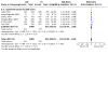
Comparison 8: Analysis by cannulation device, Outcome 4: Primary cannulation success in 'non‐cross‐over' studies
Subgroup analysis according to cannulation device could not be performed as all 'non‐cross‐over' studies used a sphincterotome for both cannulation techniques.
Subgroup analyses according to the involvement of trainees in cannulation
'Cross‐over' and 'non‐cross‐over' studies
Trainees were allowed to start cannulation in five studies (Bailey 2008; Gruchy 2007; Kawakami 2012; Kobayashi 2013; Nambu 2011). If cannulation was unsuccessful after a predefined cannulation time limit, the experienced endoscopists would take over the procedure. In other studies (Apostolopoulos 2005; Artifon 2007; Katsinelos 2008; Lee 2009; Lella 2004; Masci 2015), experienced endoscopists performed all procedures. In one study (Apostolopoulos 2005), trainees handled the guidewire. Four studies (Mangiavillano 2007; Mangiavillano 2011; Savadkoohi 2012; Zhang 2007) did not provide information on whether trainees were involved in cannulation.
Post‐ERCP pancreatitis (PEP)
Six studies with involvement of only experienced endoscopists in cannulation reported the rates of PEP for all randomized participants (Apostolopoulos 2005; Artifon 2007; Katsinelos 2008; Lee 2009; Lella 2004; Masci 2015), comprising a total of 894 participants in the guidewire‐assisted cannulation technique and 888 in the contrast‐assisted cannulation technique groups. There was no important heterogeneity among the studies (P = 0.26, I² = 23%). Unweighted pooled rates of PEP were 2.9% for the guidewire‐assisted cannulation technique and 9.1% for the contrast‐assisted cannulation technique. In studies with involvement of only experienced endoscopists, the pooled RR for PEP was 0.34, 95% CI 0.20 to 0.58, P < 0.0001 (Analysis 9.1).
9.1. Analysis.

Comparison 9: Analysis by trainee involvement in cannulation, Outcome 1: Post‐ERCP pancreatitis in all studies
Five studies had involvement of trainees in cannulation and reported the rates of PEP (Bailey 2008; Gruchy 2007; Kawakami 2012; Kobayashi 2013; Nambu 2011). Unweighted pooled rates of PEP were 4.4% for the guidewire‐assisted cannulation technique and 5.0% for the contrast‐assisted cannulation technique. There was no important heterogeneity among the studies (P = 0.38, I² = 5%). In studies with involvement of trainees in cannulation, the pooled RR for PEP was 0.93, 95% CI 0.60 to 1.46, P = 0.76 (Analysis 9.1).
Four studies did not provide information on whether trainees were involved in cannulation (Mangiavillano 2007; Mangiavillano 2011; Savadkoohi 2012; Zhang 2007), and comprised a total of 553 participants in the guidewire‐assisted cannulation technique and 391 in the contrast‐assisted cannulation technique groups. There was no important heterogeneity among the studies (P = 0.86, I² = 0%). Unweighted pooled rates of PEP were 4.0% for the guidewire‐assisted cannulation technique and 9.7% for the contrast‐assisted cannulation technique. In studies that did not provide information on whether trainees were involved in cannulation, the pooled RR for PEP was 0.44, 95% CI 0.26 to 0.73, P = 0.001 (Analysis 9.1).
There was evidence of a difference between the two subgroups (studies with versus without trainee involvement in cannulation) (test for subgroup differences P = 0.004; I² = 87.7%) or for the three subgroups (test for subgroup differences P = 0.009; I² = 78.5%) for the outcome of PEP, favouring the guidewire‐assisted cannulation technique when only experienced endoscopists were involved in cannulation.
Primary cannulation success
Six studies had involvement of only experienced endoscopists in cannulation and reported the primary cannulation success rates for all randomized participants (Apostolopoulos 2005; Artifon 2007; Katsinelos 2008; Lee 2009; Lella 2004; Masci 2015), comprising a total of 894 participants in the guidewire‐assisted cannulation technique and 888 in the contrast‐assisted cannulation technique groups. Unweighted pooled primary cannulation success rates were 86.4% for the guidewire‐assisted cannulation technique and 76.1% for the contrast‐assisted cannulation technique. There was considerable heterogeneity among the studies (P < 0.00001, I² = 94%). In studies that had involvement of only experienced endoscopists in cannulation, the pooled RR for primary cannulation success was 1.13, 95% CI 0.96 to 1.34, P = 0.15 (Analysis 9.2).
9.2. Analysis.

Comparison 9: Analysis by trainee involvement in cannulation, Outcome 2: Primary cannulation success in all studies
Five studies had involvement of trainees in cannulation (Bailey 2008; Gruchy 2007; Kawakami 2012; Kobayashi 2013; Nambu 2011). All except one study (Gruchy 2007) reported primary cannulation success rates. Unweighted pooled primary cannulation rates were 77.2% for the guidewire‐assisted cannulation technique and 75.0% for the contrast‐assisted cannulation technique. There was no important heterogeneity among the studies (P = 0.40, I² = 0%). In studies that had involvement of trainees in cannulation, the pooled RR for primary cannulation success was 1.02, 95% CI 0.96 to 1.08, P = 0.56 (Analysis 9.2).
Four studies did not provide information on whether trainees were involved in cannulation (Mangiavillano 2007; Mangiavillano 2011; Savadkoohi 2012; Zhang 2007). Three studies reported primary cannulation success rates (Mangiavillano 2007; Savadkoohi 2012; Zhang 2007). The primary cannulation success rates were 94.7% for the guidewire‐assisted cannulation technique and 90.5% for the contrast‐assisted cannulation technique. The pooled RR for primary cannulation success was 1.01, 95% CI 0.99 to 1.04, P = 0.32 (Analysis 9.2).
There was no evidence of a difference between the two subgroups (studies with versus without trainee involvement in cannulation) (test for subgroup differences P = 0.24; I² = 28.3%) or three subgroups (test for subgroup differences P = 0.43; I² =0%) for the outcome of primary cannulation success.
'Non‐cross‐over' studies
Post‐ERCP pancreatitis (PEP)
Among the seven 'non‐cross‐over' studies, experienced endoscopists performed all procedures in four studies (Apostolopoulos 2005; Artifon 2007; Lee 2009; Lella 2004), and three studies did not provide information on whether trainees were involved in cannulation (Mangiavillano 2007; Savadkoohi 2012; Zhang 2007). No 'non‐cross‐over' studies had involvement of trainees in cannulation.
All four 'non‐cross‐over' studies with involvement of only experienced endoscopists in cannulation reported the rates of PEP for all randomized participants (Apostolopoulos 2005; Artifon 2007; Lee 2009; Lella 2004), comprising a total of 567 participants in the guidewire‐assisted cannulation technique and 563 in the contrast‐assisted cannulation technique. There was no important heterogeneity among the studies for this analysis (P = 0.73, I² = 0%). Unweighted pooled rates of PEP for participants were 1.6% for the guidewire‐assisted cannulation technique and 8.7% for the contrast‐assisted cannulation technique. In 'non‐cross‐over' studies with involvement of only experienced endoscopists in cannulation, the pooled RR for PEP was 0.21, 95% CI 0.10 to 0.41, P < 0.00001 (Analysis 9.3).
9.3. Analysis.
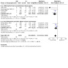
Comparison 9: Analysis by trainee involvement in cannulation, Outcome 3: Post‐ERCP pancreatitis in 'non‐cross‐over' studies
Three 'non‐cross‐over' studies did not provide information on whether trainees were involved in cannulation (Mangiavillano 2007; Savadkoohi 2012; Zhang 2007), and comprised a total of 507 participants in the guidewire‐assisted cannulation technique and 349 in the contrast‐assisted cannulation technique groups. Unweighted pooled rates of PEP for participants were 3.9% for the guidewire‐assisted cannulation technique and 9.7% for the contrast‐assisted cannulation technique. The pooled RR for PEP was 0.43, 95% CI 0.25 to 0.74, P = 0.002 (Analysis 9.3).
There was no evidence of a difference between the two subgroups (studies with trainee involvement in cannulation versus studies with uncertain trainee involvement in cannulation) for the outcome of PEP (test for subgroup differences: P = 0.09; I² = 64.4%).
Primary cannulation success
All four 'non‐cross‐over' studies with involvement of only experienced endoscopists in cannulation reported primary cannulation success rates for all randomized participants (Apostolopoulos 2005; Artifon 2007; Lee 2009; Lella 2004), comprising a total of 567 participants in the guidewire‐assisted cannulation technique and 563 in the contrast‐assisted cannulation technique. Unweighted pooled primary cannulation success rates were 89.2% for the guidewire‐assisted cannulation technique and 83.5% for the contrast‐assisted cannulation technique. There was considerable heterogeneity among the studies for this analysis (P < 0.0001, I² = 87%). The pooled RR for primary cannulation success was 1.06, 95% CI 0.93 to 1.21, P = 0.37 (Analysis 9.4).
9.4. Analysis.

Comparison 9: Analysis by trainee involvement in cannulation, Outcome 4: Primary cannulation success in 'non‐cross‐over' studies
Three 'non‐cross‐over' studies did not provide information on whether trainees were involved in cannulation (Mangiavillano 2007; Savadkoohi 2012; Zhang 2007), and comprised a total of 507 participants in the guidewire‐assisted cannulation technique and 349 in the contrast‐assisted cannulation technique groups. Unweighted pooled primary cannulation success rates were 94.5% for the guidewire‐assisted cannulation technique and 90.5% for the contrast‐assisted cannulation technique. The pooled RR for primary cannulation success was 1.01, 95% CI 0.99 to 1.04, P = 0.33 (Analysis 9.4).
There was no evidence of a difference between the two subgroups (studies with trainee involvement in cannulation versus studies with uncertain trainee involvement in cannulation) for the outcome of primary cannulation success (test for subgroup differences: P = 0.47; I² = 0%).
Discussion
This updated systematic review and meta‐analysis included 15 randomized controlled trials (RCTs) that assessed the clinical effectiveness and safety of the guidewire‐assisted cannulation technique compared to the contrast‐assisted cannulation technique in the common bile duct (CBD) for the prevention of post‐endoscopic retrograde cholangiopancreatography (ERCP) pancreatitis (PEP) and for achieving selective biliary cannulation. This has been a complex area to review systemically due to the variability in trial designs. These include permission versus non‐permission of technique 'cross‐over' when the randomized technique failed, the use of precut sphincterotomy as a rescue technique, the variable definitions used by the primary studies for difficult cannulation before technique 'cross‐over' or resorting to precut sphincterotomy, the use of prophylactic pancreatic duct (PD) stents, the different cannulation devices used between intervention arms, the variable number and experience of endoscopists, and the involvement of trainees in cannulation. Furthermore, studies have used different criteria to diagnose and grade the severity of PEP.
To standardize the definitions of PEP and to allow comparability between trials, we determined a priori that PEP, as defined by the consensus definition (Cotton 1991) or the revised Atlanta Classification (Banks 2013), to be the most important primary outcome. According to the consensus definition, PEP was defined by the presence of abdominal pain characteristic of pancreatitis associated with a serum amylase level of at least three times above the upper limit of normal at 24 hours after the procedure, together with an unplanned hospital stay or an extension of a planned hospital stay by at least two days (Cotton 1991). We also defined the severity of PEP based on the consensus criteria (Cotton 1991) depending on the number of days of hospitalisation and local complications secondary to pancreatitis as mild (hospital stay of up to three days), moderate (hospital stay for 4 to 10 days) and severe (hospital stay for more than 10 days with a significant complication). Although it appears the consensus definition (abdominal pain associated with a serum amylase level at least three times above the upper limit of normal at 24 hours after ERCP) was used by most studies to define PEP (Apostolopoulos 2005; Artifon 2007; Bailey 2008; Gruchy 2007; Katsinelos 2008; Kawakami 2012; Kobayashi 2013; Lee 2009; Mangiavillano 2011; Masci 2015; Nambu 2011; Zhang 2007), procedure‐related hospital stay was specifically stated as part of the diagnostic criteria by only one study (Gruchy 2007). In one study (Lella 2004), a serum amylase level of at least five times the upper limit of normal was used, which may have reduced the apparent rate of PEP. In four studies (Artifon 2007; Kobayashi 2013; Savadkoohi 2012; Zhang 2007), all participants were admitted for overnight observation after ERCP. As a result, participants were more likely to undergo laboratory and radiological evaluation of abdominal pain as opposed to being discharged home following ERCP. This may have increased the apparent rates of PEP in three studies (Artifon 2007; Kobayashi 2013; Savadkoohi 2012). Two studies (Mangiavillano 2007; Savadkoohi 2012) did not specify the criteria for the diagnosis of PEP. Severity of PEP was graded using the consensus criteria in eight studies (Bailey 2008; Katsinelos 2008; Kawakami 2012; Kobayashi 2013; Lee 2009; Masci 2015; Nambu 2011; Zhang 2007). Two studies (Apostolopoulos 2005; Artifon 2007) graded severity using the Ranson's criteria (Ranson 1974) and the Balthazar grading system (Balthazar 1990). Two studies (Lella 2004; Mangiavillano 2007) did not specify the criteria used for severity assessment. Three other studies did not provide outcome data regarding the severity of PEP (Gruchy 2007; Mangiavillano 2011; Savadkoohi 2012). No studies used the revised Atlanta Classification in diagnosing or grading the severity of PEP (Banks 2013).
Because the definitions and grading of severity of PEP were variable between studies, we decided to accept the definitions used by the primary studies for this review. We acknowledge that the variable definitions used by the primary studies are likely to introduce heterogeneity in the analyses. However, the definition of PEP still remains a controversial issue. The consensus definition (Cotton 1991) or the revised Atlanta Classification (Banks 2013) have not been adopted widely, and varying definitions of PEP have been used both in research and in clinical practice. In our review, the incidence of PEP ranged from 2.0% (Lella 2004) to 12.6% (Savadkoohi 2012) among the included studies (Table 4). This varying incidence of PEP may be attributable to a combination of factors, differences in patient populations (case‐mix), techniques performed during the procedure, endoscopic expertise, definitions of PEP used, methods of data collection, and completeness of follow‐up and assessment (Freeman 2004a; Testoni 2002).
Summary of main results
Post‐ERCP pancreatitis (PEP)
Post‐endoscopic retrograde cholangiopancreatography (ERCP) pancreatitis (PEP) is the primary outcome of this review. We found moderate‐certainty evidence that the guidewire‐assisted cannulation technique probably reduces the risk of PEP compared to the conventional contrast‐assisted cannulation technique. Heterogeneity in this analysis could be explained mainly by differences in trial design ('cross‐over' versus 'non‐cross‐over' to the alternative technique if the randomized technique failed). Other factors such as the use of a prophylactic pancreatic duct (PD) stent, cannulation devices (sphincterotome versus standard catheter), and involvement of trainees in cannulation also contributed to heterogeneity, but these factors could be confounded by trial design due to overlap in comparison groups between the trial design and these subgroups. Most information was obtained from studies with high risk of bias for blinding of participants and personnel (the endoscopists). This may have an impact on the rates of PEP, depending on the preference and expertise of the endoscopists performing the procedure.
Severity of post‐ERCP pancreatitis (PEP)
The severity of PEP is an important clinical outcome as it correlates with mortality, complications, and length of hospital stay. We found low‐certainty evidence that the guidewire‐assisted cannulation technique may reduce the risk of mild PEP compared to the contrast‐assisted technique. As well, we found low‐certainty evidence that the guidewire‐assisted cannulation technique may reduce the risk of moderate or severe PEP compared to the contrast‐assisted cannulation technique. However, the event rates for moderate and severe PEP were low, resulting in wide confidence intervals around the estimates.
Primary cannulation success
Primary cannulation success is an important benchmark of successful ERCP. A high primary cannulation success rate reduces the risk of difficult cannulation, repeated cannulation attempts, and the need for precut sphincterotomy, all of which have been reported as independent procedure‐related risk factors for PEP (Freeman 2001). We found low‐certainty evidence that the guidewire‐assisted cannulation technique may result in an increase in primary cannulation success compared to the contrast‐assisted cannulation technique. The substantial heterogeneity in this analysis was unexplained. As well, the lack of blinding of endoscopists may introduce bias due to the endoscopist's experience and preference of techniques.
Secondary cannulation success
Secondary cannulation success is defined as successful cannulation after 'cross‐over' to the alternative technique when the randomized technique failed. This is an important outcome as a high secondary cannulation success rate may reduce the need for precut sphincterotomy or the risk of a failed procedure. Among 'cross‐over' studies, we found very low‐certainty evidence that the guidewire‐assisted cannulation technique may reduce the 'cross‐over' rates, but may have little to no effect on the secondary cannulation rates compared to the contrast‐assisted cannulation technique, but the evidence is very uncertain.
Overall cannulation success
Overall cannulation success is an important outcome as failed procedures usually necessitate repeat ERCP or a radiological or a surgical procedure, which carry additional costs and risks (Perdue 2004). We found moderate‐certainty evidence that the guidewire‐assisted cannulation technique probably results in little to no difference in the overall cannulation success rates compared to the contrast‐assisted cannulation technique. However, this outcome is difficult to interpret because the overall effect could be diluted by 'cross‐over' studies and the use of precut as a rescue technique.
The need for precut sphincterotomy
The need for precut sphincterotomy is an important clinical outcome as it has been reported to be associated with an increased risk of complications including PEP, bleeding, and perforation (Cennamo 2010; Freeman 2001; Masci 2003). We found moderate‐certainty evidence that the guidewire‐assisted cannulation technique probably reduces the need for precut sphincterotomy slightly compared to the contrast‐assisted cannulation technique.
Inadvertent PD cannulation or injection
Inadvertent PD manipulation (cannulation or injection) has been reported to be associated with an increased risk of PEP (Masci 2003; Vandervoort 2002). We found very low‐certainty evidence that the guidewire‐assisted cannulation technique may result in little to no difference in the risk of inadvertent PD manipulation (cannulation or injection) compared to the contrast‐assisted cannulation technique.
ERCP‐related complications
With regard to safety endpoints, we found low‐certainty evidence that the guidewire‐assisted cannulation technique may result in little to no difference in the risk of post‐sphincterotomy bleeding compared to the contrast‐assisted cannulation technique. The risk of bleeding appeared to be low and most bleeding episodes either stopped spontaneously or with medical or endoscopic therapies. As well, we found very low‐certainty evidence that the guidewire‐assisted cannulation technique may have little to no effect on the risk of perforation compared to the contrast‐assisted cannulation technique, but the evidence is very uncertain. Finally, we found moderate‐certainty evidence that the guidewire‐assisted cannulation technique probably results in little to no difference in mortality. Overall, the risks of perforation, cholangitis, and mortality appeared to be very low.
Summary of findings on subgroup analyses
Meta‐analyses on subgroups of the studies were performed to explore sources of heterogeneity. Meta‐regression was not performed given the small number of included studies. We prespecified all subgroup analyses based on scientific rationale. Due to the observational nature of subgroup analyses, the following results should be considered hypothesis‐generating for further testing rather than evidence that should change practice. Furthermore, differences between subgroups, particularly those that correspond to differences between studies, need to be interpreted cautiously since chance variation between subgroups is inevitable. The rationale for and the limitations of the analyses are discussed followed by a summary of the findings on each subgroup analysis.
Trial design
Endoscopic trials usually involve the comparison of an established technique with a new technique. 'Cross‐over' between interventions is not uncommon due to unforeseen technical challenges or endoscopic findings. This perceived need for 'cross‐over' may be motivated by the moral imperative to avoid the potential adverse consequences of failed ERCP, complications, and the need for repeat ERCP, percutaneous transhepatic cholangiography, or surgery. This 'cross‐over' design, however, should not be confused with trials in which all participants are randomized to a sequence of treatments or interventions. In this review, technique 'cross‐over' was permitted in eight included studies (Bailey 2008; Gruchy 2007; Katsinelos 2008; Kawakami 2012; Kobayashi 2013; Mangiavillano 2011; Masci 2015; Nambu 2011); participants were permitted to 'cross‐over' to the alternative technique if the randomized technique failed to achieve biliary cannulation within a predefined cannulation limit. Among 'cross‐over' studies, the percentage of participants requiring 'cross‐over' to the alternative technique ranged from 14.9%(Kobayashi 2013) to 32.2% (Katsinelos 2008). These 'cross‐over' studies are at risk for contamination due to the unavoidable carry‐over effects in the subgroup of participants who received the alternative technique after failing the assigned technique. Hence, an observed effect cannot be attributed to the randomized intervention alone. Furthermore, the 'cross‐over' effect can substantially reduce the power of a trial to find an overall treatment difference. There is also the concern of differential procedural 'cross‐over' bias. In some studies, there were disproportionately larger numbers of participants who required 'cross‐over' from the contrast‐assisted cannulation technique to the guidewire‐assisted cannulation technique (Bailey 2008; Katsinelos 2008; Masci 2015) than vice versa. This may well be due to lower primary cannulation success with the contrast‐assisted cannulation technique than with the guidewire‐assisted cannulation technique. Alternatively, it is conceivable that this may also reflect the preference or expertise of the endoscopist, and the trial results may therefore be biased. Nevertheless, restricting analyses to participants who did not 'cross‐over' would certainly produce biased results because those who required 'cross‐over' were likely to be more difficult to cannulate and therefore carried a higher risk of PEP than those who did not 'cross‐over'. We, therefore, decided a priori to investigate trial design as a potential source of heterogeneity by performing subgroup analysis ('cross‐over' versus 'non‐cross‐over' studies).
In our prespecified subgroup analysis according to trial design, we found significant subgroup differences between 'non‐cross‐over' and 'cross‐over' studies for the outcome of PEP suggesting trial design is an important source of heterogeneity.
Publication type
The inclusion of unpublished data in meta‐analyses is controversial. There is empirical evidence to suggest that published studies are more likely to report significant or clinically favourable results than unpublished studies (Dickersin 1987; Easterbrook 1991; Eyding 2010). This is likely to be due to studies with negative results not being submitted for publication rather than being rejected after submission. When unfavourable results of clinical trials are not published, meta‐analyses and systematic reviews that are based only on published data may overestimate the treatment effects. On the other hand, unpublished studies may be of lower methodological quality than published studies (Cook 1993), and their inclusion may therefore compromise the validity of a meta‐analysis. However, previous studies have found no significant methodological differences between published and unpublished studies (Dickersin 1987; Easterbrook 1991). To ameliorate the effects of publication bias, we included both published and unpublished studies in our meta‐analysis. We also decided a priori to investigate publication type as a potential source of heterogeneity by comparing the results of published and unpublished studies. However, subgroup analysis according to publication type did not show any significant subgroup difference.
Risk of bias
The success of randomization depends on two interrelated processes, random sequence generation and allocation concealment. Inadequate sequence generation has been shown to yield exaggerated estimates of intervention effects compared with trials with adequate sequence generation (Als‐Nielsen 2004; Kjaergard 2001; Schulz 2002). This is because selection bias can arise due to selective enrolment and non‐enrolment of participants into a study if the sequence generation is not truly random. Concealment of allocation is important in protecting the merit of randomization. Without concealment of allocation, investigators may systematically influence group allocation. Empiric studies have shown that inadequate allocation concealment can also lead to exaggerated estimates of treatment effects (Kjaergard 2001; Schulz 1995) but with scope for bias in either direction. In addition, inadequate reporting has been associated with biased treatment estimates (Moher 1998; Schulz 1995). We, therefore, performed prespecified subgroup analyses according to random sequence generation (unclear versus low risk of bias) and allocation concealment (unclear versus low risk of bias). However, subgroup analyses according to risk of bias (random sequence generation or allocation concealment) did not show any significant subgroup differences.
Precut sphincterotomy
Precut sphincterotomy is often used after conventional methods of biliary cannulation have failed. Although the use of precut sphincterotomy may improve the cannulation success rate, prospective studies have suggested that it is an independent risk factor for post‐ERCP complications including PEP (Cennamo 2010; Freeman 2001; Masci 2003). However, it remains controversial whether precut alone or the repeated attempts at cannulation prior to precut is the culprit factor in the development of PEP (Testoni 2011). We were not able to perform meta‐analysis based on individual patient‐level data because none of the studies reported individual patient data. Subgroup data were provided by three 'non‐cross‐over' studies (Apostolopoulos 2005; Artifon 2007; Lee 2009). We, therefore, performed prespecified between‐study subgroup analysis according to the permission of precut sphincterotomy for all included studies and within‐study subgroup analysis for the studies that provided subgroup data (Apostolopoulos 2005; Artifon 2007; Lee 2009). However, subgroup analyses according to the permission of precut sphincterotomy did not show any significant subgroup differences.
Inadvertent guidewire insertion or contrast injection into the PD (inadvertent PD manipulation)
Several mechanisms have been postulated for the prevention of PEP with the guidewire‐assisted cannulation technique. They include facilitating selective biliary cannulation, limiting papillary trauma, and minimizing inadvertent contrast injection into the main PD or the papilla itself (submucosal injection) and, thereby, reducing the possibility of mechanical, chemical, and hydrostatic injury to the pancreas when compared to the contrast‐assisted cannulation technique. However, inadvertent guidewire insertion into the PD (especially when performed repeatedly) may result in injury to the papilla or the PD, increasing the likelihood of PEP. Moreover, it remains unclear whether inadvertent guidewire insertion into the PD is safer than inadvertent contrast injection into the PD with regard to PEP. We were not able to perform meta‐analysis based on individual patient‐level data because none of the studies reported individual patient data. Subgroup data were provided by four 'non‐cross‐over' studies (Artifon 2007; Lee 2009; Lella 2004; Mangiavillano 2007) and one 'cross‐over' study (Nambu 2011). We, therefore, performed prespecified within‐study subgroup analysis for the studies that provided subgroup data according to inadvertent guidewire cannulation or contrast injection into the PD (inadvertent PD manipulation). However, subgroup analyses according to inadvertent PD manipulation did not show any significant subgroup differences.
PD stent
Outflow tract oedema caused by cannulation trauma to the papilla, inadvertent PD manipulation, and contrast injection may cause obstruction to the flow of pancreatic secretions with subsequent acute pancreatic inflammation. It has been postulated that a stent placed across the injured outflow tract may help to maintain the flow of pancreatic secretions and reduce the intraductal pressure after ERCP. Indeed, PD stent placement in high‐risk patients has been found to significantly reduce the risk of PEP (Choudhary 2011). We were not able to perform meta‐analysis based on individual patient‐level data because none of the studies reported individual patient data. Separate subgroup data according to the use of a PD stent were also not reported by any of the included studies. We, therefore, performed a prespecified between‐study subgroup analysis according to the permission of a PD stent for all included studies. Subgroup analyses according to the permission of the use of PD stent showed significant subgroup differences. However, there was a significant overlap between trial design and the use of PD stents. All studies that permitted the use of PD stents were 'cross‐over' studies, whereas all except one study (Nambu 2011) that did not permit the use of PD stents were 'non‐cross‐over' studies. Hence, trial design rather than the use of PD stents may be a more important source of heterogeneity for the outcome of PEP.
Cannulation device
Biliary cannulation is best performed from below with an upward view of the papilla and with the cannulation device in line with the axis of the common bile duct (CBD) towards the 11 o'clock position (Freeman 2005). Standard catheters are limited in their ability to vary the angle of approach to the papilla independent of the endoscope to gain biliary access. The tip of the sphincterotome, however, can be adjusted to give preferential upward angulation for selective biliary cannulation. Indeed, RCTs have found the use of a sphincterotome to be superior to that of a standard catheter for achieving selective biliary cannulation, with a significant reduction in cannulation times and in the number of attempts required for selective biliary cannulation (Cortas 1999; Schwacha 2000). Whether the use of a sphincterotome for cannulation results in less PEP is not clear. We, therefore, performed prespecified between‐study subgroup analyses according to the use of cannulation device (sphincterotome versus standard catheter). However, subgroup analysis according to cannulation device did not show any significant subgroup differences.
Involvement of trainees in cannulation
Trainee participation in the procedure has been shown to be a significant risk factor for the development of PEP (Cheng 2006). This increased risk is possibly due to multiple cannulation attempts by trainees. The findings of the UK National Confidential Enquiry into Patient Outcomes and Death relating to ERCP suggested that trainees with experience of > 200 ERCPs had an unsupervised cannulation rate of 66%; this fell to 40% for those with experience of < 200 ERCPs (Williams 2007a). This is in contrast to a cannulation success rate of over 90% in experienced endoscopists. We, therefore, performed prespecified between‐study subgroup analyses according to the involvement of trainees in cannulation. Subgroup analyses according to the involvement of trainees in cannulation showed significant subgroup differences for the outcome of PEP. However, there was a significant overlap between trial design and the involvement of trainees in cannulation. All studies with the involvement of trainees in cannulation were 'cross‐over' studies, whereas all but two studies (Katsinelos 2008; Masci 2015) with involvement of only experienced endoscopists were 'non‐cross‐over' studies. Hence, trial design rather than the involvement of trainees may be a more important source of heterogeneity for the outcome of PEP.
Overall completeness and applicability of evidence
This systematic review was designed to include trials from around the world regardless of publication status or language of publication. All studies identified by the search could be retrieved in full. Moreover, we were able to obtain unpublished data from authors of primary studies, including data of a completed trial that has been published in abstract format only as an interim analysis (Gruchy 2007). Hence, we believe this review is comprehensive, and the results reflect the available evidence for the guidewire‐assisted cannulation technique compared to the conventional contrast‐assisted cannulation technique for the prevention of post‐endoscopic retrograde cholangiopancreatography (ERCP) pancreatitis (PEP). Most studies defined PEP as abdominal pain characteristic of pancreatitis associated with a rise in serum amylase level of at least three times the upper limit of normal at 24 hours after the procedure according to the consensus criteria (Cotton 1991). The participants included in this meta‐analysis had intact papilla and required endoscopic retrograde cholangiopancreatography (ERCP) for a variety of pancreaticobiliary diseases including common bile duct (CBD) stones, pancreaticobiliary malignancies, sphincter of Oddi dysfunction (SOD), and idiopathic recurrent pancreatitis. Participants included were of a wide age range (18 to 96 years). There were equal proportions of males and females. Studies were conducted in high‐volume, tertiary care settings (nine single‐centre studies and six multicenter studies). Procedures were performed by either single or multiple experienced endoscopists, with or without the involvement of trainees. Therefore, the generalizability of findings to low‐volume centres with less expertise in ERCP, especially in the use of the guidewire technique, may be limited. Cannulation was carried out with either a sphincterotome (in most studies) or a catheter using either contrast or a 0.035‐inch hydrophilic guidewire. Precut sphincterotomy or 'cross‐over' to the alternative cannulation technique was permitted if the randomized technique failed due to difficult cannulation in most studies. Pancreatic duct (PD) stents were used for prophylaxis of PEP in some studies. No studies reported the use of prophylactic nonsteroidal anti‐inflammatory drugs (NSAIDs). The findings of significant subgroup differences according to the use of PD stents and involvement of trainees in cannulation should be interpreted with caution keeping in mind the significant overlap with trial design. Overall, we found moderate‐certainty evidence that the guidewire‐assisted cannulation technique probably reduces the risk of PEP compared to the conventional contrast‐assisted cannulation technique.
In summary, the evidence found by this review addressed the review question very well. The results of this review are applicable to most people undergoing ERCP for biliary or pancreatic diseases performed by endoscopists in high‐volume, tertiary care settings. Although it is unlikely that in clinical practice biliary cannulation is performed with either technique alone, the guidewire‐assisted cannulation technique appears to be superior to the contrast‐assisted cannulation technique considering the certainty of evidence and the balance of benefits and harms. However, the routine use of guidewires in biliary cannulation will be dependent on local expertise, availability, and cost.
Quality of the evidence
The certainty (or quality) of evidence for the outcome of post‐endoscopic retrograde cholangiopancreatography (ERCP) pancreatitis (PEP) was judged to be moderate due to serious study limitations. This is because most information was obtained from studies with high risk of bias for blinding of participants and personnel (the endoscopists) (Apostolopoulos 2005; Artifon 2007; Bailey 2008; Gruchy 2007; Katsinelos 2008; Kawakami 2012; Kobayashi 2013; Lee 2009; Lella 2004; Mangiavillano 2007; Mangiavillano 2011; Masci 2015; Nambu 2011; Savadkoohi 2012; Zhang 2007). Lack of binding of the endoscopist may have an impact on cannulation success and PEP depending on the experience, expertise, and preference of the endoscopist performing the procedure. One study (Gruchy 2007) was considered as being at high risk of bias for incomplete outcome data as 25% of participants were lost to follow‐up. Nevertheless, the results remained robust with the exclusion of this only high risk of bias study for incomplete outcome data (Gruchy 2007). Furthermore, the robustness of the results during sensitivity analyses would support the overall certainty of evidence and conclusions reached by this review. There was significant heterogeneity for the outcome of PEP, which could be explained by differences in trial design. Therefore, the certainty of evidence for the outcome of PEP was not downgraded because of inconsistency or heterogeneity. Other factors such as the use of prophylactic pancreatic duct (PD) stents, cannulation devices, and involvement of trainees in cannulation also contribute to heterogeneity, but these factors could be confounded by trial design due to the overlap in comparison groups between trial design and these subgroups.
We judged the certainty of evidence to be low for severity of PEP (mild, moderate, or severe) due to serious risk of bias and inconsistency; low for primary cannulation success due to serious risk of bias and inconsistency; very low for secondary cannulation success due to serious risk of bias, inconsistency, and imprecision; moderate for overall cannulation success due to serious risk of bias; moderate for the need for precut sphincterotomy due to serious risk of bias; and very low for inadvertent PD cannulation/injection due to serious risk of bias, inconsistency, and imprecision. For ERCP complications, we judged the certainty of evidence to be low for post‐sphincterotomy bleeding due to serious risk of bias and imprecision; very low for perforation due to serious risk of bias, inconsistency, and imprecision; and moderate for mortality due to serious risk of bias.
The Summary of Findings Table (Table 1) provides detailed descriptions and justification for the downgrading of the certainty of evidence.
Potential biases in the review process
Our comprehensive and systematic literature search of published studies, unpublished data sources, and ongoing studies in major conference proceedings, ClinicalTrials.gov, and the WHO International Clinical Trials Registry Platform (ICTRP) portal should have minimized the likelihood of missing relevant evidence. We contacted all authors of included studies for missing outcome data by email, and we were successful in obtaining missing data for six studies (Apostolopoulos 2005; Bailey 2008; Gruchy 2007; Kobayashi 2013; Lee 2009; Nambu 2011). Translation of one Chinese study was also performed for data extraction (Zhang 2007). Indeed, our systematic review has identified more eligible studies than all other published systematic reviews to date (Cennamo 2009; Cheung 2009; De Moura 2016; Ma 2016; Shao 2009).
We explored small‐study effects (a tendency for the intervention effects estimated in smaller studies to differ from those estimated in larger studies), of which non‐reporting bias is one potential cause, using funnel plots (Figure 4). Visual inspection of the funnel plot suggests asymmetry with a gap in the bottom right side of the graph. This impression is partially caused by one study (Lella 2004) at the bottom left of the most common effect. Although this study (Lella 2004) had the third‐largest sample size (N = 400) among the included studies, it had the highest standard error due to loss of statistical power resulting from low event rates (zero event in the guidewire‐assisted cannulation arm). The low event rates may be due to the use of a more stringent definition of post‐endoscopic retrograde cholangiopancreatography (ERCP) pancreatitis (PEP) (amylase more than 5 times the upper limit of normal) (Lella 2004) compared to the other studies (amylase more than 3 times the upper limit of normal). In addition, the asymmetry may be due to a lack of negative studies with high standard error and low statistical power. Funnel plot asymmetry tests, however, showed discordant results with a positive Egger test (P = 0.04) and negative Harbord‐Egger test (P = 0.25).
4.

Funnel plot of comparison: 1 Guidewire‐assisted cannulation vs contrast‐assisted cannulation, Main analysis, outcome: 1.1 Post‐ERCP pancreatitis (ITT)
Funnel plot asymmetry cannot, however, be considered to be proof of non‐reporting bias in a meta‐analysis (Sterne 2000). True clinical or methodological heterogeneity between studies may also lead to funnel plot asymmetry (Sterne 2000; Higgins 2021). It has been recognized that the funnel plot itself is inappropriate in the presence of significant heterogeneity (Ioannidis 2007; Terrin 2003). This is because the funnel plot is based on the premise that studies come from a single underlying population and all studies estimate a single true effect (Light 1984). In the presence of significant heterogeneity, it is highly likely that studies are in fact estimating a range of effects rather than a single true effect (Terrin 2003). Furthermore, the application of funnel plot asymmetry tests such as the Egger (Egger 1997) and the Harbord‐Egger (Harbord 2006) tests to detect non‐reporting bias is inappropriate or not meaningful in the presence of significant heterogeneity, and may lead to false‐positive claims for non‐reporting bias (Harbord 2006; Ioannidis 2007), although the Harbord‐Egger test has better properties than the Egger test (Harbord 2006). Also, when the average event rate per trial is low, these tests may give false‐positive results (Sterne 2000). The trim and fill method for adjusting for non‐reporting bias has also been shown to spuriously adjust the estimate of the overall treatment effect when the studies are heterogeneous (Terrin 2003). Indeed, there is moderate heterogeneity for the outcome of PEP in our review that could be explained by mainly differences in trial design ('cross‐over' versus 'non‐cross‐over'). In addition, other factors such as the use of a prophylactic pancreatic duct (PD) stent, cannulation devices (sphincterotome versus standard catheter), and involvement of trainees in cannulation also contributed to heterogeneity, but these factors could have been confounded by trial design due to overlap in comparison groups between trial design and these subgroups. For the primary outcome of PEP in our review, closer inspection of the funnel plot reveals that the 'non‐cross‐over' studies are scattered to the left of the most common effect, and most of the 'cross‐over' studies are scattered to the right, suggesting that trial design (a source of heterogeneity) may be an important source of funnel plot asymmetry (Figure 5). We were not able to pool the studies separately according to trial design to test for non‐reporting bias (or publication bias) because each group would have fewer than 10 trials (Ioannidis 2007). Based on the reasons outlined above, the application of funnel plot and asymmetry tests should be considered inappropriate or not meaningful for assessing non‐reporting bias in this meta‐analysis.
5.

Funnel plot of comparison: 2 Analysis according to trial design, outcome: 2.1 Post‐ERCP pancreatitis
A potential limitation of this review would be the variable definitions and grading of severity of PEP used by some trials (Apostolopoulos 2005; Artifon 2007; Lella 2004). The heterogeneity of criteria used to define PEP and classify its severity may make direct comparisons of these trials difficult. However, the definition of PEP still remains a controversial issue. It is well recognized that the rise in serum amylase may vary considerably without any clinical significance (Testoni 2000). However, patients with hyperamylasaemia post‐procedure (even with mild pain) are more likely to be carefully monitored with a prolonged hospital stay in both research and clinical settings. This adds to the confusion in the definition and evaluation of PEP, especially when procedure‐related hospital stay is part of the definition of PEP according to the consensus criteria (Cotton 1991). Nevertheless, the variable definitions used by the included studies likely reflect 'real world' practices with a highly variable incidence of PEP depending on the definition criteria adopted (Testoni 2000).
Another potential limitation of this review is the inclusion of 'cross‐over' studies in the main analysis, which may have diluted the treatment effect of the guidewire‐assisted cannulation technique for the prevention of PEP. Nevertheless, we still found moderate‐certainty evidence that the guidewire‐assisted cannulation technique probably reduces the risk of PEP compared to the contrast‐assisted cannulation technique with the inclusion of 'cross‐over' studies. In addition, our subgroup analyses confirmed that trial design was a significant source of heterogeneity.
Finally, the inclusion of predominantly unclear risk of bias studies for allocation concealment and blinding of outcome assessment, and high risk of bias studies for blinding of participants and personnel (the endoscopists) in the analyses may have biased our effect estimates. In particular, blinding of participants and outcome assessors is essential for reducing bias in the case of PEP when there is some degree of subjectivity in the interpretation of pancreatic pain. Lack of binding of the endoscopist may also have an impact on cannulation success and PEP depending on the experience, expertise, and preference of the endoscopist performing the procedure.
Agreements and disagreements with other studies or reviews
There have been five fully published systematic reviews (Cennamo 2009; Cheung 2009; De Moura 2016; Ma 2016; Shao 2009) on this topic that compared the guidewire‐assisted cannulation technique with the conventional contrast‐assisted cannulation technique for the prevention of post‐endoscopic retrograde cholangiopancreatography (ERCP) pancreatitis (PEP). There are some notable differences between these reviews and our review. Due to inadequate information presented in abstract format, we were not able to contrast the methodologies and findings of five other meta‐analyses published only in conference proceedings (Choudhary 2009; Choudhary 2010b; Epstein 2009; Inaganti 2013; Madhoun 2009) with our review.
The first systematic review by Shao and colleagues (Shao 2009) included four fully published randomized controlled trials (RCTs) (Artifon 2007; Bailey 2008; Lella 2004; Lee 2009). It failed to show a significant association between the use of the guidewire‐assisted cannulation technique and reduction of PEP (RR 0.34, 95% CI 0.10 to 1.17; P = 0.09). Subgroup analysis, however, showed a benefit of the guidewire‐assisted cannulation technique in trials without 'cross‐over' design (RR 0.20, 95% CI 0.09 to 0.40; P < 0.00001). First, three potentially eligible trials were not included in this meta‐analysis, a fully published RCT (Katsinelos 2008) and two trials (Gruchy 2007; Mangiavillano 2007) published in abstract format. In particular, the latter two trials (Gruchy 2007; Mangiavillano 2007) should have been identified through their search of the 2006 to 2008 proceedings of the American Gastroenterological Digestive Disease Week (published in Gastroenterology and Gastrointestinal Endoscopy) and the United European Gastroenterology Week (published in Gut and Endoscopy). Because of the small number of studies included, this meta‐analysis (Shao 2009) may have been underpowered to detect a clinically important difference between the two cannulation techniques. All three trials (Gruchy 2007; Katsinelos 2008; Mangiavillano 2007) were included in our meta‐analysis. Second, it is important to highlight that there were some discrepancies in the rates of PEP for one trial (Bailey 2008) between this review (Shao 2009) and our review. For this 'cross‐over' trial (Bailey 2008), Shao and colleagues restricted their analyses to participants who did not 'cross‐over' to the alternative cannulation technique. The PEP rates for Bailey 2008 were reported to be 10/202 in the guidewire‐assisted cannulation group and 7/211 in the contrast‐assisted cannulation group before 'cross‐over' of the cannulation technique (data requested from the primary authors by Shao 2009). Based on the publication by Bailey and colleagues (Bailey 2008), and with confirmation from the authors of the primary study, PEP occurred in 29/413 participants, 16/202 in the guidewire arm and 13/211 in the contrast arm with 50 participants crossed over to guidewire and 22 participants crossed over to contrast. Therefore, it would appear the PEP rates for Bailey 2008 should be 10/180 in the guidewire‐assisted cannulation group and 7/161 in the contrast‐assisted cannulation group before 'cross‐over'. We included all participants with PEP in our main analysis and also in our subgroup analyses according to trial design based on intention‐to‐treat (ITT) (16/215 in the guidewire‐assisted cannulation technique versus 13/215 in the contrast‐assisted cannulation technique) (Bailey 2008). In order to avoid bias, we decided a priori not to restrict our analyses to participants who did not 'cross‐over' because those who required 'cross‐over' were likely to be more difficult to cannulate and therefore carried a higher risk of PEP than those who did not 'cross‐over'.
The second systematic review by Cennamo and colleagues (Cennamo 2009) included five fully published RCTs (Artifon 2007; Bailey 2008; Katsinelos 2008; Lee 2009; Lella 2004). Two potentially eligible trials published in abstract format (Gruchy 2007; Mangiavillano 2007) were not included in this meta‐analysis. It concluded that the guidewire‐assisted cannulation technique significantly increases the primary cannulation rate (OR 2.05, 95% CI 1.27 to 3.31) and reduces the risk of PEP (OR 0.23, 95% CI 0.13 to 0.41) compared with the contrast‐assisted cannulation technique. However, the conclusion was based on the exclusion of the two 'cross‐over' trials (Bailey 2008; Katsinelos 2008) from the analysis of the PEP outcome. The results may therefore be biased towards a reduction of PEP. Instead, we included all studies in the main analyses and explored subgroup differences according to trial design in our systematic review.
The third systematic review by Cheung and colleagues (Cheung 2009) included seven RCTs (five 'non‐cross‐over' trials and two 'cross‐over' trials) (Artifon 2007; Bailey 2008; Gruchy 2007; Katsinelos 2008; Lee 2009; Lella 2004; Mangiavillano 2007). It was decided a priori that the analysis of PEP would be performed separately by trial design. The review concluded that there was a significant reduction in PEP when using the guidewire‐assisted cannulation technique compared with the contrast‐assisted cannulation technique (RR 0.38, 95% CI 0.19 to 0.76) among 'non‐cross‐over' trials only. One trial (Gruchy 2007), published in abstract format, was included under 'non‐cross‐over' trials. We included this trial (Gruchy 2007) under 'cross‐over' trials after confirming with the authors of the primary study that this was in fact a 'cross‐over' study by design. In addition, one potentially eligible trial (Apostolopoulos 2005), published in abstract format, was not included in this meta‐analysis. This trial (Apostolopoulos 2005) should have been identified through their search of the 2004 to 2008 conference abstracts (Digestive Disease Week, American College of Gastroenterology, British Society of Gastroenterology, and United European Gastroenterology Week). This meta‐analysis (Cheung 2009) also concluded that there was no significant reduction of precut with the guidewire‐assisted cannulation technique compared with the contrast‐assisted cannulation technique (RR 0.57, 95% CI 0.29 to 1.11). With a larger number of included studies, we found moderate‐certainty evidence that the guidewire‐assisted cannulation technique probably reduces the need for precut sphincterotomy slightly compared to the contrast‐assisted cannulation technique (RR 0.79, 95% CI 0.64 to 0.96; P = 0.02).
The fourth systematic review by de Moura and colleagues (De Moura 2016) included nine RCTs (Artifon 2007; Bailey 2008; Katsinelos 2008; Kawakami 2012; Kobayashi 2013; Lee 2009; Lella 2004; Nambu 2011; Savadkoohi 2012). The authors planned to include only 'non‐cross‐over' trials in the meta‐analysis as they claimed that "mixing 'non‐cross‐over' studies with 'cross‐over' studies will cause a loss in statistical power difference in the overall efficacy of the intervention and an inability to distinguish charitable or noxious effects related to the intervention". However, they had erroneously included five 'cross‐over' trials in the meta‐analysis (Bailey 2008; Katsinelos 2008; Kawakami 2012; Kobayashi 2013; Nambu 2011). As well, they had excluded two 'non‐cross‐over' trials that were published in abstract format (Apostolopoulos 2005; Mangiavillano 2007) and missed a 'non‐cross‐over' trial that was published in Chinese (Zhang 2007). The review concluded that the guidewire‐assisted cannulation technique, when compared to the conventional contrast technique, reduces the risk of PEP (risk difference (RD) 0.03, 95% CI 0.01 to 0.05) and increases primary cannulation success (RD 0.07, 95% CI 0.03 to 0.12). Despite the methodological limitations, the results of this review (De Moura 2016) are in agreement with our review which included both 'cross‐over' and 'non‐cross‐over' trials. We decided a priori to investigate trial design as a potential source of heterogeneity. In our prespecified subgroup analysis according to trial design, we found significant subgroup differences between 'non‐cross‐over' and 'cross‐over' trials for the outcome of PEP suggesting that heterogeneity in this analysis could be explained by differences in trial design.
The fifth systematic review by Ma and colleagues (Ma 2016) was published in Chinese with an abstract translated into English. This review included all nine RCTs (Artifon 2007; Bailey 2008; Katsinelos 2008; Kawakami 2012; Kobayashi 2013; Lee 2009; Lella 2004; Masci 2015; Nambu 2011) that were included in our current review. In addition, a study (Zhang 2007) published in Chinese was included in this review (Ma 2016). This Chinese study (Zhang 2007) was not identified in the literature search of our previous review as it was published in a non‐indexed journal. In our current review, we included this Chinese study (Zhang 2007). Additionally, we included five more studies, four were in abstract format (Apostolopoulos 2005; Gruchy 2007; Mangiavillano 2007; Mangiavillano 2011) and one was a fully published study (Savadkoohi 2012) that should have been identified through their literature search. This review (Ma 2016) also concluded that the guidewire‐assisted cannulation technique had a lower risk of PEP (RR 0.54, 95% CI 0.41 to 0.71) and a higher primary cannulation success rate (RR 1.04, 95% CI 1.01 to 1.06) compared to contrast‐assisted cannulation. Due to the concerns of the scientific quality of studies published in a non‐indexed journal, we performed a post hoc sensitivity analysis with restriction to English‐language studies and found that the results remained robust for the outcome of PEP with the exclusion of the only non‐indexed study (Zhang 2007).
Authors' conclusions
Implications for practice.
With the increasing availability of safer and less invasive diagnostic modalities including magnetic resonance cholangiopancreatography (MRCP) and endoscopic ultrasound (EUS), endoscopic retrograde cholangiopancreatography (ERCP) has become primarily a therapeutic procedure for a wide spectrum of biliary and pancreatic disorders. Contrast‐assisted cannulation is the conventional method commonly used to achieve selective deep biliary cannulation. When primary attempts with contrast‐assisted cannulation fail, guidewires are sometimes used as a secondary technique to facilitate biliary cannulation. Increasingly, guidewires are used as a primary cannulation technique despite conflicting evidence to support this practice (Löhr 2012).
The present analysis found that the guidewire‐assisted cannulation technique probably reduces the risk of post‐endoscopic retrograde cholangiopancreatography (ERCP) pancreatitis (PEP) compared to the conventional contrast‐assisted cannulation technique (moderate‐certainty evidence). In addition, the guidewire‐assisted cannulation technique is associated with greater primary cannulation success (low‐certainty evidence), less precut sphincterotomy (moderate‐certainty evidence), and no increase in ERCP‐related complications (low‐certainty evidence for post‐sphincterotomy bleeding; very low‐certainty evidence for perforation; moderate‐certainty evidence for mortality). The balance of harms and benefits is therefore in favour of the guidewire‐assisted cannulation technique. Our results suggest that the guidewire‐assisted cannulation technique reduces PEP by minimizing contrast injection into the pancreatic duct (PD) and by limiting papillary trauma. Although it is unlikely that in clinical practice biliary cannulation is performed with either technique alone, our results support the guidewire‐assisted cannulation technique as the superior first‐line primary cannulation technique. However, the routine use of guidewires in biliary cannulation will be dependent on local expertise, availability, and cost. Moreover, it remains to be proven whether the use of the guidewire‐assisted cannulation technique is cost‐effective in terms of prevention of PEP in the context of other pharmacologic or non‐pharmacologic interventions (for example, rectal indomethacin, PD stent). Nevertheless, the cost of the guidewires may be partly offset by easier cannulation and less use of precut sphincterotomy, which necessitates the use of another device (for example, a needle‐knife). In the era of therapeutic ERCP, the guidewire‐assisted cannulation technique using a sphincterotome may be considered superior to the contrast‐assisted cannulation technique considering guidewires have become essential in maintaining ductal access during therapeutic manoeuvres (for example, stent placement, stone extraction).
Implications for research.
This review has highlighted the need for further research on the optimal cannulation techniques for the prevention of post‐endoscopic retrograde cholangiopancreatography (ERCP) pancreatitis (PEP).
Standardized definitions are important for adequate communication in clinical practice and for research. There are currently two definitions of PEP (Banks 2013; Cotton 1991). In the consensus definition (Cotton 1991), PEP is defined as clinical pancreatitis with amylase at least three times the upper limit of normal at more than 24 hours after the procedure, requiring hospitalisation or prolongation of planned admission. The consensus definition grades the severity of PEP based on the length of hospitalisation. In the 2012 revised Atlanta Classification (Banks 2013), the diagnosis of PEP requires two of three features: 1) abdominal pain consistent with acute pancreatitis, 2) serum amylase or lipase greater than three times the upper limit of normal, and 3) characteristic findings of acute pancreatitis on abdominal imaging. This classification defines severity based on the presence or absence of organ failure and of local or systemic complications (Banks 2013). This definition, however, was not developed specifically for PEP, but for all‐cause acute pancreatitis (Banks 2013). The correlation between these two definitions seems to be poor in a prospective study (Artifon 2010). In a retrospective study, the revised Atlanta Classification was found to be superior to the consensus criteria in predicting mortality in patients with PEP (Smeets 2019). Increasingly, the Atlanta Classification is used in research studies. The European Society of Gastrointestinal Endoscopy (ESGE) guideline stated that both definitions of PEP may be used, but neither of these is ideal in the setting of PEP (Dumonceau 2014; Dumonceau 2020). In this systematic review, most studies used the consensus definition in diagnosing and grading the severity of PEP, and none used the revised Atlanta Classification. The value of a standardized definition for clinical outcomes lies in its ability to provide consistency across studies that can facilitate the evaluation of the safety and effectiveness of endoscopic procedures. Toward this end, the definition and reporting of PEP will need to be updated and standardized to reflect the current knowledge. To improve the adoption of these standardized definitions by clinicians and researchers, there is a need to validate them in prospective clinical studies.
There are different variations of the guidewire‐assisted cannulation technique. Some endoscopists prefer the insertion of a sphincterotome into the ampulla during cannulation, while others use the non‐touch technique of probing the bile duct with the guidewire. While some endoscopists prefer an assistant to handle the guidewire, others prefer to handle the guidewire themselves. Recently, the short guidewire system has allowed endoscopists greater control of the wire during cannulation (ASGE Technology Committee 2007). Theoretically, the short guidewire systems may lead to faster device exchange, less use of fluoroscopy, reduced procedure time, decreased sedation requirements, improved wire stability, and increased endoscopist control of the wire with less dependence on support staff (Reddy 2009). Furthermore, the physician‐controlled guidewire‐assisted cannulation technique has the potential to decrease papillary trauma and the risk of PEP. However, there are limited data on the ease of use and efficacy of the short guidewire systems (Draganov 2010). There is a need for further research on the efficacy and safety of these various guidewire‐assisted cannulation techniques.
Although our analysis suggested a benefit of the guidewire‐assisted cannulation technique in reducing the risk of PEP, our review also highlights the paucity of cost and cost‐effectiveness data in the context of other pharmacologic or non‐pharmacologic interventions (for example, rectal indomethacin, pancreatic duct (PD) stent, aggressive intravenous hydration). Future randomized controlled trials (RCTs) should include data on cost and resource utilization. In addition, decision analyses and economic evaluations may help identify the most cost‐effective strategy for the prevention of PEP.
'Cross‐over' effect can substantially reduce the power of a trial to find an overall treatment difference. However, 'cross‐over' between techniques is not uncommon both in clinical trials and in clinical practice due to unforeseen technical challenges or endoscopic findings. The perceived need for 'cross‐over' is often motivated by the moral imperative to avoid the potential adverse consequences of failed ERCP and complications, and the need for repeat ERCP, percutaneous transhepatic cholangiography, or surgery. The optimal solution to this problem is to avoid 'cross‐over' design or to keep 'cross‐over' or the use of the rescue technique to a reasonable minimum. However, 'cross‐over' of technique may be unavoidable due to ethical concerns (Holubkov 2009). Future trials should explicitly report trial design ('cross‐over' versus 'non‐cross‐over' of technique), criteria for technique 'cross‐over', and outcome pertaining to participants with and without 'cross‐over' to allow assessment of potential bias.
The endoscopist’s expertise, case volume, and case‐mix have been considered to be potential factors that can influence the outcome of ERCP. Concerns have been raised about the potential impact of a new intervention over time (learning curve) on RCTs by distorting comparisons (Cook 2004). Failure to control for the learning curve effects may underestimate the treatment effect of any new intervention. In addition, variation in trainee involvement in one intervention arm compared to another may occur as technically challenging interventions are more likely to be performed by experienced endoscopists than by trainees. This differential involvement of trainees between arms implies a bias against the intervention with more trainee involvement. Furthermore, failure to maintain a consistently high quality of procedures may dilute any important treatment differences and may have an impact on patient outcomes. One solution is to avoid trainees altogether in RCTs. However, this would be impractical since most RCTs are done at academic centres. Another solution is to define competency thresholds and standards (based on procedural numbers and competency thresholds) for all endoscopists prior to their participation in RCTs. In addition, some degree of standardization in techniques may improve comparability between trials.
What's new
| Date | Event | Description |
|---|---|---|
| 29 October 2021 | New citation required but conclusions have not changed | Updated literature search (3 new studies were identified and updated data for 1 RCT); information and references were updated in the Background. |
| 14 June 2021 | New search has been performed | An updated search was performed on February 26 2021. Two new studies published in 2012 and in 2015 were identified. A study in Chinese published in 2007 was identified from a Chinese systematic review published in 2016. A conference abstract in 2010 that was included in our previous version was published in full in 2013. So, we updated the data. In total, we included three more RCTs and updated one RCT, compared to the published version in 2012. |
History
Protocol first published: Issue 2, 2012 Review first published: Issue 12, 2012
Acknowledgements
Cochrane Gut supported the authors in the development of this systematic review.
The following people conducted the editorial process for this article:
Co‐ordinating Editor/Sign‐off Editor (final editorial decision): Professor Morris Gordon, Cochrane Gut ‐ UK, University of Central Lancashire
Managing Editor (selected peer reviewers, collated peer‐reviewer comments, provided editorial guidance to authors, edited the article): Ghazaleh Aali, Cochrane Gut Group ‐ UK, University of Central Lancashire
Copy Editor (copy‐editing and production): Anne Lethaby, University of Auckland, Auckland, NZ
Peer‐reviewers (provided comments and recommended an editorial decision): Dr. Marianna Arvanitaki, Erasme University Hospital, Brussels, Belgium (clinical review); Ms. Sarah Rhodes, Centre for Biostatistics, University of Manchester (statistical review); Dr. Farhad Shokraneh, University College London (search review).
The 'Methods' section of this review was based on a standard template used by the Cochrane Gut Group.
The authors would also like to acknowledge the following authors for kindly providing additional data on their trials: Dr. Periklis Apostolopoulos (NIMTS Hospital, Athens, Greece), Dr. Tomoko Nambu (Toho University Ohashi Medical Center, Tokyo, Japan), Dr. Do Hyun Park (University of Ulsan College of Medicine, Asian Medical Center, Seoul, South Korea), Dr. Go Kobayashi (Sendai City Medical Center, Sendai, Japan), Dr. Michael J Bourke (Westmead Hospital, Sydney, Australia), and Dr. Stephen Gruchy (Dalhousie University, Halifax, Canada). The authors would also like to acknowledge Teo Quay*, Cochrane Gut Group for providing support for authors. All listed authors provided data for the previously published version of the review and no new data was obtained for this update.
*Teo Quay is a member of Cochrane Gut‐Canada, and provided support for authors, but she was not otherwise involved in the editorial process or decision‐making for this article.
Appendices
Appendix 1. CENTRAL (via Ovid Evidence‐Based Medicine Reviews Database (EBMR)) search strategy
ERCP.mp. or Cholangiopancreatography, Endoscopic Retrograde/
(endoscop* adj2 retrograd* adj2 (cholangiopancreatograph* or cholangio‐pancreatograph*)).tw,kw.
Sphincterotomy, Endoscopic/
((endoscop* adj3 sphincterotom*) or EST).tw,kw.
papillotom*.tw,kw. or papillotomy/
rendezvous.tw,kw.
or/1‐6
exp Pancreatitis/
pancreatitis.tw,kw.
complications.tw,kw.
or/8‐10
7 and 11
(guidewir* or wireguid* or guided‐wir* or guide‐wir* or wire‐guid*).tw,kw.
(guid* and wir*).tw,kw.
(PDW or PGW or DGW or DGT).tw,kw.
or/13‐15
12 and 16
Appendix 2. MEDLINE (via Ovid) search strategy
ERCP.mp. or Cholangiopancreatography, Endoscopic Retrograde/
(endoscop* adj2 retrograd* adj2 (cholangiopancreatograph* or cholangio‐pancreatograph*)).tw,kw.
Sphincterotomy, Endoscopic/
((endoscop* adj3 sphincterotom*) or EST).tw,kw.
papillotom*.tw,kw. or papillotomy/
rendezvous.tw,kw.
or/1‐6
exp Pancreatitis/
pancreatitis.tw,kw.
complications.tw,kw.
or/8‐10
7 and 11
(guidewir* or wireguid* or guided‐wir* or guide‐wir* or wire‐guid*).tw,kw.
(guid* and wir*).tw,kw.
(PDW or PGW or DGW or DGT).tw,kw.
or/13‐15
12 and 16
randomized controlled trial.pt.
controlled clinical trial.pt.
random*.ab.
trial.ab.
groups.ab.
or/18‐22
17 and 23
exp animals/ not humans/
24 not 25
Note: Lines 18‐25. RCT filter: “Cochrane Highly Sensitive Search Strategy for identifying randomized trials in MEDLINE: sensitivity‐maximising version (2008 revision); Ovid format”. We made the following minor revisions: we used “random*.ab.” instead of “randomized.ab” or “randomly.ab.” to capture word variations such as “randomized, randomization, random”; we removed “drug therapy.fs.” from the above filter as this review is not related to drug therapy.
Appendix 3. Embase (via Ovid) search strategy
ERCP.mp. or endoscopic retrograde cholangiopancreatography/
(endoscop* adj2 retrograd* adj2 (cholangiopancreatograph* or cholangio‐pancreatograph*)).tw,kw.
endoscopic sphincterotomy/
((endoscop* adj3 sphincterotom*) or EST).tw,kw.
papillotom*.tw,kw. or exp endoscopic papillotomy/
rendezvous.tw,kw.
or/1‐6
exp Pancreatitis/
pancreatitis.tw,kw.
complications.tw,kw.
or/8‐10
7 and 11
(guidewir* or wireguid* or guided‐wir* or guide‐wir* or wire‐guid*).tw,kw.
(guid* and wir*).tw,kw.
(PDW or PGW or DGW or DGT).ti,ab.
or/13‐15
12 and 16
random:.tw.
placebo:.mp.
double‐blind:.tw.
or/18‐20
exp animal/ not human/
21 not 22
17 and 23
Note: Lines 18‐20. RCT filter. Hedge Best balance of sensitivity and specificity filter for identifying randomized trials in Embase. https://hiru.mcmaster.ca/hiru/HIRU_Hedges_EMBASE_Strategies.aspx
Appendix 4. CINAHL (via EBSCO) search strategy
(MH "Cholangiopancreatography, Endoscopic Retrograde") OR TX (endoscop* N2 retrograd* N2 (cholangiopancreatograph* or cholangio‐pancreatograph*) )
TX endoscopic retrograde cholangiopancreatography OR ERCP
TX endoscopic N2 sphincterotom*
TX papillotom* OR rendezvous
1 or 2 or 3 or 4
(MH "Pancreatitis+") OR TX pancreatitis
5 and 6
MH "treatment outcomes+" OR MH "experimental studies+" or random*
7 and 8
Note: line 8. Wong 2006, "therapy studies" filter ‐ small drop in sensitivity with a substantive gain in specificity version. PMID: 16773925
Appendix 5. ClinicalTrials.gov search strategy (advanced search)
Advanced search:
Intervention/treatment: (endoscopic retrograde cholangiopancreatography OR ERCP OR endoscopic sphincterotomy OR papillotomy OR rendezvous) AND (guidewire OR guide wire)
Outcome: pancreatitis
Study type: Interventional studies (clinical trials)
Appendix 6. WHO ICTRP search strategy (standard search)
(endoscopic retrograde cholangiopancreatography OR ERCP OR endoscopic sphincterotomy OR papillotomy OR rendezvous) AND (pancreatitis) AND (guidewire OR guide wire)
Data and analyses
Comparison 1. Guidewire‐assisted cannulation versus contrast‐assisted cannulation, main analysis.
| Outcome or subgroup title | No. of studies | No. of participants | Statistical method | Effect size |
|---|---|---|---|---|
| 1.1 Post‐ERCP pancreatitis (ITT) | 15 | 4426 | Risk Ratio (M‐H, Random, 95% CI) | 0.51 [0.36, 0.72] |
| 1.2 Post‐ERCP pancreatitis (per‐protocol) | 15 | 4307 | Risk Ratio (M‐H, Random, 95% CI) | 0.51 [0.36, 0.73] |
| 1.3 Severity of post‐ERCP pancreatitis | 12 | Risk Ratio (M‐H, Random, 95% CI) | Subtotals only | |
| 1.3.1 Mild post‐ERCP pancreatitis | 12 | 3819 | Risk Ratio (M‐H, Random, 95% CI) | 0.47 [0.26, 0.83] |
| 1.3.2 Moderate post‐ERCP pancreatitis | 12 | 3819 | Risk Ratio (M‐H, Random, 95% CI) | 0.76 [0.38, 1.52] |
| 1.3.3 Severe post‐ERCP pancreatitis | 12 | 3819 | Risk Ratio (M‐H, Random, 95% CI) | 0.69 [0.27, 1.81] |
| 1.4 Primary cannulation success (with the randomised technique before technique 'cross‐over' or precut) | 13 | 3962 | Risk Ratio (M‐H, Random, 95% CI) | 1.06 [1.01, 1.12] |
| 1.5 Need for 'cross‐over' to the alternative technique (in 'cross‐over' studies) | 5 | 1576 | Risk Ratio (M‐H, Random, 95% CI) | 0.65 [0.43, 0.98] |
| 1.6 Secondary cannulation success (after technique 'cross‐over' in 'cross‐over' studies) | 5 | 341 | Risk Ratio (M‐H, Random, 95% CI) | 0.81 [0.53, 1.26] |
| 1.7 Overall cannulation success | 15 | 4426 | Risk Ratio (M‐H, Random, 95% CI) | 1.01 [1.00, 1.03] |
| 1.8 The need for precut sphincterotomy | 10 | 2849 | Risk Ratio (M‐H, Random, 95% CI) | 0.79 [0.64, 0.96] |
| 1.9 Inadvertent pancreatic duct injection or cannulation | 8 | 2524 | Risk Ratio (M‐H, Random, 95% CI) | 0.88 [0.76, 1.01] |
| 1.10 Post‐sphincterotomy bleeding | 7 | 2122 | Risk Ratio (M‐H, Random, 95% CI) | 0.87 [0.49, 1.54] |
| 1.11 Perforation | 8 | 2522 | Risk Ratio (M‐H, Random, 95% CI) | 0.93 [0.11, 8.23] |
Comparison 2. Analysis according to trial design.
| Outcome or subgroup title | No. of studies | No. of participants | Statistical method | Effect size |
|---|---|---|---|---|
| 2.1 Post‐ERCP pancreatitis | 15 | 4426 | Risk Ratio (M‐H, Random, 95% CI) | 0.51 [0.36, 0.72] |
| 2.1.1 'Non‐cross‐over' studies | 7 | 1986 | Risk Ratio (M‐H, Random, 95% CI) | 0.33 [0.21, 0.50] |
| 2.1.2 'Cross‐over' studies | 8 | 2440 | Risk Ratio (M‐H, Random, 95% CI) | 0.74 [0.52, 1.06] |
Comparison 3. Analysis by publication type.
| Outcome or subgroup title | No. of studies | No. of participants | Statistical method | Effect size |
|---|---|---|---|---|
| 3.1 Post‐ERCP pancreatitis | 15 | 4426 | Risk Ratio (M‐H, Random, 95% CI) | 0.51 [0.36, 0.72] |
| 3.1.1 Full text | 11 | 3632 | Risk Ratio (M‐H, Random, 95% CI) | 0.55 [0.36, 0.82] |
| 3.1.2 Abstract | 4 | 794 | Risk Ratio (M‐H, Random, 95% CI) | 0.34 [0.15, 0.73] |
Comparison 4. Analysis by risk of bias.
| Outcome or subgroup title | No. of studies | No. of participants | Statistical method | Effect size |
|---|---|---|---|---|
| 4.1 Post‐ERCP pancreatitis according to random sequence generation | 15 | 4426 | Risk Ratio (M‐H, Random, 95% CI) | 0.51 [0.36, 0.72] |
| 4.1.1 Low risk of bias for random sequence generation | 9 | 3157 | Risk Ratio (M‐H, Random, 95% CI) | 0.51 [0.30, 0.86] |
| 4.1.2 Unclear risk of bias for random sequence generation | 6 | 1269 | Risk Ratio (M‐H, Random, 95% CI) | 0.50 [0.31, 0.82] |
| 4.2 Post‐ERCP pancreatitis according to allocation concealment | 15 | 4426 | Risk Ratio (M‐H, Random, 95% CI) | 0.51 [0.36, 0.72] |
| 4.2.1 Low risk of bias for allocation concealment | 6 | 1846 | Risk Ratio (M‐H, Random, 95% CI) | 0.61 [0.38, 0.97] |
| 4.2.2 Unclear risk of bias for allocation concealment | 9 | 2580 | Risk Ratio (M‐H, Random, 95% CI) | 0.42 [0.25, 0.71] |
Comparison 5. Analysis according to precut sphincterotomy.
| Outcome or subgroup title | No. of studies | No. of participants | Statistical method | Effect size |
|---|---|---|---|---|
| 5.1 Post‐ERCP pancreatitis in all studies that did or did not permit precut sphincterotomy | 15 | 4426 | Risk Ratio (M‐H, Random, 95% CI) | 0.51 [0.36, 0.72] |
| 5.1.1 Studies permitted precut | 13 | 3826 | Risk Ratio (M‐H, Random, 95% CI) | 0.54 [0.38, 0.76] |
| 5.1.2 Studies did not permit precut | 1 | 400 | Risk Ratio (M‐H, Random, 95% CI) | 0.06 [0.00, 1.01] |
| 5.1.3 No information provided for precut | 1 | 200 | Risk Ratio (M‐H, Random, 95% CI) | 0.33 [0.07, 1.61] |
| 5.2 Post‐ERCP pancreatitis in 'non‐cross‐over' studies among patients who did or did not undergo precut sphincterotomy | 3 | 730 | Risk Ratio (M‐H, Random, 95% CI) | 0.44 [0.25, 0.75] |
| 5.2.1 Patients had precut | 3 | 117 | Risk Ratio (M‐H, Random, 95% CI) | 0.31 [0.08, 1.18] |
| 5.2.2 Patients did not have precut | 3 | 613 | Risk Ratio (M‐H, Random, 95% CI) | 0.42 [0.19, 0.92] |
Comparison 6. Analysis according to inadvertent guidewire insertion or contrast injection into the PD.
| Outcome or subgroup title | No. of studies | No. of participants | Statistical method | Effect size |
|---|---|---|---|---|
| 6.1 Post‐ERCP pancreatitis in all studies among patients with and without inadvertent PD manipulation | 5 | 1372 | Risk Ratio (M‐H, Random, 95% CI) | 0.33 [0.19, 0.58] |
| 6.1.1 Patients with inadvertent PD manipulation | 5 | 484 | Risk Ratio (M‐H, Random, 95% CI) | 0.28 [0.11, 0.71] |
| 6.1.2 Patients without inadvertent PD manipulation | 5 | 888 | Risk Ratio (M‐H, Random, 95% CI) | 0.37 [0.19, 0.73] |
| 6.2 Post‐ERCP pancreatitis in 'non‐cross‐over' studies among patients with and without inadvertent PD manipulation | 4 | 1200 | Risk Ratio (M‐H, Random, 95% CI) | 0.31 [0.17, 0.57] |
| 6.2.1 Patients with inadvertent PD manipulation | 4 | 377 | Risk Ratio (M‐H, Random, 95% CI) | 0.19 [0.06, 0.58] |
| 6.2.2 Patients without inadvertent PD manipulation | 4 | 823 | Risk Ratio (M‐H, Random, 95% CI) | 0.38 [0.19, 0.78] |
Comparison 7. Analysis according to the use of PD stent.
| Outcome or subgroup title | No. of studies | No. of participants | Statistical method | Effect size |
|---|---|---|---|---|
| 7.1 Post‐ERCP pancreatitis in all studies that did or did not permit the use of PD stent | 15 | 4426 | Risk Ratio (M‐H, Random, 95% CI) | 0.51 [0.36, 0.72] |
| 7.1.1 Studies permitted PD stent | 6 | 2180 | Risk Ratio (M‐H, Random, 95% CI) | 0.78 [0.52, 1.18] |
| 7.1.2 Studies did not permit PD stent | 4 | 902 | Risk Ratio (M‐H, Random, 95% CI) | 0.24 [0.13, 0.47] |
| 7.1.3 No information provided for the use of PD stent | 5 | 1344 | Risk Ratio (M‐H, Random, 95% CI) | 0.41 [0.25, 0.68] |
| 7.2 Post‐ERCP pancreatitis in 'non‐cross‐over' studies that did or did not permit the use of PD stent | 5 | 1330 | Risk Ratio (M‐H, Random, 95% CI) | 0.22 [0.12, 0.42] |
| 7.2.1 Studies did not permit PD stent | 3 | 730 | Risk Ratio (M‐H, Random, 95% CI) | 0.22 [0.11, 0.45] |
| 7.2.2 No information provided for the use of PD stent | 2 | 600 | Risk Ratio (M‐H, Random, 95% CI) | 0.20 [0.04, 1.03] |
Comparison 8. Analysis by cannulation device.
| Outcome or subgroup title | No. of studies | No. of participants | Statistical method | Effect size |
|---|---|---|---|---|
| 8.1 Post‐ERCP pancreatitis in all studies | 15 | 4426 | Risk Ratio (M‐H, Random, 95% CI) | 0.52 [0.37, 0.72] |
| 8.1.1 Sphincterotome in both arms | 10 | 3166 | Risk Ratio (M‐H, Random, 95% CI) | 0.40 [0.25, 0.64] |
| 8.1.2 Standard catheter in both arms | 2 | 535 | Risk Ratio (M‐H, Random, 95% CI) | 0.87 [0.43, 1.77] |
| 8.1.3 Sphincterotome with guidewire versus standard catheter with contrast | 2 | 494 | Risk Ratio (M‐H, Random, 95% CI) | 0.80 [0.38, 1.70] |
| 8.1.4 Studies that did not provide details about cannulation device used in either arm | 2 | 231 | Risk Ratio (M‐H, Random, 95% CI) | 0.56 [0.25, 1.26] |
| 8.2 Primary cannulation success in all studies | 13 | 3962 | Risk Ratio (M‐H, Random, 95% CI) | 1.06 [1.01, 1.12] |
| 8.2.1 Sphincterotome in both arms | 9 | 2790 | Risk Ratio (M‐H, Random, 95% CI) | 1.04 [1.00, 1.09] |
| 8.2.2 Standard catheter in both arms | 2 | 535 | Risk Ratio (M‐H, Random, 95% CI) | 1.25 [0.86, 1.82] |
| 8.2.3 Sphincterotome with guidewire versus standard catheter with contrast | 2 | 494 | Risk Ratio (M‐H, Random, 95% CI) | 1.00 [0.89, 1.11] |
| 8.2.4 Studies that did not provide details about cannulation device used in either arm | 1 | 143 | Risk Ratio (M‐H, Random, 95% CI) | 1.00 [0.79, 1.25] |
| 8.3 Post‐ERCP pancreatitis in 'non‐cross‐over' studies | 6 | 1843 | Risk Ratio (M‐H, Random, 95% CI) | 0.28 [0.17, 0.45] |
| 8.3.1 Sphincterotome in both arms | 6 | 1843 | Risk Ratio (M‐H, Random, 95% CI) | 0.28 [0.17, 0.45] |
| 8.4 Primary cannulation success in 'non‐cross‐over' studies | 6 | 1843 | Risk Ratio (M‐H, Random, 95% CI) | 1.03 [0.99, 1.08] |
| 8.4.1 Sphincterotome in both arms | 6 | 1843 | Risk Ratio (M‐H, Random, 95% CI) | 1.03 [0.99, 1.08] |
Comparison 9. Analysis by trainee involvement in cannulation.
| Outcome or subgroup title | No. of studies | No. of participants | Statistical method | Effect size |
|---|---|---|---|---|
| 9.1 Post‐ERCP pancreatitis in all studies | 15 | 4426 | Risk Ratio (M‐H, Random, 95% CI) | 0.51 [0.36, 0.72] |
| 9.1.1 ERCP performed by experienced endoscopists | 6 | 1782 | Risk Ratio (M‐H, Random, 95% CI) | 0.34 [0.20, 0.58] |
| 9.1.2 ERCP performed by trainees first then by experienced endoscopists (trainee involvement) | 5 | 1700 | Risk Ratio (M‐H, Random, 95% CI) | 0.93 [0.60, 1.46] |
| 9.1.3 No information provided about trainee involvement | 4 | 944 | Risk Ratio (M‐H, Random, 95% CI) | 0.44 [0.26, 0.73] |
| 9.2 Primary cannulation success in all studies | 13 | 3962 | Risk Ratio (M‐H, Random, 95% CI) | 1.06 [1.01, 1.12] |
| 9.2.1 ERCP performed by experienced endoscopists | 6 | 1782 | Risk Ratio (M‐H, Random, 95% CI) | 1.13 [0.96, 1.34] |
| 9.2.2 ERCP performed by trainees first then by experienced endoscopists (trainee involvement) | 4 | 1324 | Risk Ratio (M‐H, Random, 95% CI) | 1.02 [0.96, 1.08] |
| 9.2.3 No information provided about trainee involvement | 3 | 856 | Risk Ratio (M‐H, Random, 95% CI) | 1.01 [0.99, 1.04] |
| 9.3 Post‐ERCP pancreatitis in 'non‐cross‐over' studies | 7 | 1986 | Risk Ratio (M‐H, Random, 95% CI) | 0.33 [0.21, 0.50] |
| 9.3.1 ERCP performed by experienced endoscopists | 4 | 1130 | Risk Ratio (M‐H, Random, 95% CI) | 0.21 [0.10, 0.41] |
| 9.3.2 No information provided about trainee involvement | 3 | 856 | Risk Ratio (M‐H, Random, 95% CI) | 0.43 [0.25, 0.74] |
| 9.4 Primary cannulation success in 'non‐cross‐over' studies | 7 | 1986 | Risk Ratio (M‐H, Random, 95% CI) | 1.03 [0.99, 1.08] |
| 9.4.1 ERCP performed by experienced endoscopists | 4 | 1130 | Risk Ratio (M‐H, Random, 95% CI) | 1.06 [0.93, 1.21] |
| 9.4.2 No information provided about trainee involvement | 3 | 856 | Risk Ratio (M‐H, Random, 95% CI) | 1.01 [0.99, 1.04] |
Characteristics of studies
Characteristics of included studies [ordered by study ID]
Apostolopoulos 2005.
| Study characteristics | ||
| Methods | Single‐centre, RCT. Enrolment period: unclear. Endoscopist(s): all procedures were performed by 2 experienced endoscopists. Guidewire was handled by 2 GI interns with more than 2 years of training (information provided by authors). | |
| Participants | Country: Greece. 123 patients with suspected choledocholithiasis | |
| Interventions |
|
|
| Outcomes | PEP; successful cannulation of the CBD; inadvertent PD cannulation/injection; asymptomatic hyperamylasaemia; cholangitis; mortality | |
| Notes |
|
|
| Risk of bias | ||
| Bias | Authors' judgement | Support for judgement |
| Random sequence generation (selection bias) | Unclear risk | "The patients were randomized into two groups." Conference proceeding; no further information was provided. |
| Allocation concealment (selection bias) | Unclear risk | Conference proceeding; no information was provided. |
| Blinding of participants and personnel (performance bias) All outcomes | High risk | Conference proceeding; no information was provided. Endoscopists could not be blinded. |
| Blinding of outcome assessment (detection bias) All outcomes | Unclear risk | Conference proceeding; no information was provided. |
| Incomplete outcome data (attrition bias) All outcomes | Low risk | PEP data were reported in the per‐protocol sample. Participants who failed cannulation and underwent precut were excluded from the analysis of the primary study. Additional outcome data of these participants were provided by the authors of the primary study. We performed our analysis based on the ITT principle and used PP data in the sensitivity analysis. |
| Selective reporting (reporting bias) | Low risk | Reported all important outcomes |
| Other bias | Low risk | No other risk of bias |
Artifon 2007.
| Study characteristics | ||
| Methods | Multicentre (three tertiary care hospitals in Sao Paulo, Brazil), RCT. Enrolment period: July 2002 to October 2003. Endoscopist(s): a single experienced endoscopist performed all procedures. | |
| Participants | Country: Brazil. 300 patients undergoing ERCP for a biliary indication | |
| Interventions |
|
|
| Outcomes | PEP; severity of PEP; ease of CBD cannulation (assessed by attempts required for CBD cannulation: easy [0 to 3 attempts], moderate [4 to 6 attempts], difficult [7 to 10 attempts]); rates of precut; inadvertent PD cannulation/injection; change in amylase/lipase/CRP levels over 24 hours; complications (bleeding, perforation) | |
| Notes |
|
|
| Risk of bias | ||
| Bias | Authors' judgement | Support for judgement |
| Random sequence generation (selection bias) | Low risk | "by using block randomization" |
| Allocation concealment (selection bias) | Low risk | "Following intubation of the duodenum and identification of the ampulla, a numbered envelope was drawn from a set of sealed envelopes containing the allocation on a card and the endoscopist was informed about the participant’s group assignment. The assignment was recorded by an independent staff member." |
| Blinding of participants and personnel (performance bias) All outcomes | High risk | In the abstract, the study was described as "a single centre, blinded, randomized trial". However, it was unclear as to who was blinded in the study. Endoscopist could not be blinded. |
| Blinding of outcome assessment (detection bias) All outcomes | Unclear risk | It is unclear whether the outcome assessor was blinded. "A research assistant recorded patient and procedure‐related data prospectively at the time of ERCP" and "A research assistant carried out subsequent outcome assessments at follow‐up visit or by telephone interview and chart review." Following ERCP, all participants were admitted for overnight observation. As a result, participants were more likely to undergo laboratory and radiological evaluation of abdominal pain as opposed to being discharged home following ERCP. Results were therefore susceptible to detection bias if outcome assessors were not blinded. |
| Incomplete outcome data (attrition bias) All outcomes | Low risk | No participants were lost to follow‐up. PEP data reported based on ITT sample |
| Selective reporting (reporting bias) | Low risk | Reported all planned outcomes |
| Other bias | Low risk | More women in the guidewire‐assisted cannulation group than in the contrast‐assisted cannulation group (39.3% vs. 27.3%, P = 0.03). This is likely a chance finding. |
Bailey 2008.
| Study characteristics | ||
| Methods | Single‐centre, RCT. Enrolment period: August 2003 to April 2006. Endoscopist(s): two experienced endoscopists supervised procedures performed by a dedicated ERCP training fellow. The fellow commenced the procedure in the majority of cases (77.5%). | |
| Participants | Country: Australia. 430 patients with an intact papilla who were referred for ERCP | |
| Interventions |
|
|
| Outcomes | PEP; cannulation success; time to successful cannulation; the number of cannulation attempts; the number of inadvertent PD cannulations or injections; independent predictors of PEP and adjusted odds ratios from multiple logistic regression | |
| Notes |
|
|
| Risk of bias | ||
| Bias | Authors' judgement | Support for judgement |
| Random sequence generation (selection bias) | Low risk | "randomized using a computer‐generated randomization program" |
| Allocation concealment (selection bias) | Unclear risk | No information was provided. |
| Blinding of participants and personnel (performance bias) All outcomes | High risk | No information was provided. Endoscopists could not be blinded. "The proceduralist was informed of which treatment had been assigned immediately after commencement of the procedure." |
| Blinding of outcome assessment (detection bias) All outcomes | Unclear risk | No information was provided. "All patients were assessed clinically at the bedside before discharge from the endoscopy unit"; "All patients were asked to have serum collected for amylase and lipase levels the day after ERCP"; and "Telephone interviews were performed by the endoscopy fellow on day 1 and day 30 after ERCP". |
| Incomplete outcome data (attrition bias) All outcomes | Low risk | No participants were lost to follow‐up. Excluded 17 randomized participants (13 vs. 4) from the final analysis, primarily because of the presence of unsuspected prior sphincterotomy or surgically altered anatomy. ITT sample was used in our analysis. |
| Selective reporting (reporting bias) | Low risk | Reported all planned outcomes |
| Other bias | Low risk | No other risk of bias |
Gruchy 2007.
| Study characteristics | ||
| Methods | Single‐centre, RCT. This study was an interim analysis of an ongoing trial presented in 2007 as a conference proceeding. We contacted the primary author and obtained data of the competed trial in April 2012. Enrolment period: unclear. Endoscopist(s): unclear. Authors contacted: There was one trainee with "minimal involvement in procedures". | |
| Participants | Country: Canada. In the conference proceeding, 216 patients underwent their first ERCP and had not previously been diagnosed with pancreatitis. Completed trial data were obtained from authors: a total of 376 participants were randomized. | |
| Interventions |
|
|
| Outcomes | PEP; successful cannulation of the CBD; bleeding, perforation, infection; cannulation success rate and incidence of 'cross‐over' | |
| Notes |
|
|
| Risk of bias | ||
| Bias | Authors' judgement | Support for judgement |
| Random sequence generation (selection bias) | Unclear risk | "randomly assigned"; conference proceeding'; no further information provided. Authors contacted: "randomization performed by the research office". However, the groups appeared to be highly unbalanced in terms of numbers of randomization (241 in the guidewire‐assisted cannulation versus 135 participants in the contrast‐assisted cannulation). This raises concerns whether the method used to generate the random sequence was truly random. |
| Allocation concealment (selection bias) | Unclear risk | Conference proceeding; no information provided |
| Blinding of participants and personnel (performance bias) All outcomes | High risk | Conference proceeding; no information provided. Endoscopists could not be blinded. |
| Blinding of outcome assessment (detection bias) All outcomes | Unclear risk | No information was provided. Authors contacted: each participant received blood work pre‐ERCP and 24 hrs post‐ERCP. A research nurse obtained a follow‐up phone call at 24 hrs and 30 days. |
| Incomplete outcome data (attrition bias) All outcomes | High risk | Information obtained from authors: 23.7 % (57/241) participants in the guidewire‐assisted cannulation group and 26.6% (36/135) participants in the contrast‐assisted cannulation group were lost to follow‐up (dropouts). The reasons for 'dropouts' included: unable to obtain blood work and unable to reach for follow‐up phone call. It is unclear how many of these 'dropouts' had PEP because participants with PEP could be admitted to other hospitals. The authors of the primary study, therefore, performed per‐protocol analyses on the data. However, we performed all our analyses based on the ITT principle. As it was unclear how many of the 'dropouts' had successful cannulation or PEP, analyzing the data of this study based on ITT may have underestimated both the cannulation success rates and the PEP rates. |
| Selective reporting (reporting bias) | Low risk | Reported all planned outcomes |
| Other bias | Unclear risk | The authors stated that "analysis was based on ITT sample" in the conference abstract. However, the percentage provided for PEP incidence for each group could not be translated into participant numbers based on ITT analysis. We contacted the authors and obtained data for the completed trial. However, data were analyzed and reported only in the PP sample. |
Katsinelos 2008.
| Study characteristics | ||
| Methods | Multicentre (two tertiary referral centres in Thessaloniki and Larissa, Greece), RCT. Enrolment period: June 2006 to December 2006. Endoscopist(s): all procedures were performed by two experienced endoscopists. | |
| Participants | Country: Greece. 332 patients referred for therapeutic ERCP | |
| Interventions |
|
|
| Outcomes | PEP; successful cannulation of the CBD (within a period of 20 minutes); cannulation time; number of attempts at CBD cannulation; inadvertent PD cannulation/injection; complication rates (bleeding, perforation) | |
| Notes |
|
|
| Risk of bias | ||
| Bias | Authors' judgement | Support for judgement |
| Random sequence generation (selection bias) | Unclear risk | "Randomization was prepared by a biostatistician." Unclear how the random sequence was generated |
| Allocation concealment (selection bias) | Low risk | "A trainee in gastroenterology who was not participating in the study carried out the randomization based on an opaque envelope system." |
| Blinding of participants and personnel (performance bias) All outcomes | High risk | "It was impossible for the endoscopist to be blinded." It was unclear whether participants or personnel were blinded. No information was provided. |
| Blinding of outcome assessment (detection bias) All outcomes | Unclear risk | No information was provided. "Patients were observed after the procedure for symptoms such as abdominal pain, nausea, and fever. Plain abdominal radiographs and CT scans were obtained in patients with post‐procedure symptoms." It was unclear whether participants were discharged home or admitted for observation. |
| Incomplete outcome data (attrition bias) All outcomes | Low risk | No participants were lost to follow‐up. PEP reported based on ITT sample |
| Selective reporting (reporting bias) | Low risk | Reported all planned outcomes |
| Other bias | Low risk | No other risk of bias |
Kawakami 2012.
| Study characteristics | ||
| Methods | Multi‐centre (15 referral endoscopy units), RCT with a 2 x 2 factorial design. Enrolment period: September 2009 to March 2010. Endoscopist(s): multi‐endoscopists with mixed operator expertise (low, moderate/high) performing or directly supervising dedicated ERCP fellows | |
| Participants | 400 consecutive patients with naive papillae who were candidates for ERCP | |
| Interventions | Participants were assigned to four groups according to both cannulation device type (catheter or sphincterotome) and method (contrast or guidewire):
All procedures were performed by using a 15‐degree backward oblique angle duodenoscope with an elevator function (Olympus). Guidewire: a hydrophilic hard‐type 0.035‐inch guidewire (Jagwire, Boston Scientific) Catheter: a variety of catheters (Olympus; Boston Scientific; MTW, Endoskopie) Sphincterotome: a single type of sphincterotome (Olympus) |
|
| Outcomes | Success rate of selective bile duct cannulation within 10 minutes; selective bile duct cannulation time; fluoroscopy time for selective bile duct cannulation; number of attempts at bile duct cannulation; number of PD opacifications; number of inadvertent PD insertions; use of precut; final success rate of selective bile duct cannulation; and complications including PEP, hyperamylasaemia, and ampulla of Vater perforation; univariate and multivariate analyses to identify risk factors for failure of selective bile duct cannulation | |
| Notes |
|
|
| Risk of bias | ||
| Bias | Authors' judgement | Support for judgement |
| Random sequence generation (selection bias) | Low risk | "Randomization of patients was performed according to a computer‐generated schedule."; Randomization was performed just before the ERCP procedure, with stratification by each endoscopy unit." |
| Allocation concealment (selection bias) | Low risk | "The patients were enrolled via a dedicated website and the method of selective bile duct cannulation identified just after enrolment."; "The person generating the randomization schedule was not involved in determining patient eligibility, administering treatment, or determining the outcome." |
| Blinding of participants and personnel (performance bias) All outcomes | High risk | "Non–double‐blind study" |
| Blinding of outcome assessment (detection bias) All outcomes | Unclear risk | No information was provided. "Serum amylase was measured 24 hours after ERCP." It was unclear whether patients were discharged home or admitted for observation. |
| Incomplete outcome data (attrition bias) All outcomes | Low risk | No participants lost to follow‐up. PEP reported based on ITT sample |
| Selective reporting (reporting bias) | Low risk | Reported all planned outcomes |
| Other bias | Low risk | No other risk of bias |
Kobayashi 2013.
| Study characteristics | ||
| Methods | Multicenter (six tertiary referral centres and three university hospitals), RCT. Enrolment period: April 2008 to March 2009. Endoscopist(s): A total of 34 endoscopists, 22 of whom had ERCP experience of more than 500 cases, were involved. | |
| Participants | Country: Japan. 322 patients with indications for ERCP requiring selective biliary cannulation | |
| Interventions |
|
|
| Outcomes | PEP; ease of cannulation of the CBD; successful cannulation of the CBD; time required for cannulation of the CBD; inadvertent PD cannulation/injection | |
| Notes |
|
|
| Risk of bias | ||
| Bias | Authors' judgement | Support for judgement |
| Random sequence generation (selection bias) | Low risk | "Sealed envelope method (randomization was prepared by a third party)" |
| Allocation concealment (selection bias) | Low risk | "Sealed envelope method (randomization was prepared by a third party)" |
| Blinding of participants and personnel (performance bias) All outcomes | High risk | No detailed information provided. It was impossible for the endoscopist to be blinded. |
| Blinding of outcome assessment (detection bias) All outcomes | Unclear risk | No detailed information provided |
| Incomplete outcome data (attrition bias) All outcomes | Low risk | PEP reported in ITT sample; it seemed no participants were lost to follow‐up. |
| Selective reporting (reporting bias) | Low risk | Reported prespecified outcomes |
| Other bias | Low risk | No other risk of bias |
Lee 2009.
| Study characteristics | ||
| Methods | Single‐centre, RCT. Enrolment period: June 2006 to May 2007. Endoscopist(s): all procedures were performed by a single experienced endoscopist. | |
| Participants | Country: Korea. 300 consecutive patients with native papilla and pancreaticobiliary disease who were candidates for therapeutic biliary ERCP | |
| Interventions |
|
|
| Outcomes | PEP; successful cannulation of the CBD; hyperamylasaemia; inadvertent PD cannulation/injection; use of needle‐knife sphincterotomy; risk factors for PEP; procedure‐related complications (bleeding, perforation); mortality | |
| Notes |
|
|
| Risk of bias | ||
| Bias | Authors' judgement | Support for judgement |
| Random sequence generation (selection bias) | Low risk | Randomization "by means of computer‐generated numbers" |
| Allocation concealment (selection bias) | Unclear risk | No information was provided. |
| Blinding of participants and personnel (performance bias) All outcomes | High risk | No information was provided. Endoscopists could not be blinded. |
| Blinding of outcome assessment (detection bias) All outcomes | Unclear risk | No information was provided. |
| Incomplete outcome data (attrition bias) All outcomes | Low risk | No participant was lost to follow‐up. PEP data reported in ITT sample. "The serum amylase level was measured before ERCP and 24 hours thereafter". It was unclear whether participants were discharged home or admitted for observation. |
| Selective reporting (reporting bias) | Low risk | Reported all planned outcomes |
| Other bias | Low risk | No other risk of bias |
Lella 2004.
| Study characteristics | ||
| Methods | Single‐centre, RCT. Enrolment period: September 2000 to December 2002. Endoscopist(s): a single experienced endoscopist performed all procedures. | |
| Participants | Country: Italy. 400 consecutive patients with pancreatic and biliary disease who were candidates for therapeutic ERCP | |
| Interventions |
|
|
| Outcomes | PEP; hyperamylasaemia; successful cannulation of the CBD; mortality; ease of cannulation; number of cannulations: duration of the procedure; number of inadvertent PD cannulations/injections | |
| Notes |
|
|
| Risk of bias | ||
| Bias | Authors' judgement | Support for judgement |
| Random sequence generation (selection bias) | Low risk | "using random numbers generated by a computer program." |
| Allocation concealment (selection bias) | Unclear risk | No information was provided. |
| Blinding of participants and personnel (performance bias) All outcomes | High risk | No information was provided. Endoscopists could not be blinded. |
| Blinding of outcome assessment (detection bias) All outcomes | Unclear risk | No information was provided. "The presence/absence of pancreatic‐like pain was recorded by an endoscopy staff member who was unaware of the serum amylase and white blood cell count values before the procedure and at 2, 4, 8, and 24 hours afterwards". However, it was unclear whether the staff member was blinded to the assigned intervention. "Patients with serum amylase more than 5 times the upper limit of normal remained under observation in hospital until 48 hours after ERCP, whereas, all others were discharged within 24 hours after ERCP." |
| Incomplete outcome data (attrition bias) All outcomes | Low risk | No participant was lost to follow‐up. |
| Selective reporting (reporting bias) | Low risk | Reported all planned outcomes |
| Other bias | Low risk | No other risk of bias |
Mangiavillano 2007.
| Study characteristics | ||
| Methods | Single‐centre, RCT. Enrolment period: unclear. Endoscopist(s): unclear | |
| Participants | Country: Italy. 200 patients with biliary disease submitted to ERCP | |
| Interventions |
|
|
| Outcomes | PEP (reported according to type of duct cannulated or failed cannulation) | |
| Notes |
|
|
| Risk of bias | ||
| Bias | Authors' judgement | Support for judgement |
| Random sequence generation (selection bias) | Unclear risk | Conference proceeding; no information was provided. |
| Allocation concealment (selection bias) | Unclear risk | Conference proceeding; no information was provided. |
| Blinding of participants and personnel (performance bias) All outcomes | High risk | Conference proceeding; no information was provided. Endoscopists could not be blinded. |
| Blinding of outcome assessment (detection bias) All outcomes | Unclear risk | Conference proceeding; no information was provided. |
| Incomplete outcome data (attrition bias) All outcomes | Low risk | PEP reported in ITT sample. All participants were accounted for with no loss to follow‐up. |
| Selective reporting (reporting bias) | Low risk | Reported all important outcomes. Information pertaining to successful cannulation and inadvertent PD cannulation presented in the table was unclear. |
| Other bias | Low risk | No other risk of bias |
Mangiavillano 2011.
| Study characteristics | ||
| Methods | Multi‐centre, RCT. Enrolment period: unclear. Endoscopist(s): unclear, but likely multiple endoscopists in multiple centres | |
| Participants | Country: Italy. 88 PEP high‐risk patients (no definition provided for high‐risk patients) | |
| Interventions |
|
|
| Outcomes | PEP; post‐ERCP 24‐h serum amylase; number of CBD cannulation attempts; technical success (successful cannulation of the CBD) | |
| Notes |
|
|
| Risk of bias | ||
| Bias | Authors' judgement | Support for judgement |
| Random sequence generation (selection bias) | Unclear risk | Conference proceeding; no information was provided. |
| Allocation concealment (selection bias) | Unclear risk | Conference proceeding; no information was provided. |
| Blinding of participants and personnel (performance bias) All outcomes | High risk | Conference proceeding; no information was provided. Endoscopists could not be blinded. |
| Blinding of outcome assessment (detection bias) All outcomes | Unclear risk | Conference proceeding; no information was provided. |
| Incomplete outcome data (attrition bias) All outcomes | Low risk | PEP was reported in ITT sample; no participants were lost to follow‐up. |
| Selective reporting (reporting bias) | Low risk | Reported all important outcomes |
| Other bias | Low risk | No other risk of bias |
Masci 2015.
| Study characteristics | ||
| Methods | Multi‐centre (8 Italian tertiary referral endoscopy centres), RCT. Enrolment period: June 2012 to December 2013. Endoscopist(s): 11 endoscopists with experience of more than 150 ERCPs per year | |
| Participants | Country: Italy. 320 consecutive patients with pancreaticobiliary disease who had been referred for ERCP, had an intact papilla and one or more risk factors for the development of PEP (CBD diameter < 10 mm, previous episode of acute pancreatitis or recurrent acute pancreatitis, sphincter of Oddi dysfunction type 1, female sex). The diagnosis of a pancreaticobiliary disorder was based on clinical and laboratory data and findings on computed tomography, magnetic resonance imaging, and/or endoscopic ultrasound. | |
| Interventions |
|
|
| Outcomes | Primary outcome: Rate of successful biliary cannulation and incidence of PEP Secondary outcomes: incidence of bleeding, incidence of perforation, number of attempts at biliary cannulation, and number of unintentional PD cannulations/opacifications |
|
| Notes |
|
|
| Risk of bias | ||
| Bias | Authors' judgement | Support for judgement |
| Random sequence generation (selection bias) | Low risk | Before ERCP, enrolled participants were randomly allocated to one of two groups on the basis of a sealed envelope method used by a physician who was not involved in performing ERCP or in the critical care of the participants. |
| Allocation concealment (selection bias) | Low risk | Before ERCP, enrolled participants were randomly allocated to one of two groups on the basis of a sealed envelope method used by a physician who was not involved in performing ERCP or in the critical care of the participants. |
| Blinding of participants and personnel (performance bias) All outcomes | High risk | No detailed information was provided. It was impossible for the endoscopist to be blinded. |
| Blinding of outcome assessment (detection bias) All outcomes | Unclear risk | Data from consecutive participants were collected in a prospective manner through a standardized case report form and centralized at a co‐ordinating centre for analysis and interpretation. However, it was unclear whether the outcome assessor was blinded. |
| Incomplete outcome data (attrition bias) All outcomes | Low risk | PEP was reported in the ITT sample; it seemed no participants were lost to follow‐up. |
| Selective reporting (reporting bias) | Low risk | Reported prespecified outcomes |
| Other bias | Low risk | No other risk of bias |
Nambu 2011.
| Study characteristics | ||
| Methods | Single‐centre, RCT. Enrolment period: July 2007 to December 2009. Endoscopist(s): Multiple endoscopists. First, a trainee endoscopist attempted the cannulation and if it was not successful, an expert endoscopist tried. | |
| Participants | Country: Japan. 172 ERCP patients with native papilla undergoing cholangiography, bile or tissue sampling from the gallbladder or the bile duct, or treatment of biliary diseases | |
| Interventions |
All procedures were performed by using a 15‐degree backward oblique angle duodenoscope with an elevator function (Olympus). |
|
| Outcomes | PEP; successful cannulation of the CBD; time to achieve successful deep cannulation; number of cannulation attempts; number of accidental PD insertions; amount of contrast medium used; post‐ERCP complications other than pancreatitis | |
| Notes |
|
|
| Risk of bias | ||
| Bias | Authors' judgement | Support for judgement |
| Random sequence generation (selection bias) | Low risk | "divided randomly just prior to ERCP into two groups: biliary cannulation by wire‐guided cannulation or standard cannulation with contrast injection using a sealed envelope method by a physician who was not involved in performing the endoscopic procedure or in the critical care of the patient" |
| Allocation concealment (selection bias) | Low risk | "divided randomly using a sealed envelope method by a physician who was not involved in performing the endoscopic procedure or in the critical care of the patient" |
| Blinding of participants and personnel (performance bias) All outcomes | High risk | No information was provided. Endoscopists could not be blinded. |
| Blinding of outcome assessment (detection bias) All outcomes | Unclear risk | No information was provided. "Following each procedure, we monitored patients for subjective symptoms such as abdominal pain and nausea and conducted physical examinations of the abdomen. Blood samples collected 2 h after ERCP were used to measure complete blood count and serum amylase level, and those collected after 18 h were used to measure complete blood count, hepatobiliary enzymes, serum amylase, lipase, pancreatic amylase, and CRP." It is unclear whether participants were discharged home or admitted for observation post‐procedure. |
| Incomplete outcome data (attrition bias) All outcomes | Low risk | No participant was lost to follow‐up. 2 cases of bilio‐duodenal fistula were excluded from the contrast‐assisted group in the analysis. We used the ITT sample in our analysis. |
| Selective reporting (reporting bias) | Low risk | Reported all planned outcomes. But for some outcomes (median time for deep cannulation, median number of endoscopic procedures, median number of accidental contrast injections or guidewire insertions into the PD), no raw data were provided ("no significant differences were reported"). |
| Other bias | Low risk | No other risk of bias |
Savadkoohi 2012.
| Study characteristics | ||
| Methods | Single‐centre, RCT. Enrolment period: April 2010 to March 2011. Endoscopist(s): unclear | |
| Participants | Country: Iran. 143 patients who needed ERCP. Patients with "no successful cannulation" or sphincterotomy were excluded. We assumed patients with prior failed cannulation were excluded from the study. | |
| Interventions |
|
|
| Outcomes | Primary outcome: PEP Secondary outcome: hyperamylasaemia |
|
| Notes |
|
|
| Risk of bias | ||
| Bias | Authors' judgement | Support for judgement |
| Random sequence generation (selection bias) | Unclear risk | Only mentioned "randomly" |
| Allocation concealment (selection bias) | Unclear risk | No information was provided. |
| Blinding of participants and personnel (performance bias) All outcomes | High risk | No detailed information provided. It was impossible for the endoscopist to be blinded. |
| Blinding of outcome assessment (detection bias) All outcomes | Unclear risk | No detailed information provided |
| Incomplete outcome data (attrition bias) All outcomes | Low risk | PEP reported in ITT sample. It seemed no participants were lost to follow‐up. |
| Selective reporting (reporting bias) | Low risk | Reported prespecified outcomes |
| Other bias | Low risk | No other risk of bias |
Zhang 2007.
| Study characteristics | ||
| Methods | Single‐centre, RCT. Enrolment period: May 2002 to August 2006. Endoscopist(s): all procedures were performed by the same physician. The article was published in Chinese. | |
| Participants | Country: China. 513 consecutive patients undergoing ERCP. Patients with chronic pancreatitis or history of sphincterotomy were excluded. | |
| Interventions |
|
|
| Outcomes | Primary outcome: PEP Secondary outcome: hyperamylasaemia |
|
| Notes |
|
|
| Risk of bias | ||
| Bias | Authors' judgement | Support for judgement |
| Random sequence generation (selection bias) | Low risk | Computer generated, 2:1 ratio |
| Allocation concealment (selection bias) | Unclear risk | No information was provided. |
| Blinding of participants and personnel (performance bias) All outcomes | High risk | All procedures were performed by the same endoscopist. No blinding was reported. |
| Blinding of outcome assessment (detection bias) All outcomes | Unclear risk | No information was provided. |
| Incomplete outcome data (attrition bias) All outcomes | Low risk | PEP was reported in the ITT sample. It seemed no participants were lost to follow‐up. |
| Selective reporting (reporting bias) | Low risk | Reported prespecified outcomes |
| Other bias | Low risk | No other risk of bias |
CBD: common bile duct CC: contrast‐assisted cannulation CRP: C‐reactive protein CT: computed tomography ERCP: endoscopic retrograde cholangiopancreatography GI: gastrointestinal GW: guidewire ITT: intention‐to‐treat NKS: needle knife sphincterotomy NSAIDs: non‐steroidal anti‐inflammatory drugs PD: pancreatic duct PEP: post‐ERCP pancreatitis PP: per‐protocol RCT: randomized controlled trial vs.: versus WGC: wire‐guided cannulation
Characteristics of excluded studies [ordered by study ID]
| Study | Reason for exclusion |
|---|---|
| Angsuwatcharakon 2010 | Inappropriate intervention: double‐guidewire cannulation versus precut sphincterotomy. Preliminary report (conference presentation) of Angsuwatcharakon 2012 |
| Angsuwatcharakon 2012 | Inappropriate intervention: double‐guidewire cannulation versus precut sphincterotomy |
| Artifon 2005 | Preliminary report (conference presentation) of Artifon 2007 |
| Bailey 2006a | Preliminary report (conference presentation) of Bailey 2008 |
| Bailey 2006b | Post hoc analysis of two RCTs aimed to assess the effect of needle knife sphincterotomy (NKS) |
| Bailey 2006c | Preliminary report (conference presentation) of Bailey 2008 |
| Balderas 2011 | Inappropriate intervention: double‐guidewire technique versus pancreatic duct stent, not an RCT |
| Bassan 2018 | Inappropriate interventions: 0.025‐inch versus 0.035‐inch guidewires |
| Cennamo 2009 | A meta‐analysis of 5 RCTs that compared primary biliary cannulation and post‐ERCP pancreatitis rates with the wire‐guided method and the standard cannulation technique |
| Cha 2011 | Inappropriate intervention: double‐guidewire versus transpancreatic precut sphincterotomy in difficult biliary cannulation |
| Cheung 2009 | A meta‐analysis of 7 RCTs that compared guidewire‐guided with conventional contrast‐guided bile duct cannulation for the prevention PEP |
| Choudhary 2009 | A meta‐analysis of 6 RCTs that compared guidewire with conventional methods for cannulation rate and PEP |
| Choudhary 2010a | A meta‐analysis of 7 RCTs that compared pancreatic guidewire use for deep biliary cannulation with conventional guidewire use without pancreatic cannulation |
| Choudhary 2010b | A meta‐analysis of 7 RCTs that compared guidewires with conventional methods for cannulation rate and PEP |
| Cote 2010 | Inappropriate intervention: pancreatic duct guidewire vs. pancreatic duct stent |
| De Moura 2016 | A meta‐analysis of 9 RCTs that compared guidewire‐assisted cannulation with conventional contrast methods for prevention of PEP |
| De Tejada 2007 | Inappropriate intervention: double‐guidewire technique versus standard cannulation technique. Preliminary report and duplicate data of De Tejada 2009 |
| De Tejada 2009 | Inappropriate intervention: double‐guidewire technique versus standard cannulation technique |
| Epstein 2009 | A systematic review of 3 RCTs that compared wire‐guided cannulation with conventional contrast injection in ERCP |
| Gon 2016 | Inappropriate interventions: modified small J shaped‐tip guidewire versus conventional straight‐tip guidewire |
| Inaganti 2013 | A meta‐analysis of 10 RCTs that compared guidewire cannulation with conventional contrast cannulation in preventing PEP |
| Ito 2008 | Inappropriate intervention: double‐guidewire technique, not an RCT |
| Ito 2010 | Not an RCT |
| Kamata 2011 | Not an RCT |
| Lee 2004 | A case series, not an RCT |
| Ma 2016 | A meta‐analysis in Chinese of 10 RCTs that compared wire‐guided biliary cannulation and conventional cannulation for the prevention of PEP |
| Madhoun 2009 | A meta‐analysis of 5 RCTs that compared wire‐guided cannulation technique with conventional cannulation as a strategy to reduce PEP |
| Maeda 2003 | Inappropriate intervention: pancreatic duct guidewire versus persistence with a conventional catheter in difficult cases of selective bile duct cannulation |
| Mariani 2012 | Not an RCT |
| Nakai 2011 | Not an RCT |
| Nambu 2009 | Preliminary report (conference presentation) of Nambu 2011 |
| Park 2008 | Preliminary report (conference presentation) of Lee 2009 |
| Pereira‐Lima 2021 | Inappropriate interventions: hybrid technique (guidewire and contrast) versus exclusive guidewire cannulation |
| Shao 2009 | A meta‐analysis of 4 RCTs that compared wire‐guided cannulation with conventional contrast‐assisted cannulation for the incidence of PEP |
| Trifan 2011 | Not an RCT |
| Zheng 2010 | Inappropriate intervention: double‐guidewire versus standard cannulation technique |
ERCP: endoscopic retrograde cholangiopancreatography NKS: needle knife sphincterotomy PEP: post‐ERCP pancreatitis RCT: randomized controlled trials vs.: versus
Differences between protocol and review
In the protocol, both the primary outcome of post‐endoscopic retrograde cholangiopancreatography (ERCP) pancreatitis (PEP) and the secondary outcome of severity of PEP were defined by the consensus definition (Cotton 1991). Because the definitions of PEP and grading of severity of PEP were variable between studies, we decided to accept the definitions used by the primary studies for this review.
Among all trials and within trials that did not permit technique 'cross‐over' ('non‐cross‐over' trials), subgroup analysis for primary cannulation success was not performed for precut sphincterotomy as primary cannulation success was defined as successful cannulation with the randomized technique prior to 'cross‐over' or the use of rescue technique.
In the current version, we made the following changes:
In the previous version, the primary outcome measure was post‐ERCP pancreatitis (PEP), as defined by the primary studies. If different definitions of PEP were provided by the same study, the consensus definition (Cotton 1991) was used for the assessment of this outcome. In the current version, we also accepted the revised Atlanta Classification (Banks 2013) for assessment of this outcome if different definitions of PEP were provided by the same study. However, no study used the revised Atlanta Classification (Banks 2013) to diagnose PEP.
In the previous version, one of the secondary outcomes was severity of PEP, as defined by the primary studies. If different definitions of severity of PEP were provided by the same study, the consensus criteria (Cotton 1991) was used for assessment of this outcome. In the current version, we also accepted the revised Atlanta Classification (Banks 2013) for assessment of this outcome if different definitions of PEP were provided by the same study. However, no study used the revised Atlanta Classification (Banks 2013) in grading the severity of PEP.
In the previous version, we assessed heterogeneity using the Chi² test (P < 0.15, significant heterogeneity) and I² statistic (> 25%, substantial heterogeneity) using a random‐effects model along with visual inspection of forest plots. In the current version, we assessed heterogeneity using the Chi² test (P < 0.10, significant heterogeneity) and I² statistic (> 50%, substantial heterogeneity) using a random‐effects model along with visual inspection of forest plots as per the updated Cochrane guidance. We also followed the updated guidance of the Cochrane Handbook in the interpretation of heterogeneity using the wording of "not important, moderate, substantial, and considerable".
We changed the risk of bias for random sequence generation from unclear to low risk of bias for Nambu 2011 in the drop‐down list. In the previous version, an unintentional error was made in the drop‐down selection when we picked unclear instead of low risk of bias for random sequence generation for this study (Nambu 2011). Our assessment of low risk of bias for random sequence generation for this study was correct (Nambu 2011). Therefore, no change was made to the text.
We updated the risk of bias assessment for Kobayashi 2013 based on the updated full publication. In the previous version of the review, we were unable to assess random sequence allocation, allocation concealment, and blinding of outcome assessment as no information was provided in the abstract. The full publication provided information that allowed us to make judgement on random sequence allocation and allocation concealment. We have assessed both domains as low risk of bias based on the updated information. Unfortunately, no information was provided in the full publication to allow judgement for blinding of outcome assessment.
We updated the data on inadvertent PD manipulation for Kobayashi 2013 based on the updated full publication. In the previous version of the review, the abstract reported 74 participants with inadvertent guidewire insertion/contrast injection into the PD in the guidewire‐assisted cannulation group compared to 73 participants in the full publication. We accepted the data from the full publication for the analysis.
In the previous version, the number needed to treat (NNT) with CI was obtained from the 1/risk difference (RD). In the current version, NNT with CI were obtained from 1/(assumed comparator risk (ACR) x (1‐RR)). ACR is the risk that the outcome of interest would occur with the comparator intervention.
Due to the concerns of the scientific quality of studies published in non‐indexed journals, we performed a post hoc sensitivity analysis with restriction to English‐language studies with the exclusion of the non‐indexed study (Zhang 2007) for the outcome of PEP.
In the current version, we have added a search of ClinicalTrials.gov and the WHO International Clinical Trials Registry Platform (ICTRP) portal under Search methods for identification of studies.
In the current version, we included clarification about the planned inclusion or exclusion of cluster‐randomized and cross‐over trials under Methods ‐ Criteria for considering studies for this review ‐ Types of studies. We included trials that permitted technique 'cross‐over', in which participants were allowed to receive the alternative endoscopic technique only if the randomized technique failed. These trials are not considered conventional cross‐over trials in which all participants are randomized to a sequence of interventions rather than to an intervention. Conventional cross‐over trials can only be conducted in chronic diseases. It is not possible to conduct conventional cross‐over trials in an acute condition or short‐term illness like PEP because of the carry‐over effect from the previous intervention on to the effect of the next intervention thereby altering the results. It is also not possible to have a wash‐out period (the time required in order for an intervention to be fully washed out during a procedure like ERCP). Therefore, it was not anticipated that we would find any conventional cross‐over trials for this review. We also did not anticipate any cluster‐randomized controlled trials (RCTs), but study data would only be used if the authors had used appropriate statistical methods in taking the clustering effect into account.
In the previous version, two reviewers (YY, FT) were responsible for selection of studies, data extraction and management, and assessment of risk of bias in included studies. In the current version, two reviewers (YY, JL) were responsible for the above tasks, but a third reviewer (FT) was responsible for independently confirming the accuracy of the data and assessment.
Under the section on dealing with missing data, we have provided clarification that we contacted all authors for missing outcome data. However, we did not contact authors to obtain further information where risk of bias could not be adequately assessed using published reports, due to the concern of potential response bias and the uncertain value and validity of such practice.
In the current review, we revisited the search strategies and made several minor revisions in the updated search. Specifically, validated RCT filters were used and cited. According to Cochrane MECIR guidance, we also searched for two clinical trials registers: ClinicalTrials.gov and WHO ICTRP.
Under Assessment of Risk of Bias in included studies, we have added description of other potential sources of bias that are relevant to this review including baseline imbalance between groups of participants, differential diagnostic activity, study changes due to interim results, deviations from the study protocol, and inappropriate administration of an intervention or having co‐intervention(s).
Contributions of authors
Frances Tse: conception of the review, design of the review, co‐ordination of the review, collection of data for the review, assessment of the risk of bias in the included studies, analysis of data, assessment of the certainty in the body of evidence, interpretation of data, writing of the review.
Jasmine Liu: search and selection of studies for including in the review, collection of data for the review, assessment of the risk of bias in the included studies, analysis of data, interpretation of data, writing of the review.
Yuhong Yuan: search and selection of studies for including in the review, collection of data for the review, assessment of the risk of bias in the included studies, analysis of data, assessment of the certainty in the body of evidence, interpretation of data, writing of the review.
Paul Moayyedi: conception of the review, design of the review, interpretation of data, writing of the review.
Grigorios I Leontiadis: conception of the review, design of the review, interpretation of data, writing of the review.
Sources of support
Internal sources
-
No sources of support supplied, Other
No sources of support supplied
External sources
-
No sources of support supplied, Other
No sources of support supplied
Declarations of interest
Frances Tse: has declared that she has no conflicts of interests.
Jasmine Liu: has declared that she has no conflicts of interests.
Yuhong Yuan: has declared that she has no conflicts of interests.
Paul Moayyedi: has declared that he has no conflicts of interests.
Grigorios I Leontiadis: has declared that he has no conflicts of interests.
Frances Tse, Yuhong Yuan, Paul Moayyedi and Grigorios I Leontiadis are members of the Cochrane Gut editorial team. However, they were not involved in the editorial process and decision‐making for this article.
New search for studies and content updated (no change to conclusions)
References
References to studies included in this review
Apostolopoulos 2005 {published and unpublished data}
- Apostolopoulos P, Alexandrakis G, Liatsos C, Giannakoulopoulou E, Alogari A, Kalantzis C, et al.Guide wire-assisted selective access to the common bile duct could prevent post-ERCP pancreatitis. Endoscopy 2005;37 Suppl 1:A278. Abstract WED-E-297. [Google Scholar]
Artifon 2007 {published data only}
- Artifon EL, Sakai P, Cunha JE, Halwan B, Ishioka S, Kumar A.Guidewire cannulation reduces risk of post-ERCP pancreatitis and facilitates bile duct cannulation. American Journal of Gastroenterology 2007;102(10):2147-53. [DOI] [PubMed] [Google Scholar]
Bailey 2008 {published and unpublished data}
- Bailey AA, Bourke MJ, Williams SJ, Walsh PR, Murray MA, Lee EY, et al.A prospective randomized trial of cannulation technique in ERCP: effects on technical success and post-ERCP pancreatitis. Endoscopy 2008;40(4):296-301. [DOI] [PubMed] [Google Scholar]
Gruchy 2007 {published and unpublished data}
- Gruchy S, Macintosh DG, Farina D, Molinari M, Khaliq-kareemi M.Is the incidence of post-endoscopic cholangiopancreatography (ERCP) pancreatitis reduced in patients undergoing hydrophilic guide wire cannulation of the common bile duct (CBD) versus standard cannulation with dye injection: a randomized controlled trial. Gastroenterology 2007;132(4 Suppl 2):A 643. Abstract W1019. [Google Scholar]
Katsinelos 2008 {published data only}
- Katsinelos P, Paroutoglou G, Kountouras J, Chatzimavroudis G, Zavos C, Pilpilidis I, et al.A comparative study of standard ERCP catheter and hydrophilic guide wire in the selective cannulation of the common bile duct. Endoscopy 2008;40(4):302-7. [DOI] [PubMed] [Google Scholar]
Kawakami 2012 {published data only}
- Kawakami H, Maguchi H, Mukai T, Hayashi T, Sasaki T, Isayama H, et al.A multicenter, prospective, randomized study of selective bile duct cannulation performed by multiple endoscopists: the BIDMEN study. Gastrointestinal Endoscopy 2012;75(2):362-72. [DOI] [PubMed] [Google Scholar]
Kobayashi 2013 {published and unpublished data}
- Kobayashi G, Fujita N, Imaizumi K, Irisawa A, Suzuki M, Murakami A, et al.Wire-guided biliary cannulation technique does not reduce the risk of post-ERCP pancreatitis: multicenter randomized controlled trial. Digestive Endoscopy 2013;25(3):295-302. [DOI] [PubMed] [Google Scholar]
- Kobayashi G, Fujita N, Imaizumi K, Irisawa A, Suzuki M, Murakami A, et al.Can a guidewire cannulation technique reduce the risk of post-ERCP pancreatitis? A multicenter randomized controlled trial. Endoscopy 2010;42 Suppl 1:A89. Abstract OP409. [DOI] [PubMed] [Google Scholar]
- Kobayashi G, Fujita N, Imaizumi K, Irisawa A, Suzuki M, Murakami A, et al.Can a wire-guided cannulation technique reduce risk of post-ERCP pancreatitis and facilitate bile duct deep cannulation? Gastrointestinal Endoscopy 2010;71(5):AB109-10. Abstract 347a. [Google Scholar]
- Kobayashi G, Fujita N, Imaizumi K, Irisawa A, Suzuki M, Murakami A, et al.Risk factors for post-ERCP pancreatitis: evaluation in a prospective randomized trial comparing the guidewire cannulation technique with the conventional cannulation technique. Pancreatology 2010;10:94-5. [Google Scholar]
Lee 2009 {published and unpublished data}
- Lee TH, Park Do H, Park JY, Kim EO, Lee YS, Park JH, et al.Can wire-guided cannulation prevent post-ERCP pancreatitis? A prospective randomized trial. Gastrointestinal Endoscopy 2009;69(3 Pt 1):444-9. [DOI] [PubMed] [Google Scholar]
Lella 2004 {published data only}
- Lella F, Bagnolo F, Colombo E, Bonassi U.A simple way of avoiding post-ERCP pancreatitis. Gastrointestinal Endoscopy 2004;59(7):830-4. [DOI] [PubMed] [Google Scholar]
Mangiavillano 2007 {published data only}
- Mangiavillano B, Mariani A, Giussani A, Masci E, Testoni PA.Guidewire assisted cannulation of the papilla of Vater to reduce the risk of post-ERCP pancreatitis: a prospective randomised study. Endoscopy 2007;39 Suppl I:A373. Abstract WED-E-426. [Google Scholar]
Mangiavillano 2011 {published data only}
- Mangiavillano B, Mangano M, Limido E, Bernasconi G, Spinzi G, Teruzzi V, et al.Preliminary results on the comparison of loop-tip Cook Medical® wire vs traditional endoscopic technique cannulation in the prevention of post-ERCP pancreatitis and biliary tree access in high-risk patients. Endoscopy 2011;43 Suppl 1:A86. Abstract OP379. [Google Scholar]
Masci 2015 {published data only}
- Mangiavillano B, Limido E, Luigiano C, Mangano M, Spinzi G, Teruzzi V, et al.Interim analysis of the Italian study on the comparison of loop-tip Cook Medical® wire vs traditional endoscopic technique cannulation in the prevention of post-ERCP pancreatitis and biliary tree access in high-risk patients. Endoscopy 2012;44(Suppl 1):A141. P0174. [Google Scholar]
- Mangiavillano B, Luigiano C, Limido E, Cantu P, Mangano M, Manes G, et al.Interim analysis of the Italian study on the comparison of loop-tip Cook Medical wire vs traditional endoscopic technique cannulation in the prevention of post-ERCP pancreatitis and biliary tree access in high-risk patients. Digestive and Liver Disease 2013;45(Suppl 2):S59. [Google Scholar]
- Mangiavillano B, Mangano M, Limido E, Bernasconi G, Spinzi G, Terruzzi V, et al.Preliminary results on the comparison of loop-tip Cook Medical wire vs traditional cannulation technique in the prevention of post-ERCP pancreatitis and biliary tree access in high-risk patients. Digestive and Liver Disease 2012;44(Suppl 2):S78-9. [Google Scholar]
- Masci E, Mangiavillano B, Luigiano C, Bizzotto A, Limido E, Cantù P, et al.Comparison between loop-tip guidewire-assisted and conventional endoscopic cannulation in high risk patients. Endoscopy International Open 2015;3(5):E464-70. [DOI] [PMC free article] [PubMed] [Google Scholar]
- NCT02232451 .Comparison of loop-tip Cook Medical(R) wire vs traditional endoscopic technique cannulation in the prevention of post-ERCP pancreatitis and biliary tree access in high-risk patients . clinicaltrials.gov/show/NCT02232451 (accessed prior to 3 Feb 2022).
Nambu 2011 {published and unpublished data}
- Nambu T, Ukita T, Shigoka H, Omuta S, Maetani I.Wire-guided selective cannulation of the bile duct with a sphincterotome: a prospective randomized comparative study with the standard method. Scandinavian Journal of Gastroenterology 2011;46(1):109-15. [DOI] [PubMed] [Google Scholar]
Savadkoohi 2012 {published data only}
- Savadkoohi S, Shokri J, Savadkoohi H.Evaluation of guide wire cannulation in reduced risk of post-ERCP pancreatitis and facilitated bile duct cannulation. Caspian Journal of Internal Medicine 2012;3(1):368-71. [PMC free article] [PubMed] [Google Scholar]
Zhang 2007 {published data only}
- Zhang ZJ, Li DZ, Liu JQ, Wen XD, Wu QP.Randomize case-control study on decrease of post-ERCP pancreatitis by selective cannulation of common bile duct guided by guide wire [选择性插 管减 少逆行胰 胆管造影 术后胰腺 炎 的随机对照研究]. Chinese Journal of Digestive Endoscopy 2007;24(4):250-3. [Google Scholar]
References to studies excluded from this review
Angsuwatcharakon 2010 {published data only}
- Angsuwatcharakon P, Ridtitid W, Rerknimitr R, Kongkam P, Treeprasertsuk S, Kullavanijaya P.Pancreatic guide-wire placement assisting biliary cannulation (double guide-wire) versus precut sphincterotomy in difficult biliary cannulation: a prospective randomized study. Gastrointestinal Endoscopy 2010;71(5):AB164. [Google Scholar]
Angsuwatcharakon 2012 {published data only}
- Angsuwatcharakon P, Rerknimitr R, Ridtitid W, Ponauthai Y, Kullavanijaya P.Success rate and cannulation time between precut sphincterotomy and double-guidewire technique in truly difficult biliary cannulation. Journal of Gastroenterology and Hepatology 2012;27(2):356-61. [DOI] [PubMed] [Google Scholar]
Artifon 2005 {published data only}
- Artifon EL, Sakai P, Hondo FY, Maluf-Filho F, Monteiro-da-Cunha JE, Ishioka S.Comparison of deep bile duct cannulation with cannulatome versus cannulatome with guide wire: a prospective randomized trial. Gastrointestinal Endoscopy 2005;61(5):AB198. Abstract T1228. [Google Scholar]
Bailey 2006a {published data only}
- Bailey AA, Bourke MJ, Williams SJ, Kwan V, Lynch PM, Lee EY, et al.Guidewire cannulation improves primary success rates but does not protect against post ERCP pancreatitis: final results of a prospective randomised trial. Endoscopy 2006;38(Suppl II):A 24. Abstract OP-E-99. [Google Scholar]
Bailey 2006b {published data only}
- Bailey AA, Bourke MJ, Williams SJ, Kaffes AJ, Lee EY.Post ERCP pancreatitis and precut needle knife sphincterotomy in difficult biliary cannulation: a prospective evaluation of the relationship. Endoscopy 2006;38(Suppl II):A 24. Abstract OP-E-103. [Google Scholar]
Bailey 2006c {published data only}
- Bailey AA, Lynch PM, Lee EY, Walsh PR, Michael M, Kwan V, et al.Results of a randomised trial of cannulation technique in ERCP: effects on technical success and post ERCP pancreatitis. Gastrointestinal Endoscopy 2006;63(5):AB284. Abstract W1385. [DOI] [PubMed] [Google Scholar]
Balderas 2011 {published data only}
- Balderas V, Linsteadt J, Surapaneni SN, Echavarria J, Patel S, Rosenkranz L.Does implementation of the double wire technique and prophylactic pancreatic duct stenting reduce post (ERCP) pancreatitis in patients undergoing needle knife sphincterotomy. Gastrointestinal Endoscopy 2011;73(4 Suppl 1):AB268. [Google Scholar]
Bassan 2018 {published data only}
- Bassan MS, Sundaralingam P, Fanning SB, Lau J, Menon J, Ong E, et al.The impact of wire caliber on ERCP outcomes: a multicenter randomized controlled trial of 0.025-inch and 0.035-inch guidewires. Gastrointestinal Endoscopy 2018;87(6):1454-60. [DOI] [PubMed] [Google Scholar]
Cennamo 2009 {published data only}
- Cennamo V, Fuccio L, Zagari RM, Eusebi LH, Ceroni L, Laterza L, et al.Can a wire-guided cannulation technique increase bile duct cannulation rate and prevent post-ERCP pancreatitis?: a meta-analysis of randomized controlled trials. American Journal of Gastroenterology 2009;104(9):2343-50. [DOI] [PubMed] [Google Scholar]
Cha 2011 {published data only}
- Cha SW, Yoo YW, Kim A, Kim SH, Lee HL, Lee YJ, et al.Double guidewire technique versus transpancreatic precut sphincterotomy in difficult biliary cannulation; randomized prospective study. Gastrointestinal Endoscopy 2001;73(4 Suppl 1):AB348. Abstract Mo1445. [Google Scholar]
Cheung 2009 {published data only}
- Cheung J, Tsoi KK, Quan WL, Lau JY, Sung JJ.Guidewire versus conventional contrast cannulation of the common bile duct for the prevention of post-ERCP pancreatitis: a systematic review and meta-analysis. Gastrointestinal Endoscopy 2009;70(6):1211-9. [DOI] [PubMed] [Google Scholar]
Choudhary 2009 {published data only}
- Choudhary A, Puli SR, Ibdah JA, Bechtold ML.Guidewire use for prevention of post ERCP pancreatitis: a meta-analysis of randomized controlled trials. Gastrointestinal Endoscopy 2009;69(5):AB309. Abstract T1488. [Google Scholar]
Choudhary 2010a {published data only}
- Choudhary A, Szary NM, Arif M, Hammad HT, Thapar M, Hammoud GM, et al.Does guidewire use in the pancreatic duct during deep biliary cannulation prevent post-ERCP pancreatitis? A meta-analysis. Gastrointestinal Endoscopy 2010;71(5):AB302. Abstract T1533. [Google Scholar]
Choudhary 2010b {published data only}
- Choudhary A, Szary NM, Arif M, Hammad HT, Thapar M, Hammoud GM, et al.Does guidewire use prevent post-ERCP pancreatitis? A meta-analysis of randomized controlled trials. Gastrointestinal Endoscopy 2010;71(5):AB163. Abstract S1443. [Google Scholar]
Cote 2010 {published data only}
- Cote GA, Hovis CE, Keswani RN, Ansstas M, Edmundowicz SA, Jonnalagadda SS, et al.A randomized clinical trial comparing the use of a pancreatic duct stent or a pancreatic duct guidewire to facilitate bile duct cannulation. Gastrointestinal Endoscopy 2010;71(5):AB163-4. Abstract S1445. [Google Scholar]
De Moura 2016 {published data only}
- De Moura ET, De Moura EG, Bernardo W, Cheng S, Kondo A, De Moura DT, et al.Guide wire-a sisted cannulation versus conventional contrast to prevent pancreatitis. A systematic review and meta-analysis based on randomized control trials. Revista de Gastroenterologia del Peru 2016;36(4):308-19. [PubMed] [Google Scholar]
De Tejada 2007 {published data only}
- De Tejada AH, Calleja JL, Díaz G, Pertejo V, Espinel J, Pertejo V, et al.Double guide wire technique compared to conventional method in selected cases of difficult common bile duct cannulation in ERCP. Preliminary data from a controlled randomized study. Gastrointestinal Endoscopy 2007;65(5):AB231. Abstract M1334. [Google Scholar]
De Tejada 2009 {published data only}
- De Tejada AH, Calleja JL, Díaz G, Pertejo V, Espinel J, Cacho G, et al.Double-guidewire technique for difficult bile duct cannulation: a multicenter randomized, controlled trial. Gastrointestinal Endoscopy 2009;70(4):700-9. [DOI] [PubMed] [Google Scholar]
Epstein 2009 {published data only}
- Epstein I, Gruchy S, MacIntosh D.Wire guided cannulation versus conventional contrast injection in ERCP: a systematic review. Canadian Journal of Gastroenterology 2009;23:Abstracts TOC 64. [Google Scholar]
Gon 2016 {published data only}
- Gon K, Maetani I, Omuta S, Naruki M, Tokuhisa J, Saito M, et al.Efficacy of modified small J shaped-tip guidewire for selective biliary cannulation: a prospective randomized controlled study. Journal of Gastroenterology and Hepatology (Australia) 2016;31(Suppl 3):270. [Google Scholar]
Inaganti 2013 {published data only}
- Inaganti A, Samji NS, Komanapalli SD, Kanth R, Antillon MR, Rivera RE, et al.Guidewire cannulation in prevention of post ERCP pancreatitis. A meta-analysis of randomized controlled trials. Gastroenterology 2013;144(5 Suppl 1):S216. [Google Scholar]
Ito 2008 {published data only}
- Ito K, Fujita N, Noda Y, Kobayashi G, Obana T, Horaguchi J, et al.Pancreatic guidewire placement for achieving selective biliary cannulation during endoscopic retrograde cholangio-pancreatography. World Journal of Gastroenterology 2008;14(36):5595-600. [DOI] [PMC free article] [PubMed] [Google Scholar]
Ito 2010 {published data only}
- Ito K, Mimura T, Hara S, Kamata I, Kishimoto Y, Okano N, et al.Device and handle of difficult bile duct cannulation. Journal of Gastroenterology and Hepatology 2010;25 :A30. [Google Scholar]
Kamata 2011 {published data only}
- Kamata K, Kitano M, Kudov M, Kadosaka K, Miyata T, Imai H, et al.Does wire guided cannulation reduce the risk of pancreatic disorder? Journal of Gastroenterology and Hepatology 2011;26:38. [Google Scholar]
Lee 2004 {published data only}
- Lee YT, Chan FK, Sung JJ.Selective biliary cannulation aided by pancreatic duct guided technique (PDGT). Endoscopy 2004;36 Suppl I:A160. Abstract TUE-E-27. [Google Scholar]
Ma 2016 {published data only}
- Ma Q, Yao YD, Hou XX, Wang MC, Wei ZG, Tian JH, et al.The efficiency of different biliary cannulation in endoscopic retrograde cholangiopancreatography: a meta-analysis. Zhonghua Nei Ke Za Zhi 55;11:836-68. [DOI] [PubMed] [Google Scholar]
Madhoun 2009 {published data only}
- Madhoun M, Te CC, Stoner J, Maple JT.Does wire-guided cannulation prevent post-ERCP pancreatitis? A meta-analysis. Gastrointestinal Endoscopy 2009;69(5):AB132. Abstract 934. [Google Scholar]
Maeda 2003 {published data only}
- Maeda S, Hayashi H, Hosokawa O, Dohden K, Hattori M, Morita M, et al.Prospective randomized pilot trial of selective biliary cannulation using pancreatic guide-wire placement. Endoscopy 2003;35(9):721-4. [DOI] [PubMed] [Google Scholar]
Mariani 2012 {published data only}
- Mariani A, Giussani A, Di Leo M, Testoni S, Testoni PA.Guidewire biliary cannulation does not reduce post-ERCP pancreatitis compared with the contrast injection technique in low-risk and high-risk patients. Gastrointestinal Endoscopy 2012;75(2):339-46. [DOI] [PubMed] [Google Scholar]
Nakai 2011 {published data only}
- Nakai Y, Isayama H, Tsujino T, Sasahira N, Hirano K, Kogure H, et al.Impact of introduction of wire-guided cannulation in therapeutic biliary endoscopic retrograde cholangiopancreatography. Journal of Gastroenterology and Hepatology 2011;26(10):1552-8. [DOI] [PubMed] [Google Scholar]
Nambu 2009 {published data only}
- Nambu T, Ukita T, Shigoka H, Omuta S, Endo T, Maetani I.Wire-guided selective cannulation of the bile duct with a sphincterotome: a prospective randomized comparative study with the standard method: an interim report. Endoscopy 2009;41 Suppl 1:A 24. Abstract OP114. [DOI] [PubMed] [Google Scholar]
Park 2008 {published data only}
- Park DH, Lee TH, Park JY, Park JH, Lee SH, Chung IK, et al.Can wire-guided cannulation (WGC) prevent post-ERCP pancreatitis? A prospective randomized trial. Gastrointestinal Endoscopy 2008;67(5):AB328. Abstract W1617. [DOI] [PubMed] [Google Scholar]
Pereira‐Lima 2021 {published data only}
- Pereira-Lima J, Arciniegas Sanmartin ID, Watte G, Contin I, Pereira-Lima G, Quadros Onofrio FD, et al.Biliary cannulation with contrast and guide-wire versus exclusive guide-wire: a prospective, randomized, double-blind trial. Pancreatology 2021;21(2):459-65. [DOI] [PubMed] [Google Scholar]
Shao 2009 {published data only}
- Shao LM, Chen QY, Chen MY, Cai JT.Can wire-guided cannulation reduce the risk of post-endoscopic retrograde cholangiopancreatography pancreatitis? A meta-analysis of randomised controlled trials. Journal of Gastroenterology and Hepatology 2009;24(11):1710-5. [DOI] [PubMed] [Google Scholar]
Trifan 2011 {published data only}
- Trifan A, Sfarti C, Cretu M, Cojocariu C, Hutanasu C, Singeap AM, et al.Guide-wire versus conventional contrast cannulation of the common bile duct for the prevention of post-ERCP pancreatitis in patients with choledocholithiasis. Journal of Gastrointestinal and Liver Diseases 2011;20(2):149-52. [PubMed] [Google Scholar]
Zheng 2010 {published data only}
- Zheng FP, Guo YW, Tao LI, Lin XY.Double-guidewire technique for difficult bile duct cannulation in patients with biliary complications after liver transplantation. Journal of Gastroenterology and Hepatology 2010;25 Suppl 2:A51. Abstract 329. [Google Scholar]
Additional references
Akshintala 2021
- Akshintala VS, Weiland CJS, Bhullar FA, Kamal A, Kanthasamy K, Kuo A, et al.Non-steroidal anti-inflammatory drugs, intravenous fluids, pancreatic stents, or their combinations for the prevention of post-endoscopic retrograde cholangiopancreatography pancreatitis: a systematic review and network meta-analysis. Lancet Gastroenterology & Hepatology 2021;6(9):733-42. [DOI] [PubMed] [Google Scholar]
Als‐Nielsen 2004
- Als-Nielsen B, Chen W, Gluud LL, Siersma V, Hilden J, Gluud C.Are trial size and reported methodological quality associated with treatment effects? Observational study of 523 randomised trials. In: 12th International Cochrane Colloquium; 2004. Ottawa, Canada, 2004.
Artifon 2010
- Artifon EL, Chu A, Freeman M, Sakai P, Usmani A, Kumar A.A comparison of the consensus and clinical definitions of pancreatitis with a proposal to redefine post-endoscopic retrograde cholangiopancreatography pancreatitis. Pancreas 2010;39(4):530-5. [DOI] [PubMed] [Google Scholar]
ASGE Technology Committee 2007
- ASGE Technology Committee, Shah RJ, Somogyi L, Petersen BT, Tierney WM, Adler DG, et al.Short-wire ERCP systems. Gastrointestinal Endoscopy 2007;66(4):650-7. [DOI] [PubMed] [Google Scholar]
Badalov 2009
- Badalov N, Tenner S, Baillie J.The prevention, recognition and treatment of post-ERCP pancreatitis. Journal of the Pancreas 2009;10(2):88-97. [PubMed] [Google Scholar]
Balthazar 1990
- Balthazar EJ, Robinson DL, Megibow AJ, Ranson JH.Acute pancreatitis: value of CT in establishing prognosis. Radiology 1990;174:331-6. [DOI] [PubMed] [Google Scholar]
Banks 2013
- Banks PA, Bollen TL, Dervenis C, Gooszen HG, Johnson CD, Sarr MG, et al.Classification of acute pancreatitis - 2012: revision of the Atlanta classification and definitions by international consensus. Gut 2013;62(1):102-11. [DOI] [PubMed] [Google Scholar]
Bekkali 2017
- Bekkali NLH, Thomas T, Keane MG, Murray S, Joshi D, Elsayed G, et al.Preventing post-ERCP pancreatitis: the role of prophylactic pancreatic duct stenting in the rectal NSAID era. Journal of the Pancreas 2017;18(4):316-9. [PMC free article] [PubMed] [Google Scholar]
Cennamo 2010
- Cennamo V, Fuccio L, Zagari RM, Eusebi LH, Ceroni L, Laterza L, et al.Can early precut implementation reduce endoscopic retrograde cholangiopancreatography-related complication risk? Meta-analysis of randomized controlled trials. Endoscopy 2010;42(5):381-8. [DOI] [PubMed] [Google Scholar]
Cheng 2006
- Cheng CL, Sherman S, Watkins JL, Barnett J, Freeman M, Geenen J, et al.Risk factors for post-ERCP pancreatitis: a prospective multicenter study. American Journal of Gastroenterology 2006;101(1):139-47. [DOI] [PubMed] [Google Scholar]
Choksi 2015
- Choksi NS, Fogel EL, Cote GA, Romagnuolo J, Elta GH, Scheiman JM, et al.The risk of post-ERCP pancreatitis and the protective effect of rectal indomethacin in cases of attempted but unsuccessful prophylactic pancreatic stent placement. Gastrointestinal Endoscopy 2015;81:150-5. [DOI] [PubMed] [Google Scholar]
Choudhary 2011
- Choudhary A, Bechtold ML, Arif M, Szary NM, Puli SR, Othman MO, et al.Pancreatic stents for prophylaxis against post-ERCP pancreatitis: a meta-analysis and systematic review. Gastrointestinal Endoscopy 2011;73(2):275-82. [DOI] [PubMed] [Google Scholar]
Colton 2009
- Colton JB, Curran CC.Quality indicators, including complications, of ERCP in a community setting: a prospective study. Gastrointestinal Endoscopy 2009;70(3):457-67. [DOI] [PubMed] [Google Scholar]
Conn 1991
- Conn M, Goldenberg A, Concepcion L, Mandeli J.The effect of ERCP on circulating pancreatic enzymes and pancreatic protease inhibitors. American Journal of Gastroenterology 1991;86(8):1011-4. [PubMed] [Google Scholar]
Cook 1993
- Cook D, Guyatt GH, Ryan G.Should unpublished data be included in meta-analysis? Current convictions and controversies. JAMA 1993;269(21):2749-53. [PubMed] [Google Scholar]
Cook 2004
- Cook JA, Ramsay CR, Fayers P.Statistical evaluation of learning curve effects in surgical trials. Clinical Trials Journal 2004;1(5):421-7. [DOI] [PubMed] [Google Scholar]
Cortas 1999
- Cortas GA, Mehta SN, Abraham NS, Barkun AN.Selective cannulation of the common bile duct: a prospective randomized trial comparing standard catheters with sphincterotomes. Gastrointestinal Endoscopy 1999;50(6):775-9. [DOI] [PubMed] [Google Scholar]
Cotton 1991
- Cotton PB, Lehman G, Vennes J, Geenen JE, Russell RC, Meyers WC, et al.Endoscopic sphincterotomy complications and their management: an attempt at consensus. Gastrointestinal Endoscopy 1991;37(3):383-93. [DOI] [PubMed] [Google Scholar]
Dickersin 1987
- Dickersin K, Chan S, Chalmers TC, Sacks HS, Smith H Jr.Publication bias and clinical trials. Controlled Clinical Trials 1987;8(4):343-53. [DOI] [PubMed] [Google Scholar]
Draganov 2010
- Draganov PV, Kowalczyk L, Fazel A, Moezardalan K, Pan JJ, Forsmark CE.Prospective randomized blinded comparison of a short-wire endoscopic retrograde cholangiopancreatography system with traditional long-wire devices. Digestive Diseases and Sciences 2010;55(2):510-5. [DOI] [PubMed] [Google Scholar]
Dumonceau 2014
- Dumonceau JM, Andriulli A, Elmunzer BJ, Mariani A, Meister T, Deviere J, et al.Prophylaxis of post-ERCP pancreatitis: European Society of Gastrointestinal Endoscopy (ESGE) Guideline - updated June 2014. Endoscopy 2014;46(9):799-815. [DOI] [PubMed] [Google Scholar]
Dumonceau 2020
- Dumonceau JM, Kapral C, Aabakken L, Papanikolaou IS, Tringali A, Vanbiervliet G, et al .ERCP-related adverse events: European Society of Gastrointestinal Endoscopy (ESGE) Guideline. Endoscopy 2020;52(2):127-49. [DOI] [PubMed] [Google Scholar]
Easterbrook 1991
- Easterbrook PJ, Berlin JA, Gopalan R, Matthews DR.Publication bias in clinical research. Lancet 1991;337(8746):867-72. [DOI] [PubMed] [Google Scholar]
Egger 1997
- Egger M, Davey Smith G, Schneider M, Minder C.Bias in meta-analysis detected by a simple, graphical test. BMJ 1997;315(7109):629-34. [DOI] [PMC free article] [PubMed] [Google Scholar]
Elmunzer 2012
- Elmunzer BJ, Scheiman JM, Lehman GA, Chak A, Mosler P, Higgins PD, et al.A randomized trial of rectal indomethacin to prevent post-ERCP pancreatitis. New England Journal of Medicine 2012;366(15):1414-22. [DOI] [PMC free article] [PubMed] [Google Scholar]
Eyding 2010
- Eyding D, Lelgemann M, Grouven U, Härter M, Kromp M, Kaiser T, et al.Reboxetine for acute treatment of major depression: systematic review and meta-analysis of published and unpublished placebo and selective serotonin reuptake inhibitor controlled trials. BMJ 2010;341:c4737. [DOI: 10.1136/bmj.c4737] [DOI] [PMC free article] [PubMed] [Google Scholar]
Freeman 1996
- Freeman ML, Nelson DB, Sherman S, Haber GB, Herman ME, Dorsher PJ, et al.Complications of endoscopic biliary sphincterotomy. New England Journal of Medicine 1996;335(13):909-18. [DOI] [PubMed] [Google Scholar]
Freeman 2001
- Freeman ML, DiSario JA, Nelson DB, Fennerty MB, Lee JG, Bjorkman DJ, et al.Risk factors for post-ERCP pancreatitis: a prospective, multicenter study. Gastrointestinal Endoscopy 2001;54(4):425-34. [DOI] [PubMed] [Google Scholar]
Freeman 2004a
- Freeman ML, Guda NM.Prevention of post-ERCP pancreatitis: a comprehensive review. Gastrointestinal Endoscopy 2004;59(7):845-64. [DOI] [PubMed] [Google Scholar]
Freeman 2004b
- Freeman ML, Overby C, Qi D.Pancreatic stent insertion: consequences of failure and results of a modified technique to maximize success. Gastrointestinal Endoscopy 2004;59(1):8-14. [DOI] [PubMed] [Google Scholar]
Freeman 2005
- Freeman ML, Guda NM.ERCP cannulation: a review of reported techniques. Gastrointestinal Endoscopy 2005;61(1):112-25. [PMID: ] [DOI] [PubMed] [Google Scholar]
Freeman 2007
- Freeman ML.Pancreatic stents for prevention of post-endoscopic retrograde cholangiopancreatography pancreatitis. Clinical Gastroenterology and Hepatology 2007;5(11):1354-65. [DOI] [PubMed] [Google Scholar]
GRADEpro GDT [Computer program]
- GRADEpro GDT.GRADE Working Group. Hamilton (ON): GRADE Working Group, McMaster University, 2015. gradepro.org/.
Guyatt 2008
- Guyatt GH, Oxman AD, Kunz R, Vist GE, Falck-Ytter Y, Schunemann HJ, GRADE Working Group.What is 'quality of evidence' and why is it important to clinicians. British Medical Journal 2008;336:995-8. [DOI] [PMC free article] [PubMed] [Google Scholar]
Harbord 2006
- Harbord RM, Egger M, Sterne JA.A modified test for small-study effects in meta-analyses of controlled trials with binary endpoints. Statistics in Medicine 2006;25(20):3443-57. [DOI] [PubMed] [Google Scholar]
Higgins 2011
- Higgins JPT, Green S (editors).Cochrane Handbook for Systematic Reviews of Interventions. Chichester, UK: John Wiley & Sons, 2011. [Google Scholar]
Higgins 2021
- Higgins JPT, Thomas J, Chandler J, Cumpston M, Li T, Page MJ, et al (editors).Cochrane Handbook for Systematic Reviews of Interventions. Chichester, UK: John Wiley & Sons, 2021. [Google Scholar]
Holubkov 2009
- Holubkov R, Dean JM, Berger J, Anand KJ, Carcillo J, Meert K, et al.Is "rescue" therapy ethical in randomized controlled trials? Pediatric Critical Care Medicine 2009;10(4):431-8. [DOI] [PMC free article] [PubMed] [Google Scholar]
Ioannidis 2007
- Ioannidis JP, Trikalinos TA.The appropriateness of asymmetry tests for publication bias in meta-analyses: a large survey. Canadian Medical Association Journal 2007;176(8):1091-6. [DOI] [PMC free article] [PubMed] [Google Scholar]
Kiriyama 2010
- Kiriyama S, Gabata T, Takada T, Hirata K, Yoshida M, Mayumi T, et al.New diagnostic criteria of acute pancreatitis. Journal of Hepato-Biliary-Pancreatic Sciences 2010;17(1):24-36. [DOI] [PubMed] [Google Scholar]
Kjaergard 2001
- Kjaergard LL, Villumsen J, Gluud C.Reported methodologic quality and discrepancies between large and small randomized trials in meta-analyses. Annals of Internal Medicine 2001;135(11):982-9. [DOI] [PubMed] [Google Scholar]
Kobayashi 2010
- Kobayashi G, Fujita N, Imaizumi K, Irisawa A, Suzuki M, Murakami A, et al.Risk factors for Post-ERCP pancreatitis: evaluation in a prospective randomized trial comparing the guidewire cannulation technique with the conventional cannulation technique. Pancreatology 2010;10:94-5. [Google Scholar]
Kozarek 1990
- Kozarek RA.Pancreatic stents can induce ductal changes consistent with chronic pancreatitis. Gastrointestinal Endoscopy 1990;36(2):93-5. [DOI] [PubMed] [Google Scholar]
Lefebvre 2019
- Lefebvre C, Glanville J, Briscoe S, Littlewood A, Marshall C, Metzendorf MI, et al.Searching for and selecting studies. In: Higgins JPT, Thomas J, Chandler J, Cumpston M, Li T, Page MJ, et al, editors(s). Cochrane Handbook for Systematic Reviews of Interventions. 2nd edition. Chichester (UK): John Wiley & Sons, 2019:67-107. [DOI: ] [Google Scholar]
Light 1984
- Light RJ, Pillemer DB.The Science of Reviewing Research. Cambridge, MA: Harvard University Press, 1984. [Google Scholar]
Löhr 2012
- Löhr JM, Aabaken L, Arnelo U, Grönroos J, Halttunen J, Hauge T, et al.How to cannulate? A survey of the Scandinavian Association for Digestive Endoscopy (SADE) in 141 endoscopists. Scandinavian Journal of Gastroenterology 2012;47(7):861-9. [DOI: 10.3109/00365521.2012.672588] [DOI] [PubMed] [Google Scholar]
Loperfido 1998
- Loperfido S, Angelini G, Benedetti G, Chilovi F, Costan F, De Berardinis F, et al.Major early complications from diagnostic and therapeutic ERCP: a prospective multicenter study. Gastrointestinal Endoscopy 1998;48(1):1-10. [DOI] [PubMed] [Google Scholar]
Masci 2003
- Masci E, Mariani A, Curioni S, Testoni PA.Risk factors for pancreatitis following endoscopic retrograde cholangiopancreatography: a meta-analysis. Endoscopy 2003;35(10):830-4. [DOI] [PubMed] [Google Scholar]
Mazaki 2010
- Mazaki T, Masuda H, Takayama T.Prophylactic pancreatic stent placement and post-ERCP pancreatitis: a systematic review and meta-analysis. Endoscopy 2010;42(10):842-53. [DOI] [PubMed] [Google Scholar]
Moher 1998
- Moher D, Pham B, Jones A, Cook DJ, Jadad AR, Moher M, et al.Does quality of reports of randomised trials affect estimates of intervention efficacy reported in meta-analyses? Lancet 1998;352(9128):609-13. [DOI] [PubMed] [Google Scholar]
Mok 2016
- Mok SRS, Ho HC, Shah P, Patel M, Gaughan JP, Elfant AB.Lactated Ringer's solution in combination with rectal indomethacin for prevention of post-ERCP pancreatitis and readmission: a prospective randomized, double-blinded, placebo-controlled trial. Gastrointestinal Endoscopy 2017;85(5):1005-13. [DOI] [PubMed] [Google Scholar]
Njei 2020
- Njei B, McCarty TR, Muniraj T, Sharma P, Jamidar PA, Aslanian HR, et al.Comparative effectiveness of pharmacologic and endoscopic interventions for prevention of post-ERCP pancreatitis: a network meta-analysis. Endoscopy International Open 2020;8(1):E29-E40. [DOI] [PMC free article] [PubMed] [Google Scholar]
Perdue 2004
- Perdue DG, Freeman ML, ERCOST study group.Failed biliary ERCP: a prospective multicenter study of risk factors, complications and resource utilization. Gastrointestinal Endoscopy 2004;59:T1479. [Google Scholar]
Pezzilli 2002
- Pezzilli R, Romboli E, Campana D, Corinaldesi R.Mechanisms involved in the onset of post-ERCP pancreatitis. Journal of the Pancreas 2002;3(6):162-8. [PubMed] [Google Scholar]
Pocock 1989
- Pocock SJ, Hughes MD.Practical problems in interim analyses with particular regard to estimation. Controlled Clinical Trials 1989;10:2095-215. [DOI] [PubMed] [Google Scholar]
Radadiya 2019
- Radadiya D, Devani K, Arora S, Charilaou P, Brahmbhatt B, Young M, et al.Peri-procedural aggressive hydration for post endoscopic retrograde cholangiopancreatography (ERCP) pancreatitis prophylaxsis: meta-analysis of randomized controlled trials. Pancreatology 2019;19(6):819-27. [DOI] [PubMed] [Google Scholar]
Ranson 1974
- Ranson JH, Rifkind KM, Roses DF, Fink SD, Eng K, Spencer FC.Prognostic signs and the role of operative management in acute pancreatitis. Surgery, Gynecology and Obstetrics 1974;139:69-81. [PubMed] [Google Scholar]
Reddy 2009
- Reddy SC, Draganov PV.ERCP wire systems: the long and the short of it. World Journal of Gastroenterology 2009;15(1):55-60. [DOI] [PMC free article] [PubMed] [Google Scholar]
RevMan 2020 [Computer program]
- Review Manager (RevMan).Version 5.4. Copenhagen: Nordic Cochrane Centre, The Cochrane Collaboration, 2020.
Schulz 1995
- Schulz KF, Chalmers I, Hayes RJ, Altman DG.Empirical evidence of bias. Dimensions of methodological quality associated with estimates of treatment effects in controlled trials. JAMA 1995;273(5):408-12. [DOI] [PubMed] [Google Scholar]
Schulz 2002
- Schulz KF, Grimes DA.Generation of allocation sequences in randomised trials: chance, not choice. Lancet 2002;359(9305):515-9. [DOI] [PubMed] [Google Scholar]
Schunemann 2013
- Schünemann H, Brożek J, Guyatt G, Oxman A, editors.Handbook for grading the quality of evidence and the strength of recommendations using the GRADE approach (updated October 2013). GRADE Working Group, 2013. Available from gdt.guidelinedevelopment.org/app/handbook/handbook.html 2013.
Schwacha 2000
- Schwacha H, Allgaier HP, Deibert P, Olschewski M, Allgaier U, Blum HE.A sphincterotome-based technique for selective transpapillary common bile duct cannulation. Gastrointestinal Endoscopy 2000;52(3):387-91. [DOI] [PubMed] [Google Scholar]
Serrano 2019
- Serrano JPR, De Moura DTH, Bernardo WM, Ribeiro IB, Franzini TP, De Moura ETH, et al.Nonsteroidal anti-inflammatory drugs versus placebo for post-endoscopic retrograde cholangiopancreatography pancreatitis: a systematic review and meta-analysis. Endoscopy International Open 2019;7(4):E477-E486. [DOI] [PMC free article] [PubMed] [Google Scholar]
Siegel 1989
- Siegel JH, Ben-Zvi JS, Pullano W.The needle knife: a valuable tool in diagnostic and therapeutic ERCP. Gastrointestinal Endoscopy 1989;35(6):499-503. [DOI] [PubMed] [Google Scholar]
Skude 1976
- Skude G, Wehlin L, Maruyama T, Ariyama J.Hyperamylasaemia after duodenoscopy and retrograde cholangiopancreatography. Gut 1976;17(2):127-32. [DOI] [PMC free article] [PubMed] [Google Scholar]
Smeets 2019
- Smeets SJNM, Bouhouch N, Buxbaum J, Zhang H, Cho J, Verdonk RC, et al.The revised Atlanta criteria more accurately reflect severity of post-ERCP pancreatitis compared to the consensus criteria. United European Gastroenterology Journal 2019;7(4):557-64. [DOI] [PMC free article] [PubMed] [Google Scholar]
Sterne 2000
- Sterne JA, Gavaghan D, Egger M.Publication and related bias in meta-analysis: power of statistical tests and prevalence in the literature. Journal of Clinical Epidemiology 2000;53(11):1119-29. [DOI] [PubMed] [Google Scholar]
Terrin 2003
- Terrin N, Schmid CH, Lau J, Olkin I.Adjusting for publication bias in the presence of heterogeneity. Statistics in Medicine 2003;22(13):2113-26. [DOI] [PubMed] [Google Scholar]
Testoni 1999
- Testoni PA, Bagnolo F, Caporuscio S, Lella F.Serum amylase measured four hours after endoscopic sphincterotomy is a reliable predictor of postprocedure pancreatitis. American Journal of Gastroenterology 1999;94(5):1235-41. [DOI] [PubMed] [Google Scholar]
Testoni 2000
- Testoni PA, Bagnolo F, Natale C, Primignani M.Incidence of post-endoscopic retrograde-cholangiopancreatography/sphincterotomy pancreatitis depends upon definition criteria. Digestive and Liver Disease 2000;32(5):412-8. [DOI] [PubMed] [Google Scholar]
Testoni 2002
- Testoni PA.Why the incidence of post-ERCP pancreatitis varies considerably? Factors affecting the diagnosis and the incidence of this complication. Journal of the Pancreas 2002;3(6):195-201. [PubMed] [Google Scholar]
Testoni 2006
- Testoni PA.Facts and fiction in the pharmacologic prevention of post-ERCP pancreatitis: a never-ending story. Gastrointestinal Endoscopy 2006;64(5):732-4. [DOI] [PubMed] [Google Scholar]
Testoni 2010
- Testoni PA, Mariani A, Giussani A, Vailati C, Masci E, Macarri G, et al.Risk factors for post-ERCP pancreatitis in high- and low-volume centers and among expert and non-expert operators: a prospective multicenter study. American Journal of Gastroenterology 2010;105(8):1753-61. [DOI] [PubMed] [Google Scholar]
Testoni 2011
- Testoni PA, Giussani A, Vailati C, Testoni S, Di Leo M, Mariani A.Precut sphincterotomy, repeated cannulation and post-ERCP pancreatitis in patients with bile duct stone disease. Digestive and Liver Disease 2011;43(10):792-6. [DOI] [PubMed] [Google Scholar]
Udd 2010
- Udd M, Kylanpaa L, Halttunen J.Management of difficult bile duct cannulation in ERCP. World Journal of Gastrointestinal Endoscopy 2010;2(3):97-103. [DOI] [PMC free article] [PubMed] [Google Scholar]
Vandervoort 2002
- Vandervoort J, Soetikno RM, Tham TC, Wong RC, Ferrari AP Jr, Montes H, et al.Risk factors for complications after performance of ERCP. Gastrointestinal Endoscopy 2002;56(5):652-6. [DOI] [PubMed] [Google Scholar]
Varadarajulu 2006
- Varadarajulu S, Kilgore ML, Wilcox CM, Eloubeidi MA.Relationship among hospital ERCP volume, length of stay, and technical outcomes. Gastrointestinal Endoscopy 2006;64(3):338-47. [DOI] [PubMed] [Google Scholar]
Wang 2009
- Wang P, Li ZS, Liu F, Ren X, Lu NH, Fan ZN, et al.Risk factors for ERCP-related complications: a prospective multicenter study. American Journal of Gastroenterology 2009;104(1):31-40. [DOI] [PubMed] [Google Scholar]
Weiland 2021
- Weiland CJS, Smeets XJNM, Kievit W, Verdonk RC, Poen AC, Bhalla A, et al.Aggressive fluid hydration plus non-steroidal anti-inflammatory drugs versus non-steroidal anti-inflammatory drugs alone for post-endoscopic retrograde cholangiopancreatography pancreatitis (FLUYT): a multicentre, open-label, randomised, controlled trial. Lancet Gastroenterology & Hepatology 2021;6(5):350-8. [DOI] [PubMed] [Google Scholar]
Williams 2007a
- Williams EJ, Taylor S, Fairclough P, Hamlyn A, Logan RF, Martin D, et al.Are we meeting the standards set for endoscopy? Results of a large-scale prospective survey of endoscopic retrograde cholangio-pancreatography practice. Gut 2007;56(6):821-9. [DOI] [PMC free article] [PubMed] [Google Scholar]
Williams 2007b
- Williams EJ, Taylor S, Fairclough P, Hamlyn A, Logan RF, Martin D, et al.Risk factors for complication following ERCP: results of a large-scale, prospective multicenter study. Endoscopy 2007;39(9):793-801. [DOI] [PubMed] [Google Scholar]
Yadav 2002
- Yadav D, Agarwal N, Pitchumoni CS.A critical evaluation of laboratory tests in acute pancreatitis. American Journal of Gastroenterology 2002;97(6):1309-18. [DOI] [PubMed] [Google Scholar]
Yang 2017
- Yang C, Zhao Y, Li W, Zhu S, Yang H, Zhang Y, et al.Rectal nonsteroidal anti-inflammatory drugs administration is effective for the prevention of post-ERCP pancreatitis: an updated meta-analysis of randomized controlled trials. Pancreatology 2017;17(5):681-8. [DOI] [PubMed] [Google Scholar]
References to other published versions of this review
Tse 2012
- Tse F, Yuan Y, Moayyedi P, Leontiadis GI.Guidewire‐assisted cannulation of the common bile duct for the prevention of post‐endoscopic retrograde cholangiopancreatography (ERCP) pancreatitis. Cochrane Database of Systematic Reviews 2012, Issue 12. Art. No: CD009662. [DOI: 10.1002/14651858.CD009662.pub2] [DOI] [PMC free article] [PubMed] [Google Scholar]