
Figure 5. Assembly and interaction surfaces of the P5CS filament.
(A) P5CS filament assembly interface, the four P5CS protomers in one layer are colored in red, yellow, blue, and green. (B) Interaction between two adjacent γ-glutamyl phosphate reductase (GPR) domain dimers, residues F642 located at loop that interacts with P644 from another neighboring GPR domain dimer. (C) Model for hook structure interaction. (D) Enzyme activity analysis to examine P5CS wild-type or mutant proteins. All of the experiments were replicated three times (n = 3, mean ± SD).
Figure 5—source data 1. Enzymatic activity of wild-type and mutant Drosophila P5CS.
elife-76107-fig5-data1.xlsx (20.6KB, xlsx)

Figure 5—figure supplement 1. The interface of adjacent γ-glutamyl phosphate reductase (GPR) domain dimers.
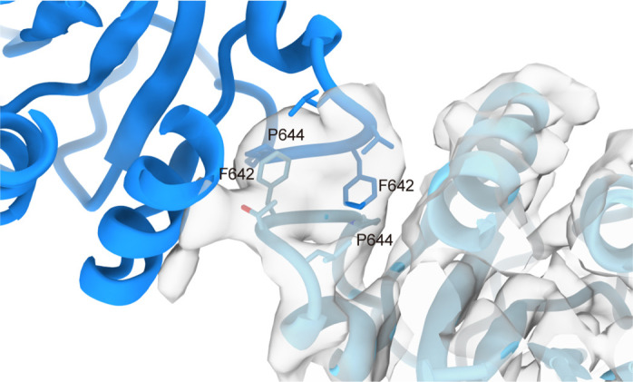
Close-up of the interaction interface, the key residues are shown as sticks, with the overlaid cryo-electron microscopy (cryo-EM) density of the glutamate kinase (GK) domain at unliganded state.
Figure 5—figure supplement 2. Negative staining of mutated P5CS.

(A) Negative stain electron microscopy micrographs of P5CSF642A mutation protein at the APO state; this mutation disrupts the filamentation of P5CS. (B) When the P5CSR124A-Glu filament was additionally incubated with ATP, depolymerization of P5CSR124A filament was observed.
Figure 5—figure supplement 3. The distance between the active sites of the glutamate kinase (GK) domain and γ-glutamyl phosphate reductase (GPR) domain.

The tetramer form of P5CS in filament; positions of each ligand are simulated in our models. G5P (pink) and ADP (brown) in the GK domain, G5P (purple), and NADPH (cyan) in the GPR domain are shown as surface representation. Distances between the G5P in the GK domain and GPR domain are indicated.
Figure 5—figure supplement 4. Sequence alignment of the representative P5CS enzymes.

The sequence alignment of P5CS sequences of Drosophila (UniProtKB: Q9VNW6), mouse (UniProtKB: Q9Z110; isoform long), human (UniProtKB: P54886; isoform long), C. elegans (UniProtKB: P54889; isoform b), and Arabidopsis (UniProtKB: P54887; isoform 1). The conserved residues are identically shaded red, and secondary structure elements are indicated above.
Figure 5—video 1. Simulated ligand-binding site of P5CS filament.
Download video file (68.1MB, mp4)
The structural models and color codes refer to Figure 5 and Figure 5—figure supplement 3.