Abstract
Objective:
To identify the main directions of growth of facial structures in subjects with hypohidrotic ectodermal dysplasia (HED).
Materials and Methods:
The 3D noninvasive facial measurements were collected in 12 subjects (6 boys, 6 girls) with HED during four assessments (at 8, 11, 12, and 15 years) using an electromagnetic digitizer. The modifications of linear distances in the upper, middle, and lower third of the face were analyzed and compared with cross-sectional data obtained in normal healthy coetaneous. For each distance, differential values between the last and the initial data were calculated individually, separately for a first (8–11 years) and a second growth period (12–15 years).
Results:
In the first time span, the growth of all facial measurements was reduced in HED subjects compared with control subjects. During this interval, most of the HED children underwent a functional and/or prosthetic treatment. During adolescence, the width and height of the lower and upper facial thirds showed a larger growth in HED subjects than in control subjects, while all facial depths and all distances in the middle facial third maintained a reduced growth.
Conclusions:
The deviation from normal facial growth of HED subjects tends to lessen with age. Functional and prosthetic appliances may have enhanced facial growth.
Keywords: Growth, Ectodermal dysplasia, Longitudinal, Face, 3D
INTRODUCTION
Craniofacial growth is the result of multiple interactions between genetic and epigenetic elements, involving both soft and hard tissue structures. During growth, the epithelium and the mesenchyme undergo a continuous development with a cascade of reciprocal inductions to finally construct an overall harmonic complex.1–3 Genetic modifications may cause abnormalities in any phase of this morphogenetic process, thus resulting in a nonharmonious facial morphology with associated functional and esthetic impairments.4,5
The craniofacial structures derived from ectoderm and neural crests—more rarely also from mesodermal and endodermal layers—are altered in subjects affected by ectodermal dysplasia (ED).
ED is a rare group of genetic syndromes inherited by autosomal recessive, autosomal dominant, or x-linked recessive transmission.3,6,7 More recently, molecular analyses have identified the mutations of genes responsible for about 50 types of ED that are involved in (1) cell adhesion, (2) transcription regulation, (3) cell-cell signaling, (4) development, and (5) other functions (eg, structural proteins, placode formation).3,6,7 Future advances in cell biology and embryogenic pathways could allow a reclassification of ectodermal dysplasia according to the functions of mutated genes.6,7
The most frequent form of ED is hypohidrotic ectodermal dysplasia (HED; OMIM 305100) characterized by hypotrichosis, hypodontia, and hypohidrosis with major manifestations in the male sex. The involved genes encode a collagenous transmembrane protein—ectodysplasin—and its two receptors, regulating the epithelial-mesenchymal interactions and the hair follicle morphogenesis.3,7 Affected patients present with a typical “aged-face” associated with prominent forehead and chin, saddle nose, everted lips, sunken cheeks, periorbital wrinkles, high-set orbits, large and low set ears, small hard tissue palate, hypoplasia of the alveolar process, and multiple agenesis of both primary and permanent teeth.4,5,8–15
Clinical management of such patients with craniofacial deformities and functional alterations should consider the quantitative assessment of the dimensions, reciprocal spatial positions, and relative proportions of the facial structures during growth to intercept deviations from the norm and possibly correct them at the appropriate time.10,15,16 Detailed knowledge of the typical growth pattern of HED subjects could provide the clinicians useful information to plan a multidisciplinary specific treatment.
Most previous reports investigated the facial features of HED by cross-sectional analyses,4,5,8,9,11–13,15 and few longitudinal studies are currently available.10,17 In their cephalometric evaluation of ED young patients, Bondarets et al.17 observed a peculiar trend of craniofacial growth towards a retrognathic maxilla and Class III sagittal relationships of the jaws. A more recent longitudinal study, performed by our research group, compared the growth of HED young patients with that of healthy reference peers and found a global reduction of all facial volumes in the syndromic subjects during childhood.10 Nevertheless, facial volumes increased their growth by time and, at the end of adolescence, the analyzed HED subjects had similar growth patterns of facial volumes compared to their reference peers and nearly double that of nonrehabilitated HED subjects from the study of Bondarets et al.17 It has been hypothesized that early orthodontic and prosthetic devices worn by the analyzed HED subjects could have improved masticatory function and promoted the growth of the middle third and lower third facial structures to levels above those found in untreated subjects with HED.10 Besides, facial volumes give an estimate of the overall structures, but they do not provide the vectorial directions of growth.11
The current study aimed to identify the actual directions of growth of the facial structures in young subjects with HED by analyzing the modifications of linear measurements in the upper, middle, and lower third of the face. The morphometric evaluation was performed noninvasively using a 3D computerized digitizer on the facial soft tissues of HED and reference subjects during an 8-year period of growth. The method has already been proved to be reliable in the quantitative assessment of craniofacial variations in both normal and syndromic patients.4,5,10,11,18
MATERIALS AND METHODS
Twelve white Italian subjects diagnosed as having HED (six boys and six girls) were analyzed. Subjects were referred for examination by the Italian National Ectodermal Dysplasia Association (ANDE). No subject had undergone any previous craniofacial surgical procedure. In all HED subjects, 3D noninvasive facial measurements and a dental formula (only erupted teeth at clinical examination excluding third molars) were collected by the same expert operator during four assessments (at 8, 11, 12, and 15 years). For the longitudinal evaluation, data were computed separately for a first (8–11 years) and a second (12–15 years) growth period.
Reference cross-sectional data were recorded in previous investigations performed by the staff of the Functional Anatomy Research Center (FARC) at the University of Milan on 160 healthy subjects of the same ethnic group (40 subjects for each age and sex subgroup). Control subjects did not have a previous history of craniofacial trauma or surgery and congenital anomalies. Part of their data had already been published.18,19
The parents or legal guardians of all the analyzed individuals gave their informed consent to participate in the analysis. All procedures were noninvasive, did not provoke damages, risks, or discomfort to the subjects, and were preventively approved by the local ethics committee in accordance with the ethical principles of the World Medical Association Declaration of Helsinki (version, 2002).
Data Collection
The data collection procedure and subsequent off-line calculations were previously published in detail.19 In summary, a single experienced operator located and marked 50 landmarks on the cutaneous facial surface of each subject. During landmark marking, the children sat relaxed with a natural head position and the teeth in an intercuspal position. For each child, this phase lasted about 5 minutes. Then, all subjects wearing removable prosthetic devices were asked to remove the appliance for data digitization lasting about 60 seconds. The x, y, and z coordinates of the facial landmarks were recorded with a computerized electromagnetic digitizer (3Draw, Polhemus Inc, Colchester, Vt) and analyzed using customized computer algorithms written by one of the authors. The reproducibility of landmark identification and digitization were previously reported and found to be reliable.10
The following soft tissue landmarks were used:
(1) Midline landmarks: tri, trichion; n, nasion; sn, subnasale; pg, pogonion.
(2) Paired landmarks (right and left side, noted r and l): exr, exl, exocanthion; tr, tl, tragion; gor, gol, gonion.
Data Analysis
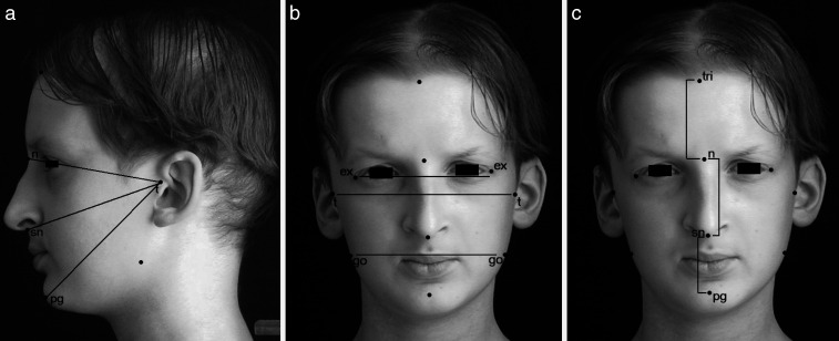
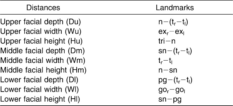
The 3D coordinates of the landmarks obtained on each subject were used to estimate linear distances (unit: mm) of depth, width, and height of the three facial thirds (Figure 1) as reported in Table 1.
Figure 1.
Facial distances in a 12-year-old HED boy. (a) Facial depths. (b) Facial widths. (c) Facial heights. The landmarks used are labeled.
Table 1.
Measurements Calculated From the Digitized Landmarks
For each facial measurement, differential values between data from the last and the initial data collection were calculated individually, separately for the first (Δ1, 8–11 years) and the second growth period (Δ2, 12–15 years). For further comparisons, differential data of both growth intervals were then normalized as a percentage of the initial values.
Mean differential normalized values were computed separately for boys and girls in each growth period. Differential normalized data were also computed for the reference subjects, using measurements obtained in boys and girls of comparable ages.
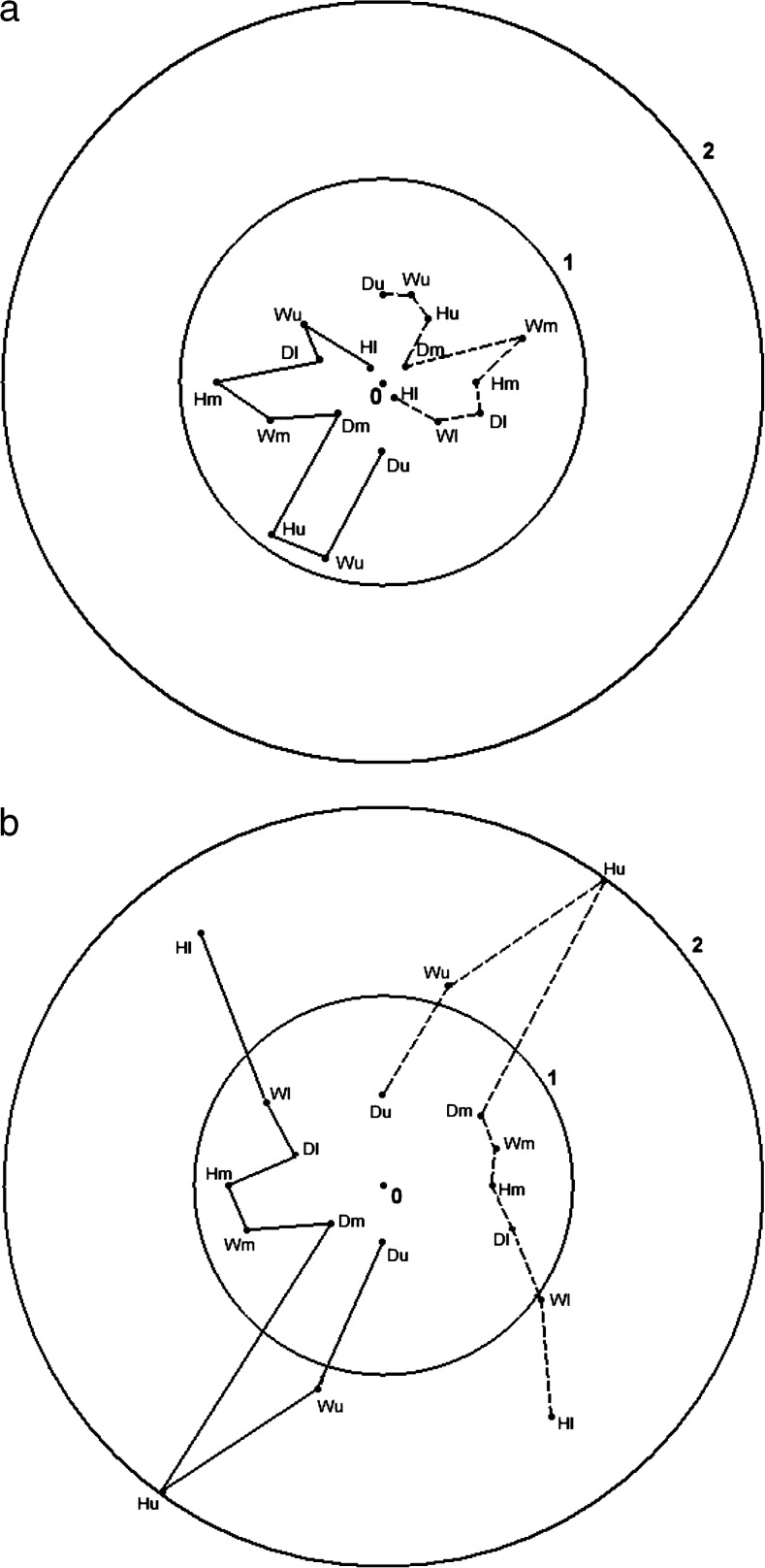
For each facial distance, the differential normalized growths (mean normalized Δ1 and Δ2) were visualized through polar or nysquit diagrams, which summarize the quantitative variation of the considered parameters of HED compared with average data obtained in reference subjects. The polar diagram provides a quantitative overview of several measuring points in one circular normalized form as all the segments approximate a circle. Therefore, diagrams were composed of two circles: the inner circle (circle 1) provided the global mean growth of the reference group, and the outer circle (circle 2) represented a 200% increment of the reference global mean growth. The origin of the axes (marked as 0) represented a null reference global mean growth.
The global differential normalized growths of HED subjects in the analyzed time span were displayed on polar diagrams separately for boys and girls. The segments included between 0 and circle 1 indicated a reduced growth of HED subjects compared with reference subjects; by contrast, segments included between circle 1 and circle 2 indicated an increased growth of HED subjects. No inferential statistics were performed because of the limited number of analyzed HED subjects.
RESULTS
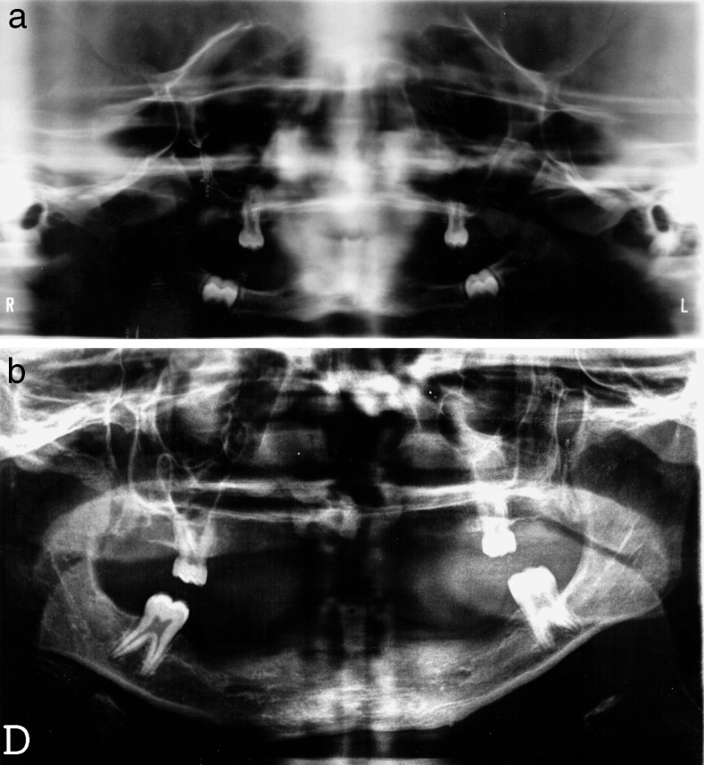
At oral examination, hypodontia was found in all HED subjects with a large variability in the number and distribution of the erupted permanent teeth. Globally, the upper central incisors were conserved in 62% of the analyzed children and had a conical shape in seven patients. Half of the HED individuals presented almost three first molars. About 25% of the HED subjects had pegged maxillary canines. Radiographic images were available only in one syndromic boy and revealed agenesis of multiple primary and permanent teeth (Figure 2).
Figure 2.
Panoramic radiograph of one HED boy at (a) 8 and (b) 11 years.
In the first growth period, the number of teeth ranged from 9 to 13 in girls and from 4 to 10 in boys (at 8 years, mean ± SD: 9.86 ± 3.02; at 11 years, mean value ± SD: 12.28 ± 5.85). In the second growth period, the number ranged from 12 to 24 in girls and from 8 to 11 in boys (at 12 years, mean ± SD: 12.83 ± 5.41; at 15 years, mean ± SD: 14.50 ± 5.16). Globally, girls with HED had more teeth than boys.
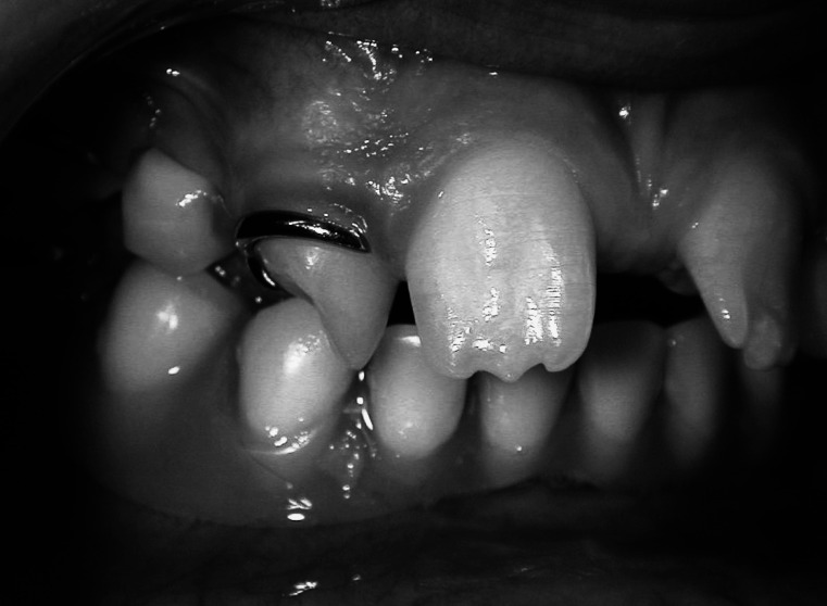
During the analyzed time span, all the syndromic subjects, except for three girls, underwent orthodontic treatment with both orthopedic and functional supports and/or prosthetic rehabilitation with partial removable dentures (Figure 3). Modification or replacement of the dental prostheses was performed annually to follow facial growth changes.
Figure 3.
Intraoral photograph in an 8-year-old girl with HED showing a complete removable mandibular prosthesis and a partial, removable maxillary denture.
To evaluate the global growth variations of facial distances of HED children and to compare them with the data of reference peers, differential values from the initial and last examinations were obtained and normalized.
In the first time interval (8–11 years), the growth of all facial measurements was reduced in HED subjects in comparison with control subjects of the same age and sex. From 12 to 15 years, upper facial third width and height increased in HED subjects compared with reference peers of both sexes, while upper facial third depth was reduced. The growth of the middle facial third was reduced in all spatial dimensions in the HED subjects. In the lower third, the growth of facial depth was reduced, while that of facial height was increased in HED subjects of both genders; by contrast, facial width growth was strictly increased in girls, but reduced in HED boys. The mean differential normalized growth of facial distances in HED subjects compared to reference subjects is visualized in Figure 4.
Figure 4.
Polar diagram illustrating the global differential normalized mean growth of the analyzed facial distances in HED and reference subjects of both genders (boys, solid line; girls, broken line) (a) during the first time interval (Δ1, 8–11 years) and (b) during the second time interval (Δ2, 12–15 years). Du indicates upper facial depth; Wu, upper facial width; Hu, anterior upper facial height; Dm, middle facial depth; Wm, middle facial width; Hm, anterior middle facial height; Dl, lower facial depth; Wl, lower facial width; and Hl, anterior lower facial height. Circle 1 provides the global mean growth of the reference subjects; circle 2 represents a 200% increment of the reference global mean growth. Origin of the axes (marked as 0) represents a null reference global mean growth.
DISCUSSION
Previous anthropometric and cephalometric cross-sectional assessments revealed individuals with HED having distinct “facies” with marked deviations from the norm.4,5,8,11–13,15,16,20,21 A recent longitudinal investigation found a global reduction of all facial volumes in the HED subjects compared with reference individuals during childhood, but some improvements in the maxillary and mandibular size were reported by time.10
In the current study, the actual directions of growth of facial thirds were calculated in HED subjects by a longitudinal 8-year quantitative evaluation using the same equipment. During childhood, the current HED boys and girls grew less than the reference peers in all analyzed facial distances, as already found in the study of facial volumes by Dellavia et al.10
At the beginning of adolescence, some modifications in the craniofacial pattern of growth occurred in the HED individuals. Forehead width and height showed a larger growth in the syndromic than in their healthy peers of both genders, in accordance with the previously reported marked growth of upper facial third volume.10 However, the measuring of forehead height in the HED subjects could be influenced by some imprecision in trichion landmark identification because of their rare, sparse, and fair hair. Besides, some reference points and distances (namely trichion and upper facial height) are difficult to define also in nonsyndromic individuals.2
During the entire growth interval, anteroposterior facial dimensions remained reduced in comparison with reference values as already reported in previous anthropometric cross-sectional evaluations.4,15 These findings suggest that diminished facial depths, with a consequently flat “aged” profile, are characteristic features of the present syndromic patients that do not modify during growth.13 Besides, the retruded position of landmark nasion determines the developing of well-described frontal bossing.10,15,16,20
The current ED-afflicted individuals had midface hypoplasia with a reduced growth of all middle facial third dimensions in both time span intervals. This result is in accordance with previous cephalometric and anthropometric data on maxillary and palatal size.8,9,13,15,17,20 In contrast, the longitudinal assessment of facial volumes growth performed by Dellavia et al.10 reported an increased growth of maxillary HED volume during adolescence. Nevertheless, there is only an apparent discordance because the maxillary alveolar process was comprised in the volume assessment of the maxilla but not in the present measurement of middle facial height. The distance between landmark subnasale and pogonion (lower facial height) involves both the maxillary and mandibular alveolar process.
Therefore, the reduced growth of the middle facial third could be attributed to diminished transversal and anteroposterior distances. In the study by Ward and Bixler,15 all facial widths were more similar to reference values, while Sforza et al.4 observed a large variability of facial distances with an overall disharmonious modification of ED faces compared with normal nonsyndromic faces.
In the lower facial third, the height increased its growth in the second analyzed time span interval with possible enhancement by dental eruption and use of functional appliances and prosthetic devices. In fact, prosthodontic therapy seemed to emphasize growth normalization as observed in implant-treated ED subjects.12,14 In the current study, the maxillary central incisors and the maxillary and mandibular first molars were the most conserved permanent teeth, even if they showed shape modifications in most HED children as found in previous investigations.12,14,21,22 In the 8-year interval, the average number of teeth of the current HED young individuals increased from 9 to 14. This factor may have induced a significant vertical growth of the alveolar bone as suggested by Johnson et al.12 who found a positive correlation between the number of maxillary missing permanent teeth and craniofacial dysmorphology in ED children. Male subjects with severe hypodontia had marked midface hypoplasia with reduced lower and total facial height.12 Similarly, Dellavia et al.9 reported larger palatal height in partially dentate than in completely edentulous 6-year-old HED boys.
The growth of lower facial width was larger in the HED than in control girls, while the growth pattern was similar in HED and in reference boys during adolescence. Besides, the HED-syndromic features appear more evident in the male sex.14 Also, it has been reported that the mandible size tends to normalize during growth in both sexes.10,13,17 The different dimensional variations between maxilla and mandible may be explained by different growth mechanisms in the two jaws: the maxilla undergoes a sutural growth, while the mandible is primarily characterized by endochondral growth at the condyles.1,13 In the ED patients, the mandibular ramus height increases over time, but the alveolar bone remains atrophic with consequent low angle vertical growth pattern.13,20
The present preliminary data confirm that early dental rehabilitation is paramount to enhance the growth potential of facial hard and soft tissues, thus permitting the attainment of more normal (and possibly more pleasant) facial features. Although facial depths and maxillary width still show a reduced growth during adolescence, a positive increase in vertical facial growth can be achieved together with improvements in speech, deglutition, and mastication. Hence, the orthodontist has to monitor functional appliances and removable dentures frequently, with continuous adaptation and replacement during growth.14,21
CONCLUSIONS
Facial growth of HED patients can be evaluated by a simple, low-cost, fast, and noninvasive examination.
The main directions of growth of facial structures were identified.
Future longitudinal analyses may quantify the effect of specific oral treatments on individual facial growth patterns.
Acknowledgments
The precious and indispensable collaboration of all patients, their families, and of the Associazione Nazionale Displasie Ectodermiche (ANDE, Italy) is gratefully acknowledged.
REFERENCES
- 1.Enlow D. H. Facial Growth. Philadelphia, Pa: WB Saunders; 1990. [Google Scholar]
- 2.Ferring V, Pancherz H. Divine proportions in the growing face. Am J Orthod Dentofacial Orthop. 2008;134:472–479. doi: 10.1016/j.ajodo.2007.03.027. [DOI] [PubMed] [Google Scholar]
- 3.Thesleff I. The genetic basis of tooth development and dental defects. Am J Med Genet. 2006;140A:2530–2535. doi: 10.1002/ajmg.a.31360. [DOI] [PubMed] [Google Scholar]
- 4.Sforza C, Dellavia C, Vizzotto L, Ferrario V. F. Variations in the facial soft-tissues of Italian individuals with ectodermal dysplasia. Cleft Palate Craniofac J. 2004;41:262–267. doi: 10.1597/03-033.1. [DOI] [PubMed] [Google Scholar]
- 5.Sforza C, Dellavia C, Goffredi M, Ferrario V. F. Soft tissue facial angles in individuals with ectodermal dysplasia: a three-dimensional noninvasive study. Cleft Palate Craniofac J. 2006;43:339–349. doi: 10.1597/05-004.1. [DOI] [PubMed] [Google Scholar]
- 6.Itin P. H, Fistarol S. K. Ectodermal dysplasias. Am J Med Genet. 2004;131C:45–51. doi: 10.1002/ajmg.c.30033. [DOI] [PubMed] [Google Scholar]
- 7.Lamartine J. Towards a new classification of ectodermal dysplasias. Clin Exp Dermatol. 2003;28:351–355. doi: 10.1046/j.1365-2230.2003.01319.x. [DOI] [PubMed] [Google Scholar]
- 8.Bondarets N, McDonald F. Analysis of the vertical facial form in patients with severe hypodontia. Am J Phys Anthropol. 2000;111:177–184. doi: 10.1002/(SICI)1096-8644(200002)111:2<177::AID-AJPA4>3.0.CO;2-8. [DOI] [PubMed] [Google Scholar]
- 9.Dellavia C, Sforza C, Malerba A, Strohmenger L, Ferrario V. F. Palatal size and shape in 6-year-old patients affected by hypohidrotic ectodermal dysplasia. Angle Orthod. 2006;76:978–983. doi: 10.2319/111105-395. [DOI] [PubMed] [Google Scholar]
- 10.Dellavia C, Catti F, Sforza C, Grandi G, Ferrario V. F. Non-invasive longitudinal assessment of facial growth in children and adolescents with hypohidrotic ectodermal dysplasia. Eur J Oral Sci. 2008;116:305–311. doi: 10.1111/j.1600-0722.2008.00550.x. [DOI] [PubMed] [Google Scholar]
- 11.Ferrario V. F, Dellavia C, Serrao G, Sforza C. Soft tissue facial areas and volumes in individuals with ectodermal dysplasia: a three-dimensional non invasive assessment. Am J Med Genet. 2004;126A:253–260. doi: 10.1002/ajmg.a.20590. [DOI] [PubMed] [Google Scholar]
- 12.Johnson E. R, Roberts M. W, Guckes A. D, Bailey L. J, Phillips C. L, Wright J. T. Analysis of craniofacial development in children with hypohidrotic ectodermal dysplasia. Am J Med Genet. 2002;112:327–334. doi: 10.1002/ajmg.10654. [DOI] [PubMed] [Google Scholar]
- 13.Lexner M. O, Bardow A, Bjorn-Jorgensen J, Hertz J. M, Almer L, Kreiborg S. Anthropometric and cephalometric measurements in X-linked hypohidrotic ectodermal dysplasia. Orthod Craniofac Res. 2007;10:203–215. doi: 10.1111/j.1601-6343.2007.00402.x. [DOI] [PubMed] [Google Scholar]
- 14.Suri S, Carmichael R. P, Tompson B. D. J Prosthet Dent. 2004;92:428–433. doi: 10.1016/j.prosdent.2004.07.014. Simultaneous functional and fixed appliance therapy for growth modification and dental alignment prior to prosthetic habilitation in hypohidrotic ectodermal dysplasia: a clinical report. [DOI] [PubMed] [Google Scholar]
- 15.Ward R. E, Bixler D. Anthropometric analysis of the face in hypohidrotic ectodermal dysplasia: a family study. Am J Phys Anthropol. 1987;74:453–458. doi: 10.1002/ajpa.1330740404. [DOI] [PubMed] [Google Scholar]
- 16.Saksena S. S, Bixler D. Facial morphometrics in the identification of gene carriers of X-linked hypohidrotic ectodermal dysplasia. Am J Med Genet. 1990;35:105–114. doi: 10.1002/ajmg.1320350120. [DOI] [PubMed] [Google Scholar]
- 17.Bondarets N, Jones R. M, McDonald F. Analysis of facial growth in subjects with syndromic ectodermal dysplasia: a longitudinal analysis. Orthod Craniofac Res. 2002;5:71–84. doi: 10.1034/j.1600-0544.2002.01159.x. [DOI] [PubMed] [Google Scholar]
- 18.Sforza C, Grandi G, Binelli M, Tommasi D. G, Rosati R, Ferrario V. F. Age- and sex-related changes in the normal human ear. Forensic Sci Int. 2009;187:110.e1–7. doi: 10.1016/j.forsciint.2009.02.019. [DOI] [PubMed] [Google Scholar]
- 19.Ferrario V. F, Sforza C, Poggio C. E, Cova M, Tartaglia G. Preliminary evaluation of an electromagnetic three-dimensional digitizer in facial anthropometry. Cleft Palate Craniofac J. 1998;35:9–15. doi: 10.1597/1545-1569_1998_035_0009_peoaet_2.3.co_2. [DOI] [PubMed] [Google Scholar]
- 20.Alcan T, Basa S, Kargül B. Growth analysis of a patient with ectodermal dysplasia treated with endosseous implants: 6-year follow-up. J Oral Rehabil. 2006;33:175–182. doi: 10.1111/j.1365-2842.2005.01566.x. [DOI] [PubMed] [Google Scholar]
- 21.Worsaae N, Jensen B. N, Holm B, Holsko J. Treatment of severe hypodontia-oligodontia—an interdisciplinary concept. Int J Oral Maxillofac Surg. 2007;36:473–480. doi: 10.1016/j.ijom.2007.01.021. [DOI] [PubMed] [Google Scholar]
- 22.Guckes A. D, Scurria M. S, King T. S, McCarthy G. R, Brahim J. S. Pattern of permanent teeth present in individuals with ectodermal dysplasia and severe hypodontia suggests treatment with dental implants. Pediatr Dent. 1998;20:278–280. [PubMed] [Google Scholar]


