Abstract
Artificial intelligence (AI) has the potential to revolutionize healthcare and dentistry. Recently, there has been much interest in the development of AI applications. Dentomaxillofacial radiology (DMFR) is within the scope of these applications due to its compatibility with image processing methods. Classification and segmentation of teeth, automatic marking of anatomical structures and cephalometric analysis, determination of early dental diseases, gingival, periodontal diseases and evaluation of risk groups, diagnosis of certain diseases, such as; osteoporosis that can be detected in jaw radiographs are among studies conducted by using radiological images. Further research in the field of AI will make great contributions to DMFR. We aim to discuss most recent AI-based studies in the field of DMFR.
Keywords: Artificial intelligence, Diagnostic imaging, Radiology, Dentistry
Core Tip: Scientists are enthusiastic about conducting artificial intelligence (AI) research related to dentomaxillofacial radiology (DMFR). Image and patient recognition are important in DMFR, however initial investment costs are still high and misdiagnosis may occur in real clinical situations. Up until now, DMFR related AI studies revealed successful results to some extent, however human physiological system is so complex that AI can be a supplementary method but not a substitution for human knowledge, capability and decision-making ability.
INTRODUCTION
In recent times, technical developments and innovation have become integral parts of clinical dentistry. Owing to recent developments in the field of artificial intelligence (AI), significant improvements may be expected in dentistry and dentomaxillofacial radiology (DMFR). AI is defined as the way, method, tool, and algorithm, that is developed for the intelligent solution of the issues encountered with computer application of intelligent thinking. They contain elements which are able to imitate human thinking, understanding, comprehension, interpretation and learning characteristics utilized for problem solving[1]. Numerous studies have been carried out in order to find solutions that utilize the latest technology to solve dental field-related issues. These studies are comprised of a wide range of objectives, including the diagnosis of caries; assessment of various pathologies; orthodontic treatment of crowded teeth and dental implant placement via robotic surgery[2-5]. In DMFR studies, this technology has come to the forefront due to its compatibility with image processing methods. Current topical examples of studies conducted on radiological images are: Classification and segmentation of teeth; automatic marking of anatomical structures and cephalometric analysis; early detection of dental diseases; gingival-periodontal diseases and evaluation of risk groups and the diagnosis of certain diseases such as osteoporosis that can be detected in jaw radiographs[6]. In dental radiology there are both theoretical and practical application examples of these specific tasks. The output gained from artificial learning is expected to reduce the daily workload of physicians as well as the rate of both false diagnosis and underdiagnosis in dental practice.
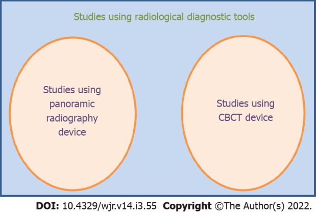
According to the radiological diagnosis tool used, we aim to present the current studies in the field of DMFR under two main headings. Current AI studies in the field of DMFR are given in Figure 1. The main study topics in DMFR related to AI are given in Table 1.
Figure 1.

Current artificial intelligence studies in the field of dentomaxillofacial radiology. CBCT: Cone beam computed tomography.
Table 1.
Main study topics in dentomaxillofacial radiology related to artificial intelligence
|
No.
|
Main study topics
|
| 1 | Localization/measurement of cephalometric landmarks |
| 2 | Diagnosis of osteoporosis |
| 3 | Classification of the maxillofacial cysts and/or tumors |
| 4 | Identification of alveolar bone resorption |
| 5 | Classification of periapical lesions |
| 6 | Diagnosis of multiple dental diseases |
| 7 | Classification of tooth types |
| 8 | Detection of dental caries |
| 9 | Classification of the stage of the lower third molar |
Some of the current AI studies using panoramic radiography devices
The most widely used radiological diagnostic tool in dentistry is the panoramic radiograph. It provides two-dimensional image and related information regarding major mandibular and maxillary jaw bones, all existing teeth and surrounding supporting tissues. Two-dimensional imaging of this region, which has a complex anatomy, causes superposition of various tissues on each other. Therefore, it is possible that panoramic radiographs can be interpreted incorrectly or incompletely in certain cases. Critical assessment of dental images is an essential portion of the diagnostic procedure in daily clinical scenarios. General evaluation by a specialist is based on tooth detection and numbering[7]. A study verified the assumption that a convolutional neural network-CNN-based method could be skilled to analyze and score tooth on panoramic images for automated dental charting objectives. The suggested method targeted at assisting dentists during their diagnostic procedures. The system’s performance level was found to be similar to the specialists’ level, which meant that the radiology specialist could use the finding gained from the technique for automated charting when solely assessment and subtle adjustments were necessary as an alternative to manual data insertion[7].
Several different studies are published on the automatic detection of odontogenic cysts and tumors[8-10]. Odontogenic cysts and tumors do not demonstrate their distinctive radiographic features until they extend to a significant dimension. The early radiographic findings of odontogenic cysts and tumors are so similar that even well trained DMFR experts cannot always accurately conduct their diagnosis. In addition, they may not reveal symptoms in advanced levels[11,12]. Because of such characteristics of odontogenic cysts and tumors, commonly observed cysts such as dentigerous cysts and odontogenic keratocytes may threaten the patient's quality of life if they are large or subsequently cause pathological fractures[13,14]. However, You Only Look Once (YOLO)-a state-of-the-art, real-time object detection system could not be only responsible for the wrong negative diagnosis in one research, which consisted of radiologically indeterminate initial pathologies and maxillary entities that even trained clinicians find difficult to accurately diagnose. As noted, some pathologies in the maxilla are hindered by low bone density and several related anatomical structures that cross with the superpositions of the panoramic image. Odontogenic keratocytes on the maxilla were not detected by both YOLO and two-thirds of clinicians, including experts and general practitioners. Surprisingly, however, there were few instances where YOLO diagnosed and accurately distinguished pathologies that clinicians could not detect[15]. The CNN YOLO detector demonstrated diagnostic effectiveness at least comparable to that of trained dentists in assessing odontogenic cysts and tumors[15]. A number of components affecting clinician ability need to be assessed in future research. It is possible that implementation of CNNs in oral and maxillofacial diagnostic imaging may reveal favorable results for clinicians[15].
Ameloblastomas and keratocystic odontogenic tumors (KCOTs) are among the most commonly observed odontogenic tumors of the jaws. Preoperative definitive detection of these lesions may help dental surgeons in treatment planning[16,17]. In another study, a CNN was created for the evaluation of ameloblastomas and KCOTs[3]. The accuracy of the CNN developed in this study was close to the accuracy of dental experts in detecting ameloblastoma and KCOTs. CNN can help reduce the workload of oral and maxillofacial surgeons by detecting ameloblastomas and KCOTs in a very short time. More research needs to be done in order to clarify and define CNN before it may be widely used in diagnostic imaging purposes[3].
In previous studies, the determination of the relationship with osteoporosis from dental panoramic radiographs was investigated by AI algorithms. In one study, 680 patients were simultaneously subjected to skeletal bone mineral density (BMD) examinations and digital panoramic radiography evaluations, and the results showed that the deep learning-based evaluation of digital panoramic radiography images could be useful and reliable in the automated screening of osteoporosis patients[18]. In another study on this subject, the effectiveness of a deep convolutional neural network (DCNN) based computer aided diagnosis (CAD) technique in osteoporosis detection on panoramic imaging was evaluated. As a result, the DCNN-based CAD technique was found to demonstrate a high level of consistency with dental radiology experts experienced in clinical osteoporosis assessment[19]. The authors suggested that a DCNN-based CAD system could provide dentists with information regarding initial diagnosis of osteoporosis and patients with asymptomatic osteoporosis may be sent to convenient medical referral for further evaluation[18,19].
In a study, a caries detection technique that used deep learning algorithms was proposed for the assessment of dental carious lesions[2]. Although the model exhibits high effectiveness in the detection of caries for both maxillary premolars and molars, this caries evaluation technique has some drawbacks. Since the study was conducted by using two dimensional images, solely interproximal and occlusal carious lesions could be detected, however; lingual and buccal carious lesions could not be detected[2].
Some of the current AI studies using cone beam computed tomography devices
Since the beginning of 2000s, cone beam computed tomography (CBCT) as a 3D imaging method has become widely used in cases where clinical examination and conventional radiographs were insufficient to reveal necessary information[20]. A CNN algorithm was created to detect periapical lesions on CBCT images. The system, which identified and enumerated teeth in volumetric data, was succeeded in diagnosing periapical lesions with 92.8% accuracy. In another study, automatic mandibular canal segmentation was performed on CBCT images with CNN developed[21]. Another area for AI is the detection of oral diseases. In a study, researchers aimed to identify and distinguish lichen planus and leukoplakia lesions with an artificial neural network trained with intraoral photographs and found promising results[22].
A 2011 study suggested that an AI technique could be useful in the automatical localization of a key landmark on CBCT images[23]. The ability to make 3D measurements for cephalometric analysis on CBCT images is an important advantage, however; the performance of automatic localization in current technique is not sufficient and effective in the clinical scenario[23]. Therefore, known techniques can be suggested for using preliminary localization of cephalometric landmarks, but manual correction is still required before further cephalometric analysis.
Limitations and future aspects
Future studies that critically assess certain issues and their clinical potential are essential. In spite of the promising performance results obtained from current AI techniques, it is mandatory to confirm the effectivenes and consistency of these techniques by using appropriate external data from new patients or collected from other dental institutions[24]. In its future goals, it can be expected not only to strengthen the effectiveness of AI techniques on par with specialists, but also to diagnose initial pathologies that are invisible to the human eye.
CONCLUSION
AI has the potential to revolutionize healthcare and dentistry. Owing to recent developments in the field of AI, scientists have become increasingly enthusiastic about conducting AI research. Image and patient recognition are important in DMFR. However, initial investment costs are currently high, and inappropriate assumptions may be made in a real-life clinical scenarios. Hitherto, DMFR-related AI studies revealed a certain degree of successful results. However, the human physiological system is exceedingly complex. As such, AI is acceptable as a supplementary method, but it cannot be seen a substitution for human knowledge, capabilities, and decision-making abilities. Additionally, the diagnostic performance of AI models may differ depending on the algorithms that are used. It is essential to validate the consistency and effectiveness of these techniques by using accurate representative images from different sources before implementing and applying these techniques to real clinical situations. With that said, further research in the field of AI has the potential to make great contributions to DMFR.
Footnotes
Conflict-of-interest statement: Authors declare no conflict of interests for this article.
Provenance and peer review: Invited article; Externally peer reviewed.
Peer-review model: Single blind
Peer-review started: March 20, 2021
First decision: July 18, 2021
Article in press: February 22, 2022
Specialty type: Radiology, nuclear medicine and medical imaging
Country/Territory of origin: Turkey
Peer-review report’s scientific quality classification
Grade A (Excellent): 0
Grade B (Very good): 0
Grade C (Good): C
Grade D (Fair): 0
Grade E (Poor): 0
P-Reviewer: Fakhradiyev I S-Editor: Fan JR L-Editor: A P-Editor: Fan JR
Contributor Information
Seyide Tugce Gokdeniz, Department of Dentomaxillofacial Radiology, Ankara University Faculty of Dentistry, Ankara 06500, Turkey.
Kıvanç Kamburoğlu, Department of Dentomaxillofacial Radiology, Ankara University Faculty of Dentistry, Ankara 06500, Turkey. dtkivo@yahoo.com.
References
- 1.Wong SH, Al-Hasani H, Alam Z, Alam A. Artificial intelligence in radiology: how will we be affected? Eur Radiol. 2019;29:141–143. doi: 10.1007/s00330-018-5644-3. [DOI] [PubMed] [Google Scholar]
- 2.Lee JH, Kim DH, Jeong SN, Choi SH. Detection and diagnosis of dental caries using a deep learning-based convolutional neural network algorithm. J Dent. 2018;77:106–111. doi: 10.1016/j.jdent.2018.07.015. [DOI] [PubMed] [Google Scholar]
- 3.Poedjiastoeti W, Suebnukarn S. Application of Convolutional Neural Network in the Diagnosis of Jaw Tumors. Healthc Inform Res. 2018;24:236–241. doi: 10.4258/hir.2018.24.3.236. [DOI] [PMC free article] [PubMed] [Google Scholar]
- 4.Faber J, Faber C, Faber P. Artificial intelligence in orthodontics. APOS Trends Orthod. 2019;9:201–205. [Google Scholar]
- 5.Woo SY, Lee SJ, Yoo JY, Han JJ, Hwang SJ, Huh KH, Lee SS, Heo MS, Choi SC, Yi WJ. Autonomous bone reposition around anatomical landmark for robot-assisted orthognathic surgery. J Craniomaxillofac Surg. 2017;45:1980–1988. doi: 10.1016/j.jcms.2017.09.001. [DOI] [PubMed] [Google Scholar]
- 6.Hwang JJ, Jung YH, Cho BH, Heo MS. An overview of deep learning in the field of dentistry. Imaging Sci Dent. 2019;49:1–7. doi: 10.5624/isd.2019.49.1.1. [DOI] [PMC free article] [PubMed] [Google Scholar]
- 7.Tuzoff DV, Tuzova LN, Bornstein MM, Krasnov AS, Kharchenko MA, Nikolenko SI, Sveshnikov MM, Bednenko GB. Tooth detection and numbering in panoramic radiographs using convolutional neural networks. Dentomaxillofac Radiol. 2019;48:20180051. doi: 10.1259/dmfr.20180051. [DOI] [PMC free article] [PubMed] [Google Scholar]
- 8.Ariji Y, Yanashita Y, Kutsuna S, Muramatsu C, Fukuda M, Kise Y, Nozawa M, Kuwada C, Fujita H, Katsumata A, Ariji E. Automatic detection and classification of radiolucent lesions in the mandible on panoramic radiographs using a deep learning object detection technique. Oral Surg Oral Med Oral Pathol Oral Radiol. 2019;128:424–430. doi: 10.1016/j.oooo.2019.05.014. [DOI] [PubMed] [Google Scholar]
- 9.Lee JH, Kim DH, Jeong SN. Diagnosis of cystic lesions using panoramic and cone beam computed tomographic images based on deep learning neural network. Oral Dis. 2020;26:152–158. doi: 10.1111/odi.13223. [DOI] [PubMed] [Google Scholar]
- 10.Cattoni F, Teté G, Calloni AM, Manazza F, Gastaldi G, Capparè P. Milled vs moulded mock-ups based on the superimposition of 3D meshes from digital oral impressions: a comparative in vitro study in the aesthetic area. BMC Oral Health. 2019;19:230. doi: 10.1186/s12903-019-0922-2. [DOI] [PMC free article] [PubMed] [Google Scholar]
- 11.Diwan A, Bhagavaldas MC, Bagga V, Shetty A. Multidisciplinary Approach in Management of a Large Cystic Lesion in Anterior Maxilla - A Case Report. J Clin Diagn Res. 2015;9:ZD41–ZD43. doi: 10.7860/JCDR/2015/13540.5992. [DOI] [PMC free article] [PubMed] [Google Scholar]
- 12.Vincent SD, Deahl ST, Johnson DL. An asymptomatic radiolucency of the posterior maxilla. J Oral Maxillofac Surg. 1991;49:1109–1115. doi: 10.1016/0278-2391(91)90147-e. [DOI] [PubMed] [Google Scholar]
- 13.Montoro JRDMC, Tavares MG, Melo DH, Franco RDL, De Mello-Filho FV, Xavier SP, Trivellato AE, Lucas AS. Ameloblastoma mandibular tratado por ressecção óssea e reconstrução imediata. Braz J Otorhinolaryngol. 2008;74:155–157. doi: 10.1016/S1808-8694(15)30768-0. [DOI] [PMC free article] [PubMed] [Google Scholar]
- 14.Ruslin M, Hendra FN, Vojdani A, Hardjosantoso D, Gazali M, Tajrin A, Wolff J, Forouzanfar T. The Epidemiology, treatment, and complication of ameloblastoma in East-Indonesia: 6 years retrospective study. Med Oral Patol Oral Cir Bucal. 2018;23:e54–e58. doi: 10.4317/medoral.22185. [DOI] [PMC free article] [PubMed] [Google Scholar]
- 15.Yang H, Jo E, Kim HJ, Cha IH, Jung YS, Nam W, Kim JY, Kim JK, Kim YH, Oh TG, Han SS, Kim H, Kim D. Deep Learning for Automated Detection of Cyst and Tumors of the Jaw in Panoramic Radiographs. J Clin Med. 2020;9 doi: 10.3390/jcm9061839. [DOI] [PMC free article] [PubMed] [Google Scholar]
- 16.Apajalahti S, Kelppe J, Kontio R, Hagström J. Imaging characteristics of ameloblastomas and diagnostic value of computed tomography and magnetic resonance imaging in a series of 26 patients. Oral Surg Oral Med Oral Pathol Oral Radiol. 2015;120:e118–e130. doi: 10.1016/j.oooo.2015.05.002. [DOI] [PubMed] [Google Scholar]
- 17.Jaeger F, de Noronha MS, Silva ML, Amaral MB, Grossmann SM, Horta MC, de Souza PE, de Aguiar MC, Mesquita RA. Prevalence profile of odontogenic cysts and tumors on Brazilian sample after the reclassification of odontogenic keratocyst. J Craniomaxillofac Surg. 2017;45:267–270. doi: 10.1016/j.jcms.2016.12.011. [DOI] [PubMed] [Google Scholar]
- 18.Lee KS, Jung SK, Ryu JJ, Shin SW, Choi J. Evaluation of Transfer Learning with Deep Convolutional Neural Networks for Screening Osteoporosis in Dental Panoramic Radiographs. J Clin Med. 2020;9 doi: 10.3390/jcm9020392. [DOI] [PMC free article] [PubMed] [Google Scholar]
- 19.Lee JS, Adhikari S, Liu L, Jeong HG, Kim H, Yoon SJ. Osteoporosis detection in panoramic radiographs using a deep convolutional neural network-based computer-assisted diagnosis system: a preliminary study. Dentomaxillofac Radiol. 2019;48:20170344. doi: 10.1259/dmfr.20170344. [DOI] [PMC free article] [PubMed] [Google Scholar]
- 20.Orhan K, Bayrakdar IS, Ezhov M, Kravtsov A, Özyürek T. Evaluation of artificial intelligence for detecting periapical pathosis on cone-beam computed tomography scans. Int Endod J. 2020;53:680–689. doi: 10.1111/iej.13265. [DOI] [PubMed] [Google Scholar]
- 21.Jaskari J, Sahlsten J, Järnstedt J, Mehtonen H, Karhu K, Sundqvist O, Hietanen A, Varjonen V, Mattila V, Kaski K. Deep Learning Method for Mandibular Canal Segmentation in Dental Cone Beam Computed Tomography Volumes. Sci Rep. 2020;10:5842. doi: 10.1038/s41598-020-62321-3. [DOI] [PMC free article] [PubMed] [Google Scholar]
- 22.Jurczyszyn K, Kozakiewicz M. Differential diagnosis of leukoplakia vs lichen planus of the oral mucosa based on digital texture analysis in intraoral photography. Adv Clin Exp Med. 2019;28:1469–1476. doi: 10.17219/acem/104524. [DOI] [PubMed] [Google Scholar]
- 23.Cheng E, Chen J, Yang J, Deng H, Wu Y, Megalooikonomou V, Gable B, Ling H. Automatic Dent-landmark detection in 3-D CBCT dental volumes. Annu Int Conf IEEE Eng Med Biol Soc. 2011;2011:6204–6207. doi: 10.1109/IEMBS.2011.6091532. [DOI] [PubMed] [Google Scholar]
- 24.Steyerberg EW. Validation of prediction models: Steyerberg E. W, Clinical Prediction Models: A Practical Approach to Development, Validation, and Updating. New York: Springer; 2010: 299-310. [Google Scholar]


