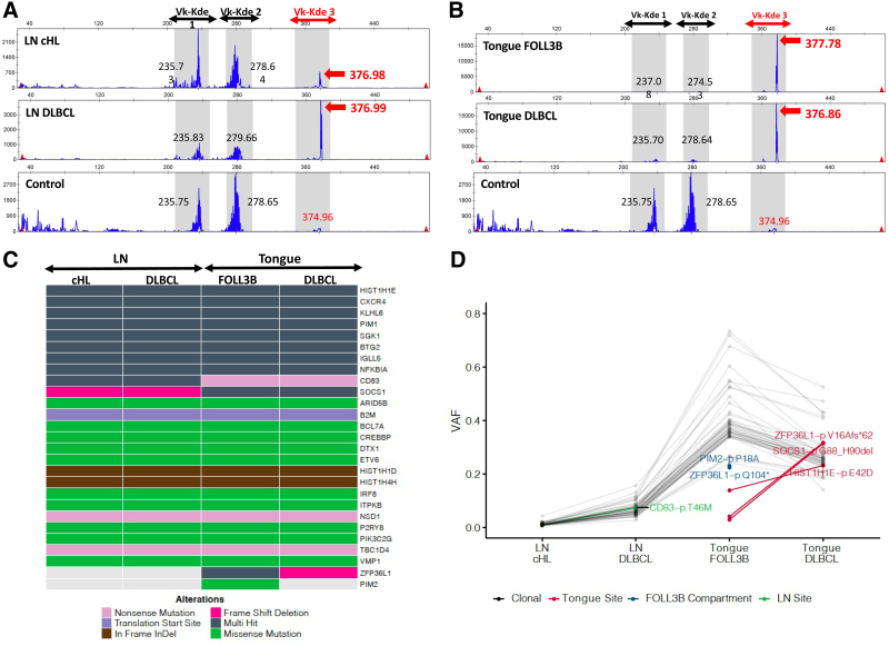
Figure 2.
Clonal relationship among the 4 tumor components. (A and B) IGK-rearrangement–based B-cell clonality analysis of composite tumor components using IGK Vk-Kde/intron-Kde BIOMED-2 primer sets in (A) the cervical LN cHL and DLBCL components, and (B) the base of tongue FOLL3B and DLBCL components. Genomic DNA was extracted from 1.0 mm core fragments obtained from regions of the paraffin-embedded tissue blocks corresponding to each highlighted tumor component. Multiplex PCR analysis was performed using IGK BIOMED-2 primer sets (IdentiClone IGK Gene Clonality Assay; Invovoscribe; Catalog # 91020021). PCR products were resolved by capillary electrophoresis and analyzed using associated software. Polyclonal control DNA (Invovoscribe, Catalog # 40920010) was used as a negative control shown in the lower panel. The IGK Vk-Kde/intron-Kde reaction yields 3 discrete collections of PCR products corresponding to the following primer sets and their anticipated PCR product size ranges: Vk-Kde 1: Vk1f/6/Vk7-Kde, 210-250 bp, Vk-Kde 2: Vk3/intron-Kde, 270-300 bp, and Vk-Kde 3 (in red): Vk2f/Vk4/Vk5-Kde, 350-390 bp. Red arrows highlight the 377 bp clonal peak identified in all tumor components in the Vk-Kde 3 window, consistent with a shared B-cell clonal origin. Black numbers provide the lengths (bp) of maximal amplitude peaks within the non-highlighted portions of the IGK Vk-Kde/intron-Kde reaction. (C and D) Targeted capture sequencing performed on the extracted genomic DNA using a customized panel of 312 lymphoma-related genes. (C) Oncoplot characterizing the type of genetic alterations detected in each tumor component. “Multi-hit” indicates more than one alteration in the same gene and are all shared except for HIST1H1E-pE42D, which is unique to the tongue biopsy. Refer to Suppl. Table S3 for individual mutations. (D) VAF of individual mutations. VAF reflects the tumor content of each sample, which is predictably lowest for the cHL component. Connected gray points indicate clonal mutations shared across all tumor components. Connected green and red points highlight mutations exclusive to the lymph node and tongue biopsies, respectively. Blue points highlight mutations unique to the FOLL3B component. cHL = classic Hodgkin lymphoma; DLBCL = diffuse large B-cell lymphoma; FOLL3B = follicular lymphoma grade 3B; LN = lymph node; PCR = polymerase chain reaction; VAF = variant allele frequency.

