The Coronavirus disease 2019 (COVID-19) pandemic has seriously affected patients with chronic lymphocytic leukemia (CLL).1 Fatalities exceeding 30% has been reported among hospitalized patients in international surveys,2,3 and in consecutively identified CLL patients.4 Additionally, many patients with CLL do not achieve seroconversion upon mRNA vaccination.5,6 We recently confirmed those observations in the course of a prospective clinical trial involving the BNT1622b2 mRNA vaccine. 7 The study included five equally sized cohorts of patients with different types of immunocompromised disorders (total n=449) and 90 healthy controls. Sixtythree percent of patients within the specific CLL cohort (n=90) seroconverted after two vaccine doses. The lowest seroconversion rate was found in patients on ibrutinib followed by those who had stopped Bruton’s tyrosine kinase inhibitor (BTKi)-therapy.7 Even though T-cell responses may occur in most patients with CLL following SARS-CoV-2 infection,4 there is still limited data on T-cell immunity following vaccination of many immunocompromised patient groups including CLL. In the first report on hematological malignancies, nine of 18 patients developed SARS-CoV-2-specific T-cell reactivity.8 Similar results were seen in a large patient group applying a whole blood interferon-g (IFN-g) release assay.9
An early study10 of T-cell responses identified IFN-g as a key cytokine produced by spike-specific CD4+ and CD8+ T cell in BNT162b1 mRNA-vaccinated individuals. We report here on T-cell immunity in patients with CLL from our above mentioned prospective clinical trial7 using IFN-g ELISpot, a validated quantitative assay to measure T-cell responses against SARS-CoV-2-specific peptides.4,11
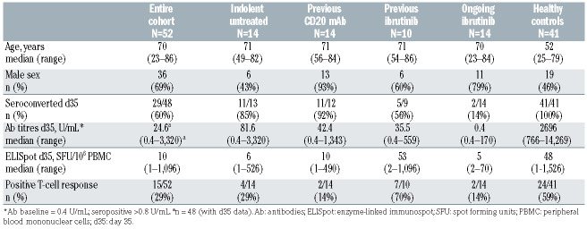
Inclusion criteria and monitoring of our vaccine clinical trial has been described earlier.7 Patients with a previous history or signs of COVID-19, or who tested positive for SARS-CoV-2 or had spike protein-specific antibodies at baseline were excluded. No patient with CLL developed break-through infection during the study or early followup. Fifty-two predefined patients from the CLL cohort (n=90) were subjected to repeated analysis of T-cell immunity against SARS-CoV-2 specific peptides. Baseline characteristics of the patients and controls are shown in Table 1. Patient groups included i) previously untreated (n=14), ii) individuals with ongoing ibrutinib therapy (n=14), iii) individuals who had stopped ibrutinib ≥2 months ago due to remission as part of another study addressing intermittent ibrutinib treatment (n=10), iv) or individuals who had received CD20 monoclonal antibody (mAb)-containing chemoimmunotherapy 6-30 months ago (of which 11 received their last dose >12 months ago) (n=14). Cellular and humoral immunity was measured at day (d) 0 (baseline), d10 (10 days after first dose), d21 and d35 (21 days after first dose, day of second dose). Seroconversion and antibody titres were analyzed as reported7 with d35 data available in 48 of the 52 predefined patients who were analyzed for T-cell immunity. Additionally, 41 of 90 healthy controls in the trial7 were preselected for T- cell analysis (Table 1).
The ELISpot assay was applied as previously described,11 using plates and reagents from a human IFNg ELISpot kit (3420-2APT-2, Mabtech). Briefly, 2.5x10⁵ peripheral blood mononuclear cells (PBMC)/well were seeded and supplemented with 0.15 μg/mL of co-stimulatory anti-CD28/CD49d (347690, BD Biosciences). Cells were stimulated for 20 hours (h) with a peptide pool covering the SARS-CoV-2 spike glycoprotein (0.5 μg/mL, LB01792; peptides&elephants) and equivalent dimethyl sulfoxide (DMSO) in unstimulated wells. Spot-forming units (SFU) were counted using the IRIS (Mabtech) automated reader system. Data points are presented as background corrected SFU/106 cells, calculated by subtracting the mean value of the corresponding duplicate unstimulated wells from the mean value of duplicate spike stimulated wells. Negative values after background correction are set to 1. The threshold for positive response corresponds to the average SFU/106 cells of unstimulated wells + 2 standard deviations (30 SFU/106 cells). Data were excluded when unstimulated wells had >100 SFU/106 cells.
All analyses were prespecified as per protocol. Data is summarized using descriptive statistics such as counts, percentages, medians and range. Categorical variables are presented as cross-tabulations and distributional differences were tested using the Chi-squared test. Significance between time points with missing values was assessed using Kruskal-Wallis test with Dunn’s posttest. Correlation analysis was done using non-parametric Spearman rank correlation. P-values <0.05 were considered significant. Graphs and associated statistical tests were performed in Prism v.9 (GraphPad Software Inc.).
Table 1.
Baseline characteristics and immunological results 2 weeks after the second dose (d35) of the mRNA BNT162b2 vaccine in relation to chronic lymphocytic leukemia patient subgroup and healthy controls.
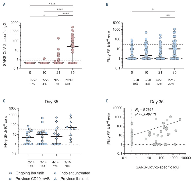
Seroconversion rates were in line with our full clinical trial report7 and are summarized in Table 1 and Figure 1A. Seroconversion occurred in 29 of 48 patients (60%) (4/52 patients had missing serology data at d35) compared to 41 of 41 (100%) of controls. The time kinetics of seroconversion are shown in Figure 1A. Only 4% and 18% of patients had seroconverted at d10 and d21, respectively, followed by 60% at d35. Patients’ and controls’ responses showed similar kinetics. There was no significant increase in SARS-CoV-2-specific immunoglobulin G (IgG) after vaccination on d10 in either group. At d21 and at d35, both groups had a significant response compared to baseline (CLL: d0 vs. d21, P<0.05, d0 vs. d35, P<0.0001; controls: d0 vs. d21, P<0.0001; d0 vs. d35, P<0.0001).
Subgroup analysis revealed that seroconversion occurred in 11 of 13 (85%) of previously untreated patients, 11 of 12 (92%) of previously CD20 mAb-treated patients, five of nine (56%) of previously ibrutinib-treated and two of 14 (14%) of patients with ongoing ibrutinib therapy. The difference between patients on or off ibrutinib was significant (P=0.036). The median antibody titer at d35 in each subgroup as above was 81.6 U/mL (range, 0.4-3,320), 42.4 U/mL (range, 0.4-1,343), 35.5 U/mL (range, 0.4-559) and 0.4 U/mL (0.4-170) respectively. This compared to a median antibody titer of 2,696 U/ml (range, 766-14,269) in controls (Table 1).
Figure 1.
Humoral and cellular immune response in chronic lymphocytic leukemia patients. Longitudinal assessment (day 0, 10, 21, 35 post-vaccination) of SARS-CoV-2-specific immunglobulin G (IgG) (A) and IFN-g T cells (B) after spike glycoprotein stimulation, with summarized number and frequency of patients tested positive. Subgroup analysis of chronic lymphocytic leukemia (CLL) patients at day 35 (C) and correlation day 35 (D). The dashed line indicates positive threshold for SARS-CoV-2-specific IgG and IFN-g spot forming units (SFU)/106 cells, 0.8 U/mL and 30 SFU/106 cells respectively. The dotted line represents the lower limit of detection of both assays. Each dot represents one patient. Rs=Spearman r value. Kruskal-Wallis test with Dunn’s correction for multiple comparisons. *P<0.05, **P< 0.01, ***P<0.001, ****P<0.0001.
Longitudinal assessment of T-cell immunity against SARS-CoV-2 spike peptides (ELISpot) is shown in Figure 1B and summarized in Table 1. At d35, 15 of 52 patients (29%) had a specific T-cell response (P<0.05 vs. baseline, Figure 1B) compared to 24 of 41 (59%) in controls (P<0.01). Pre-existing spike-cross-reactive T cells12 was observed at baseline in five of 50 patients; four patients showed no vaccine response and one patient showed a marginal increase in T-cell response (mean spot count from 44 to 70 SFU/106 PBMC at d35). A positive T-cell response was observed in nine of 50 tested patients (18%) at d10 and in six of 51 (12%) at d21.
CLL subgroup results are shown in Figure 1C and Table 1. IFN-g positivity was observed in seven of ten patients at d35 who were off ibrutinib, whereas only two of 14 patients on ibrutinib developed T-cell immunity (P<0.01). The corresponding numbers were four of 14 among previously untreated patients and two of 14 if previously treated with CD20 mAb (P<0.05 and P<0.01, respectively, vs. patients off ibrutinib).
Finally, we analyzed correlation between seroconversion and T-cell response (Figure 1D). A weak but significant correlation was observed (r=0.2861, P=0.049). Fifteen patients (29%) were double-negative i.e., neither mounted a T-cell response nor seroconverted, whereas nine (18%) came out positive in both assays. Twenty patients (39%) were positive in serology only and only three patients (2 in the off ibrutinib group) had an IFN-g response in the absence of seroconversion. Most doublenegative patients (11/15) were found among patients on ibrutinib. Double-positive patients were most frequent in those off ibrutinib (4/9). Of the 20 seroconverted patients with no T-cell response, one patient was found in the ongoing ibrutinib and one in the previously ibrutinib treated group, while eight were previously untreated and ten were previously treated with CD20 mAb.
Following natural COVID-19 infection, durable immunity including both antibodies and T cells seem to occur in most healthy individuals13 as well as in patients with CLL.4 Most healthy individuals mount T-cell responses following mRNA vaccination.14 This was reported also in patients with solid tumors.15 Lower numbers were recently reported in patients with hematological malignancies. 8,9 The present study shows that, compared to healthy controls, half as many of patients with CLL developed IFN-g T-cell response (28% vs. 59%) after two doses of mRNA vaccine. A limitation of the present T-cell assay11 is capturing of only IFN-g positive cells e.g., missing- out on other cytokine-secreting antigen-specific T cells. Despite this, we were able to capture both temporal and group dynamic changes of the SARS-CoV-2 spikespecific T-cell response, and were able to make comparison of patients with CLL with healthy controls. CLL subgroup results were driven by patients who were off ibrutinib (7/10 responded) whereas other CLL sub-groups had few T-cell responders. However, the data must be viewed with caution due to the open-label trial design and the small numbers within each subset. Thus, our subgroup analysis should be confirmed in extended studies. Even though our healthy controls were younger (median age 52 years) age did not impact on their T-cell response (data not shown). Double-negativity was found in most patients on ibrutinib who remain of major concern, suggesting that temporary cessation of BTKi may be explored onwards in future studies. Of note, Ehmsen et al.9 found T-cell responses in 26% of seronegative hematology patients whereas we found it only in three of 52 patients with CLL (6%). A third dose is currently explored in CLL and its effect on T-cell immunity, even though its additional effect on T cells was limited in solid tumors.15 CLL remain as a group of special concern in the ongoing pandemic.
Acknowledgements
We thank all patients who donated blood for this study and Leila Relander and Sonja Sönnert-Husa for technical assistance.
Funding Statement
Funding: this study was supported by grants from the SciLifeLab National COVID-19 Research Program, financed by the Knut and Alice Wallenberg Foundation, the Swedish Research Council, Region Stockholm, the Swedish Blood Cancer Foundation and Karolinska Institutet.
References
- 1.Langerbeins P, Eichhorst B. Immune dysfunction in patients with chronic lymphocytic leukemia and challenges during COVID-19 pandemic. Acta Haematol. 2021;144(5):508-518. [DOI] [PMC free article] [PubMed] [Google Scholar]
- 2.Mato AR, Roeker LE, Lamanna N, et al. Outcomes of COVID-19 in patients with CLL: a multicenter international experience. Blood. 2020;136(10):1134-1143. [DOI] [PMC free article] [PubMed] [Google Scholar]
- 3.Scarfo L, Chatzikonstantinou T, Rigolin GM, et al. COVID-19 severity and mortality in patients with chronic lymphocytic leukemia: a joint study by ERIC, the European Research Initiative on CLL, and CLL Campus. Leukemia. 2020;34(9):2354-2363. [DOI] [PMC free article] [PubMed] [Google Scholar]
- 4.Blixt L, Bogdanovic G, Buggert M, et al. Covid-19 in patients with chronic lymphocytic leukemia: clinical outcome and B- and T-cell immunity during 13 months in consecutive patients. Leukemia. 2022;36(2):476-481. [DOI] [PMC free article] [PubMed] [Google Scholar]
- 5.Herishanu Y, Avivi I, Aharon A, et al. Efficacy of the BNT162b2 mRNA COVID-19 vaccine in patients with chronic lymphocytic leukemia. Blood. 2021;137(23):3165-3173. [DOI] [PMC free article] [PubMed] [Google Scholar]
- 6.Roeker LE, Knorr DA, Thompson MC, et al. COVID-19 vaccine efficacy in patients with chronic lymphocytic leukemia. Leukemia. 2021;35(9):2703-2705. [DOI] [PMC free article] [PubMed] [Google Scholar]
- 7.Bergman P, Blennow O, Hansson L, et al. Safety and efficacy of mRNA BNT162b2 vaccine against SARS-CoV-2 in five groups of immunocompromised patients and healthy controls in a prospective open-label clinical trial. EBioMedicine. 2021;9;74:103705. [DOI] [PMC free article] [PubMed] [Google Scholar]
- 8.Monin L, Laing AG, Munoz-Ruiz M, et al. Safety and immunogenicity of one versus two doses of the COVID-19 vaccine BNT162b2 for patients with cancer: interim analysis of a prospective observational study. Lancet Oncol. 2021;22(6):765-778. [DOI] [PMC free article] [PubMed] [Google Scholar]
- 9.Ehmsen S, Asmussen A, Jeppesen SS, et al. Antibody and T cell immune responses following mRNA COVID-19 vaccination in patients with cancer. Cancer Cell. 2021;39(8):1034-1036. [DOI] [PMC free article] [PubMed] [Google Scholar]
- 10.Sahin U, Muik A, Derhovanessian E, et al. COVID-19 vaccine BNT162b1 elicits human antibody and TH1 T cell responses. Nature. 2020;586(7830):594-599. [DOI] [PubMed] [Google Scholar]
- 11.Sekine T, Perez-Potti A, Rivera-Ballesteros O, et al. Robust T cell immunity in convalescent individuals with asymptomatic or mild COVID-19. Cell. 2020;183(1):158-168. [DOI] [PMC free article] [PubMed] [Google Scholar]
- 12.Loyal L, Braun J, Henze L, et al. Cross-reactive CD4(+) T cells enhance SARS-CoV-2 immune responses upon infection and vaccination. Science. 2021;374(6564):eabh1823. [DOI] [PMC free article] [PubMed] [Google Scholar]
- 13.Cohen KW, Linderman SL, Moodie Z, et al. Longitudinal analysis shows durable and broad immune memory after SARS-CoV-2 infection with persisting antibody responses and memory B and T cells. Cell Rep Med. 2021;2(7):100354. [DOI] [PMC free article] [PubMed] [Google Scholar]
- 14.Painter MM, Mathew D, Goel RR, et al. Rapid induction of antigenspecific CD4(+) T cells is associated with coordinated humoral and cellular immunity to SARS-CoV-2 mRNA vaccination. Immunity. 2021;54(9):2133-2142. [DOI] [PMC free article] [PubMed] [Google Scholar]
- 15.Shroff RT, Chalasani P, Wei R, et al. Immune responses to two and three doses of the BNT162b2 mRNA vaccine in adults with solid tumors. Nat Med. 2021;27(11):2002-2011. [DOI] [PMC free article] [PubMed] [Google Scholar]


