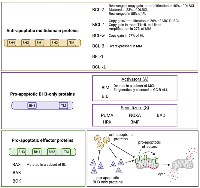
Figure 1.
The BCL-2 protein family. The three subgroups of the BCL-2 protein family and their recurrent genetic alterations are shown in the colored boxes. A schematic of the interactions occurring among the BCL-2 family members is represented in the lower right box, based on the “indirect activation” model. Overall, the antiapoptotic proteins sequester the pro-apoptotic BH3-only activators (A) and effectors, preventing the initiation of the apoptotic cascade. By contrast, the pro-apoptotic sensitizers (S) antagonize the anti-apoptotic members thus freeing the activators, which in turn trigger the polymerization of the effectors. This creates pore-like structures at the outer mitochondrial membrane which favor the release of cytochrome c (cyt c) and other apoptogenic factors. While the model shown here has been widely adopted to define the concept of apoptotic priming, recent evidence suggests that, at least in selected cancer types, BAX and BAK activation may only require that these members are freed from the anti-apoptotics, with no need of direct interaction with pro-apoptotic members. DLBCL: diffuse large B-cell lymphoma; FL: follicular lymphoma; ABC: activated B-cell; T-NHL: T-cell non-Hodgkin lymphoma; MM: multiple myeloma; HL: Hodgkin lymphoma; MCL: mantle cell lymphoma; GC-R: ALL glucocorticoid-resistant acute lymphoblastic leukemia; BL: Burkitt lymphoma.

