PLOS ONE logoLink to PLOS ONE
. 2022 Mar 31;17(3):e0265895. doi: 10.1371/journal.pone.0265895

Hemodynamic profiles by non-invasive monitoring of cardiac index and vascular tone in acute heart failure patients in the emergency department: External validation and clinical outcomes

Nicholas Eric Harrison 1,2,*, Sarah Meram 1, Xiangrui Li 3, Morgan B White 2, Sarah Henry 1, Sushane Gupta 1, Dongxiao Zhu 4, Peter Pang 2, Phillip Levy 1
Editor: Gianluigi Savarese5
PMCID: PMC8970400  PMID: 35358231

Abstract

Background

Non-invasive finger-cuff monitors measuring cardiac index and vascular tone (SVRI) classify emergency department (ED) patients with acute heart failure (AHF) into three otherwise-indistinguishable subgroups. Our goals were to validate these “hemodynamic profiles” in an external cohort and assess their association with clinical outcomes.

Methods

AHF patients (n = 257) from five EDs were prospectively enrolled in the validation cohort (VC). Cardiac index and SVRI were measured with a ClearSight finger-cuff monitor (formerly NexFin, Edwards Lifesciences) as in a previous study (derivation cohort, DC, n = 127). A control cohort (CC, n = 127) of ED patients with sepsis was drawn from the same study as the DC. K-means cluster analysis previously derived two-dimensional (cardiac index and SVRI) hemodynamic profiles in the DC and CC (k = 3 profiles each). The VC was subgrouped de novo into three analogous profiles by unsupervised K-means consensus clustering. PERMANOVA tested whether VC profiles 1–3 differed from profiles 1–3 in the DC and CC, by multivariate group composition of cardiac index and vascular tone.

Profiles in the VC were compared by a primary outcome of 90-day mortality and a 30-day ranked composite secondary outcome (death, mechanical cardiac support, intubation, new/emergent dialysis, coronary intervention/surgery) as time-to-event (survival analysis) and binary events (odds ratio, OR). Descriptive statistics were used to compare profiles by two validated risk scores for the primary outcome, and one validated score for the secondary outcome.

Results

The VC had median age 60 years (interquartile range {49–67}), and was 45% (n = 116) female. Multivariate profile composition by cardiac index and vascular tone differed significantly between VC profiles 1–3 and CC profiles 1–3 (p = 0.001, R2 = 0.159). A difference was not detected between profiles in the VC vs. the DC (p = 0.59, R2 = 0.016).

VC profile 3 had worse 90-day survival than profiles 1 or 2 (HR = 4.8, 95%CI 1.4–17.1). The ranked secondary outcome was more likely in profile 1 (OR = 10.0, 1.2–81.2) and profile 3 (12.8, 1.7–97.9) compared to profile 2. Diabetes prevalence and blood urea nitrogen were lower in the high-risk profile 3 (p<0.05). No significant differences between profiles were observed for other clinical variables or the 3 clinical risk scores.

Conclusions

Hemodynamic profiles in ED patients with AHF, by non-invasive finger-cuff monitoring of cardiac index and vascular tone, were replicated de novo in an external cohort. Profiles showed significantly different risks of clinically-important adverse patient outcomes.

Background

Acute heart failure (AHF) accounts for 1 million emergency department (ED) visits annually in the United States(US), 80% of which result in hospital admission [1,2]. AHF 30-day mortality overall (8–10% [3]) greatly exceeds the threshold of typical emergency physician (EP) risk tolerance (0.5–1%) [4], and neither EP gestalt for AHF mortality risk [5,6] nor clinical decision rules (CDRs) yet provide predictive value sufficient [1,7] to meet such low risk thresholds. Consequently, half of ED to hospital admissions for AHF involve low-risk patients for whom admission may not be necessary [1,2,810], and recent Society for Academic Emergency Medicine (SAEM) and Heart Failure Society of America(HFSA) guidelines [1] stress the importance of developing new AHF risk markers in the ED. A particular need exists for novel markers which identify low-risk AHF presentations by way of capturing the high-degree of physiologic and clinical heterogeneity between AHF patients, given relatively more established predictors of high-risk [1,11] and the high baseline ED admission rate.

Classification by hemodynamic profile is one of the oldest approaches to subgrouping the high clinical heterogeneity present among AHF patients, given that hemodynamic derangements are critical defining features of AHF pathophysiology. Hemodynamic parameters like cardiac index, vascular tone (systemic vascular resistance index {SVRI}), heart rate, blood pressure (BP), and others reflect some of the greatest physiologic heterogeneity among AHF patients [1218], and consideration of heart rate and BP are prominent features of contemporary ED AHF evaluation [14]. Cardiac index and vascular tone play an outsized role in AHF pathophysiology [14,15], yet are not generally able to be assessed in the ED. Gold standard measurement by pulmonary artery catheterization (PAC) requires specialist expertise, is highly invasive, and is employed in only 1% of contemporary AHF hospitalizations [19]. The Forrester classification of “wet-dry/warm-cold” on physical exam [20,21] is a non-invasive method for assessing cardiac index and vascular tone in AHF [1,22,23], but limited in clinical utility given a subjective nature and poor reliability, with interrater agreement of just 64% (kappa = 0.28) in ED patients [24].

Recently, a non-invasive monitor providing continuous estimation of cardiac index and vascular tone18 was described in an ED-based retrospective study of the PREMIUM (Prognostic Hemodynamic Profiling in the Acutely Ill Emergency Department Patient) registry by Nowak et. al. ClearSight (formerly “NexFin”, as it was known in this prior study; Edwards Lifesciences, Irvine, California) is an FDA-approved finger-cuff monitor which measures continuous blood pressures and pulse rates at both the radial and digital arteries. Finger-cuff monitors are attractive for ED profiling of patients by cardiac index and vascular tone, because they are non-invasive like the Forrester classification, yet provide the clinician with reproducible, continuous, and objective measurements like a PAC. Nowak et al. derived 2-dimensional hemodynamic profiles by cardiac index and vascular tone (SVRI) from the finger cuff measurements in these ED AHF patients. Profiling by these two hemodynamic variables is the same physiological construct underlying the Forrester classification, but with objective measurements guiding classification rather than highly-subjective [24] physical examination. Namely, profiling by cardiac index and vascular tone reflects that the ventricular-vascular relationship is naturally discordant, since maintenance of blood pressure requires any decrease in cardiac index to be buffered by an equal and opposite increase in vascular tone and visa-versa (i.e. Mean arterial BP = cardiac index x SVRI) [14]. Importantly, disruption of the ideal ventricular-vascular relationship, such as by nitrate metabolism, myocardial changes, neurohormonal effects, and other factors, is a key component of HF physiology [5,11,15,16,2530].

Three distinct ED AHF hemodynamic profiles by cardiac index and vascular tone were described by Nowak et al. which, critically, were not otherwise identifiable by clinical characteristics [18]. This 3-profile system observed reflected a surprisingly large degree of previously uncaptured physiologic heterogeneity [18], spanning very low and very high values in both the cardiac index and vascular tone dimensions. The prior study had 3 primary limitations identified as needing further research by the authors: 1. Finger-cuff monitors hemodynamic monitoring in AHF is novel, and the ability to reproduce this 3-profile classification needed replication in an external sample (external validation). 2. Patients were not diagnostically adjudicated as having AHF, and it is unknown whether profiling on this monitor is specific to AHF versus other diagnoses in the ED 3. The study was not powered to detect prognostic difference between profiles, and thus clinical significance of these profiles for a goal of risk-stratification is unknown.

In the current study, we sought to address these limitations. We hypothesized that 1. De novo cluster analysis of a new prospective sample of AHF patients (validation cohort, VC) would produce cardiac index/vascular tone hemodynamic profiles matching the derivation cohort(DC) [18]; 2. VC profiles would not match profiling of patients in PREMIUM with sepsis (control cohort, CC) or those with non-cardiac dyspnea (CC2), and 3. Between-profile differences in the VC would exist for both 90-day all-cause mortality (primary outcome) and a 30-day ranked composite of adverse events [31,32] (secondary outcome).

Methods

CLEAR-AHF was a multicenter prospective observational study approved as minimal-risk research by the Wayne State University (WSU) and Indiana University (IU) institutional reviews boards. The primary aim of the study was to create a multi-institutional registry of hemodynamic data in ED AHF patients, given the lack of other methods for measuring important hemodynamic parameters like cardiac index and SVRI (vascular tone) in the emergency department. A secondary aim was to validate the hemodynamic profiles derived in the pilot study [18] and determine whether these profiles were associated with clinically-important patient outcomes. Written consent was obtained from all participants. This manuscript was written to comply with the STROBE guidelines for cohort studies [33].

Study setting and participants

Patients ≥18 years old presenting to the ED were screened for enrollment 24 hours per day at five EDs in the US. Annual ED volumes range from approximately 80,000–100,000 patient visits per site. Enrollment occurred from July 2017—March 2019.

Included patients had EP suspicion of AHF and at least one of the following: dyspnea at rest or exertion, signs of AHF on chest radiograph (CXR), and/or NT-proBNP>300 pg/ml. Patients with dyspnea primarily due to other causes (by EP diagnosis), temperature >38.5°C or suspected sepsis, acute ST-elevation myocardial infarction (STEMI), pregnant women, prisoners, and those without an ejection fraction (EF) recorded within 12 months, were excluded. Two study authors with extensive AHF research experience (PP and PL), blinded to one another, performed case review to adjudicate the ED diagnosis of AHF. Disagreements were decided by discussion, and patients without diagnostically-adjudicated AHF were excluded.

Study protocol

Patients were fitted with a ClearSight finger-cuff monitor and continuous measurements recorded for 3–6 hours. Manufacturer reference range values for cardiac index and SVRI are 2.5–4.0 L/min/m2 and 1970–2390 dynes-sec/cm5/m2, respectively. Clinicians and patients were blinded to device measurements by an opaque sheet on the monitor screen to prevent bias in management decisions. Patients were excluded if they could not begin monitoring in the ED. The median time from initial IV loop diuretic to initiation of hemodynamic monitoring in the sample was 98 minutes (IQR: 29–167), and the first recorded value for cardiac index and SVRI were used for the analysis.

Research assistants used standardized data sheets to record patient demographics, medications, medical history, vital signs, clinical tests, ED treatments, ED disposition, and hospital course. Data were obtained through patient phone interview, the electronic medical record (EMR), and the reports of treating physicians. Data were recorded in REDCap (Research Electronic Data Capture; http://project-redcap.org/).

Measures

Primary measure

The primary measure of interest was each patient’s 3-level categorical hemodynamic profile, as derived in the DC [18]. Each profile is a subgroup of the multivariate distribution in two dimensions: cardiac index and vascular tone (SVRI). The first available cardiac index and vascular tone, obtained simultaneously, was used for profiling (see Analysis, Validation Parts 1–2). Finger-cuff hemodynamic monitors have >90% correlation to invasive arterial blood pressure monitoring [34], and estimate invasive cardiac index with a ~30% margin of error [18,35]. While the level of accuracy of this estimate does not imply interchangeability with invasive hemodynamic monitors [27], it is nevertheless accurate enough to be useful for initial assessment before an invasive monitoring can begin in the intensive care unit [27]. Since invasive hemodynamic monitoring is only performed in 1% of contemporary AHF hospitalizations [19] and is outside the scope of practice of EPs, a non-invasive estimate such as from a finger-cuff monitor is the only feasible measure for cardiac index in the ED. For more details on finger-cuff hemodynamic monitors, see the publication for the DC [18] and other prior literature [27].

Cluster analysis methods to subgroup patients by cardiac index and vascular tone into one of three hemodynamic profiles are described below for the VC (see Validation Part 1) and in the prior publication [18] for the DC. The CC hemodynamic profiles were obtained in an unpublished analysis by the same methods and at the same time as the DC [18], from a concurrently enrolled group of septic patients in the same registry (PREMIUM) as the DC’s AHF patients.

Hemodynamic profiling by cardiac index and vascular tone was performed de novo in the VC (see Validation Part 1), to test if profiling of the VC replicated the profiles of AHF patients in the DC [18] (hypothesis 1) and differed from the profiles of non-AHF patients in the CC (hypothesis 2). For consistency, profile numbering 1 through 3 in the VC was set to match the corresponding profiles 1–3 in the DC and CC.

Secondary measures

Other measures included numerous clinical variables: demographics, vital signs, laboratory tests performed in the ED, chest x-ray (CXR) and electrocardiogram (ECG) findings. Three validated CDRs for AHF-risk stratification in the ED were also calculated as well to place our study in context. The Emergency Heart Failure Mortality Risk Grade (EHMRG) and Get With The Guidelines HF Risk Score (GWTG-HF) have were derived [5,36] and externally validated [3739] to predict short-term AHF mortality. The STRATIFY risk score was derived [1] and validated [32] in a US ED population to predict a 30-day ranked composite of clinically-important AHF adverse events (study secondary outcome, least to most severe): 1. invasive cardiac procedure or acute coronary syndrome (IP/ACS), 2. new or emergent dialysis (NED), 3. intubation, 4. mechanical cardiac support or transplant (MCS/T), 5. death or cardiopulmonary resuscitation (D/CPR). The GWTG-HF risk score has been shown to have both short-term [36] and long-term [38,39] prognostic value for AHF mortality. EHMRG was previously demonstrated to outperform EP gestalt [5,37] for short-term AHF mortality.

Outcomes

The primary clinical outcome was mortality within 90 days of ED presentation. The secondary outcome was the hierarchical 30-day composite used in STRATIFY, described above (Secondary Measures). All events in the secondary outcome hierarchy were recorded for each patient. Multiple occurrences of the same event were treated as a single event for analysis purposes. The primary and secondary outcomes were recorded as both days-to-event (survival) and binary variables.

Study coordinators collected outcome information by EMR follow-up and telephone interview at 30 days, 90 days, 180 days, and 1 year. Both institutions have large hospital networks (4 hospitals WSU,14 hospitals IU) sharing EMR data with inpatient and outpatient services. Both also participate in state-wide healthcare information exchanges (HIEs). HIE data was used to augment patient telephone follow-up of adverse events occurring at outside hospital systems. Follow-up through the HIEs, telephone interviews, and local EMRs resulted in 100% confirmation of survival vs. death at 90 days. Records for each outcome were independently queried by two or more data abstractors blinded to the analysis.

Statistical analysis

All analyses were conducted in the R statistical programming language (http://www.r-project.org, The R Foundation, v3.6.1). Cluster analysis for the VC was performed in the ConsensusClusterPlus R package [40]. 3 sensitivity analyses were performed, with details in Supplement S1 File. Fully-runnable code and deidentified minimal data sets are included in Supplement S2S5 Files.

Validation part 1: De novo identification of hemodynamic profiles in the validation cohort

All patients were clustered de novo to ensure that the cluster algorithm assignment of hemodynamic profiles to VC patients was naïve to all patient data besides cardiac index and vascular tone, and naïve to the data and profiling in the DC [18]. We reasoned that this would help test the criterion validity of the marker of interest. First, if hemodynamic profile on the finger-cuff device represents a reproducible feature of AHF patient physiology, then profiling in the DC and VC should not appreciably differ by multivariate distribution of cardiac index and vascular tone. If they did differ, it would suggest that profiling of the DC represented random statistical noise and/or overfitting of the data rather than a reproducible physiologic feature. Second, we hypothesized the VC profiles would differ in cardiac index and vascular tone from the profiling of ED patients with sepsis in the CC. Namely, if the hemodynamic profiles in the VC represent a construct specific to AHF hemodynamics, the VC profiles should be distinguishable from 3-category profiling of a condition with markedly different hemodynamics like sepsis. Septic patients in the CC were enrolled at the same time and at the same centers as the AHF patients in the DC, as part of the PREMIUM registry.

Validation part 2: Unsupervised machine learning cluster analysis method for profiling of the validation cohort

Standard K-means clustering was used in the DC to derive the original hemodynamic profiles [18] but has two major weaknesses: 1. the data analyst must decide before clustering how many clusters (k) to subgroup the data by, and 2. clustering of the DC was performed without internal validation (i.e. using the entire cohort). Both weaknesses could potentially lead to overfitting of each class/profile to the data set. For the de novo profiling of the VC we used a machine learning tool called consensus clustering [40] to identify two-dimensional patient clusters. As in standard k-means clustering, the input is multivariate data (cardiac index and vascular tone) for each individual in the study. In consensus clustering there is no assumption of the ideal number of clusters, and internal validation to assess cluster stability is performed unsupervised through random resampling and introduction of random data perturbations [40]. The result a reduction in the chance of spurious class discovery due to overfitting of the dataset and less reliance on analyst-provided assumptions. Supplement S1 File gives further methodological details on consensus clustering and how it differs from k-means. While the DC study’s authors subjectively chose k = 3 profiles for patient classification, we allowed the learning machine to split the data into anywhere between 1–10 unique hemodynamic profiles. Nonetheless, 3–4 groups maximized the internally-validated consensus scoring based on examining consensus score dendrograms and the elbow plot of change in cumulative distribution function for each subsequent level k 1–10 (supplement S1 File).

Comparisons of VC profiling with the DC and CC profiles were first made qualitatively by superimposing scatter plots (cardiac index vs. vascular tone) of the cohorts, along with their hemodynamic profiles. Quantitative analyses were performed by PERMANOVA, in which VC profiling was compared to the DC and CC profiles for multivariable similarity of group composition by cardiac index and vascular tone. Cardiac index and vascular tone were the independent variables of the PERMANOVA models, cohort as the dependent variable, with permutations (n = 999) blocked by hemodynamic profile (1–3).

Validation part 3: Comparison of hemodynamic profiles in the validation cohort with the derivation and control cohorts

Comparisons of VC profiling with the DC and CC profiles were first made qualitatively by superimposing scatter plots (cardiac index vs. vascular tone) of the cohorts, along with their hemodynamic profiles. Quantitative analyses were performed by PERMANOVA, in which VC profiling was compared to the DC and CC profiles for multivariable similarity of group composition by cardiac index and vascular tone. In each of two PERMANOVA models (VC vs. DC, VC vs. CC), observations/patients in the comparator cohort (VC) were combined with the reference cohort (DC or CC) into a single dataset with the following variables for all observations: cardiac index, vascular tone, hemodynamic profile number assigned during clustering, and cohort name. Cardiac index and vascular tone were the independent variables of the PERMANOVA models, cohort as the dependent variable, with permutations (n = 999) blocked by hemodynamic profile (1–3). We chose α = 0.30 as the level of statistical significance for the hypotheses that cardiac index and vascular tone within each profile differed between cohorts (H1: VC vs. DC, H2: VC vs. CC). R2 for each model were reported, representing the proportion of between-cohort variance in cardiac index and vascular tone within each profile 1–3.

Comparison of validation cohort profiles by clinical features

In the DC [18], no significant (α = 0.05) difference in clinical variables was detected, suggesting that the hemodynamic profiles were not readily explained by other common clinical markers and therefore more likely to be a novel marker unto themselves. In the VC, patients were compared by profile for each clinical variable and CDR similarly. Continuous variables were compared with the non-parametric Kruskall-Wallis test or parametric ANOVA. Categorical variables were assessed with the chi-square test.

Comparison of validation cohort profiles with clinical study outcomes

The primary and secondary outcomes were assessed first by survival analysis. Kaplan-Meier curves stratified by hemodynamic profile were produced and then compared with the log-rank test. Survival for the secondary outcome was defined as freedom from any fatal or non-fatal adverse event in the hierarchy. Odds ratios (OR) with 95% confidence intervals (CI) were calculated for between-profile comparisons of the categorical parameterization of the primary (binary) and secondary (ordinal/ranked) outcomes.

Results

Participants and description of the validation cohort

Of 351 patients screened against inclusion criteria for the VC and who consented for hemodynamic monitoring, 17 did not begin monitoring in the ED, 4 did not have a recorded EF, and 78 did not have AHF on diagnostic adjudication (Fig 1). The 257 remaining patients had a median age of 60 years (interquartile range{IQR} 49–67) and 45% were female.

Fig 1. Validation cohort flow diagram.
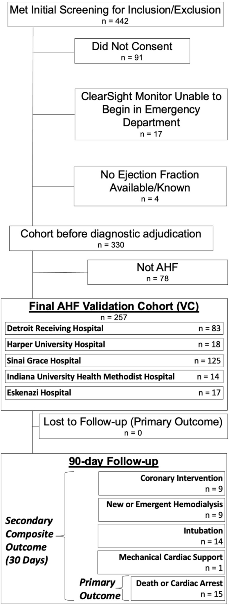
Fig 1

AHF = Acute Heart Failure.

Table 1 presents VC patient demographics, medical history, outpatient medications, initial vital signs, labs, interventions, clinical testing, risk scores, and initial values for cardiac index and vascular tone. 25% required supplemental oxygen, and 9% received a critical care intervention. 47% had HF with reduced EF (HFrEF), 79% of whom were adherent to guideline directed medical therapy. Median (IQR) for cardiac index and vascular tone were 2.40 L/min/m2 (2.00–3.08) and 3196 dynes-sec/cm5/m2 (2578–3919), respectively. Characteristics of the DC have been described previously [18].

Table 1. Clinical characteristics of patients in the validation cohort.

% (count) or median (IQR)
Patient Characteristic All Patients {n = 257} Hemodynamic Profile 1 {n = 68} Hemodynamic Profile 2 {n = 69} Hemodynamic Profile 3 {n = 120} p-value
Demographics, Medical History, and Outpatient Medications
Age (years) 60 (49–67) 59 (46–65) 62 (52–69) 59 (51–66) 0.080
Female 45% (116) 43% (29) 49% (34) 44% (53) 0.707
Body Mass Index (kg/m 2 ) 32 (26–39) 33 (28–40) 31 (25–39) 31 (25–38) 0.366
African American Race 89% (229) 85% (58) 90% (62) 91% (109) 0.490
EMS transport to ED 35% (89) 38% (26) 33% (23) 33% (40) 0.767
Hypertension 93% (240) 96% (65) 96% (66) 91% (109) 0.305
Diabetes 49% (126) 63% (43) 46% (32) 43% (51) 0.021***
Chronic Kidney Disease 38% (97) 47% (32) 33% (23) 35% (42) 0.177
Dialysis 7% (19) 10% (7) 7% (5) 6% (7) 0.531
Pulmonary Hypertension 29% (74) 29% (20) 33% (23) 26% (31) 0.544
Valvular Disease 35% (91) 35% (24) 46% (32) 29% (35) 0.059
COPD 38% (98) 35% (24) 39% (27) 39% (47) 0.854
Active Cancer 3% (7) 2% (1) 6% (4) 2% (2) 0.186
Heart Failure 93% (238) 90% (61) 93% (64) 94% (113) 0.531
HFrEF, on GDMT 37% (95) 28% (19) 44% (30) 38% (46) 0.155
HFrEF, not on GDMT 10% (25) 15% (10) 4% (3) 10% (12) 0.122
HFpEF 53% (137) 57% (39) 52% (36) 52% (62) 0.736
Ejection Fraction (%) 40 (25–59) 45 (25–60) 40 (20–50) 40 (25–60) 0.127
ACEi or ARB 48% (124) 46% (31) 55% (38) 46% (55) 0.415
Beta Blocker 70% (180) 72% (49) 70% (48) 69% (83) 0.913
Loop Diuretic 61% (157) 62% (42) 54% (37) 65% (78) 0.301
Metolazone 2% (5) 0% (0) 3% (2) 3% (3) 0.392
Antiarrhythmic 6% (15) 4% (3) 4% (3) 8% (9) 0.567
ED Initial Vitals, Labs, and Interventions
Systolic Blood Pressure (mmHg) 154 (132–178) 160 (139–177) 154 (129–175) 150 (129–180) 0.461
Diastolic Blood Pressure (mmHg) 91 (80–105) 91 (77–111) 91 (80–107) 92 (81–104) 0.933
SpO2 (%) 97 (95–99) 98 (96–99) 98 (96–99) 97 (95–98) 0.258
Respiratory Rate 20 (18–22) 20 (18–22) 18 (18–22) 20 (18–24) 0.170
Heart Rate 92 (80–105) 94 (84–105) 89 (79–101) 90 (81–106) 0.428
Sodium (mmol/L) 139 (137–142) 138 (136–141) 140 (138–142) 140 (137–142) 0.107
Potassium (mmol/L) 4.1 (3.8–4.5) 4.1 (3.8–4.4) 4.1 (3.9–4.7) 4 (3.6–4.5) 0.265
Blood Urea Nitrogen (mg/dL) 21 (15–31) 25 (16–40) 20 (16–30) 19 (15–27) 0.033***
eGFR (mL/min/1.73m 2 ) 63 (37–86) 55 (27–79) 61 (37–83) 66 (42–89) 0.129
Troponin I (ng/mL) 0.032 (0–0.07) 0 (0–0.096) 0.04 (0–0.079) 0.032 (0–0.065) 0.545
Troponin Positive 44% (113) 38% (26) 48% (33) 46% (55) 0.493
BNP (pg/mL) 1067 (414–2055) 1524 (562–2337) 1113 (443–2575) 770 (376–1967) 0.051
Supplemental O 2 25% (65) 28% (19) 19% (13) 28% (33) 0.353
IV Vasoactive, Inotrope, or PPV 9% (23) 9% (6) 7% (8) 13% (9) 0.335
Electrocardiogram (ECG) and Chest X-Ray (CXR)
Wide QRS 16% (42) 10% (7) 25% (17) 15% (18) 0.106
A-fib or A-flutter 12% (32) 6% (4) 15% (10) 15% (18) 0.362
Q Waves 9% (22) 3% (2) 15% (10) 8% (10) 0.079
Normal ECG 34% (87) 40% (27) 29% (20) 33% (40) 0.199
Alveolar Edema 19% (48) 22% (15) 16% (11) 18% (22) 0.542
Interstitial Edema 13% (34) 19% (13) 12% (8) 11% (13) 0.201
Cardiomegaly 85% (218) 88% (60) 84% (58) 83% (100) 0.647
Hyperinflated 5% (13) 3% (2) 6% (4) 6% (7) 0.661
Normal CXR 5% (14) 3% (2) 6% (4) 7% (8) 0.513
AHF Clinical Decision Rule Scores, and Earliest ED Finger-cuff Hemodynamic Measurements
STRATIFY 198 (180–226) 209 (180–252) 194 (178–219) 197 (181–223) 0.317
GWTG-HF Risk Score 31 (27–37) 31 (26–38) 33 (27–38) 31 (28–36) 0.737
EHMRG -17 (-59.4–28.9) -26 (-70.8–28) -11.5 (-44.3–41) -15.6 (-59.6–28.4) 0.309
First Cardiac Index (L/min/m 2 ) 2.4 (2.00–3.08) 3.71 (3.35–4.12) 1.81 (1.64–1.97) 2.4 (2.18–2.65) p<0.001***
First Vascular Tone {SVRI} (dynes-sec/cm5/m2) 3196 (2578–3919) 2240 (1745–2572) 4479 (4032–5260) 3180 (2801–3539) p<0.001***

Table 1 Legend—EMS = Emergency medical services; ED = Emergency department; COPD = Chronic obstructive pulmonary disease; HFrEF = Heart failure with reduced ejection fraction; HFpEF = HF with preserved EF; ACEi = Angiotensin converting enzyme inhibitor; ARB = Angiotensin receptor blocker; SpO2 = Oxygen saturation; eGFR = estimated glomerular filtration rate; BNP = Brain natriuretic peptide; O2 = oxygen; GWTG-HF = get-with-the-guidelines heart failure; EHMRG = Emergency Heart Failure Mortality Risk Grade.

EHMRG as calculated in Table 1 excludes patients who were dialysis dependent, as the EHMRG was derived and validated in a population excluding such patients. The total cohort and profile sizes without dialysis history were total cohort n = 238, profile 1 n = 61, profile 2 n = 64, profile 3 n = 113. None of the patients with dialysis history died within 90 days.

Hemodynamic profiling in the validation cohort

The consensus clustering algorithm was performed in the VC based on cardiac index and vascular tone (SVRI). Inspection of the consensus dendrograms [40] (Supplement S1 File) for each k clusters 1–10 showed the cleanest divisions to occur when the data was divided into k = 3–4 groups. Inspection of the delta area change in consensus score CDF [40] for K 1–10 (elbow plot, Supplement S1 File) show minimal improvement in area under the CDF curve for k>3. Taken together, these suggest that further divisions of the data (i.e. more profiles / increasing K) beyond k = 3 resulted primarily in data sorting at random rather than improved classification [40] by cardiac index and vascular tone in the VC. Consequently, the clustering and internally-validation performed by the consensus algorithm at k = 3 were designated hemodynamic profile 1 (n = 68), 2 (n = 69), and 3 (n = 120) in the VC.

Cardiac index, vascular tone, and clinical characteristics by profile are presented in Table 1. Profiles 1–3 resembled each other (p>0.05) for all clinical variables except BUN and history of diabetes, with each being highest in profile 1 and lowest in profile 3 (Table 1). Vascular tone and profiling overall did not appear to be a simple function of blood pressure, with no significant differences in SBP (p = 0.461) or DBP (p = 0.933) between profiles, which aligns with our prior published work on this device in an ED population of AHF patients [14] showing only moderate to low correlation of SVRI to device-estimated DBP (r = 0.587), SBP (r = 0.324), and mean arterial pressure ({MAP}, r = 0.479).

The 3 validated clinical risk scores (EHMRG, GWTG-HF, and STRATIFY) had no statistically significant difference between profiles (Table 1).

Comparison of hemodynamic profile composition in the validation cohort to the derivation and control cohorts

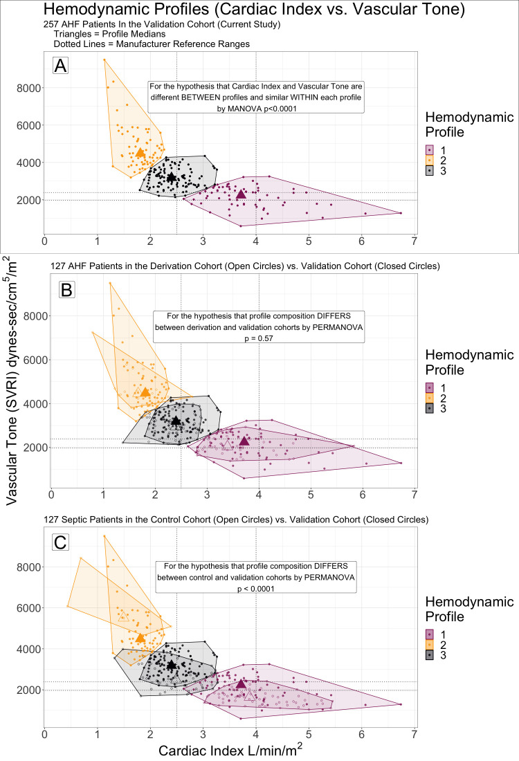
Fig 2 shows cardiac index and vascular tone for the 3 profiles in the VC alone (panel A) and compared to the DC [18] (panel B) and CC (panel C) patients’ hemodynamic profiles. Multivariate statistical comparison by PERMANOVA did not show the profiles in the DC [18] and VC to be significantly different at the prespecified α = 0.3 threshold (Fig 2B, p = 0.59, R2 = 0.016). A significant difference (PERMANOVA p = 0.001, R2 = 0.159) in cardiac index and vascular tone was present between the VC and CC profiles (Fig 2C).

Fig 2. Hemodynamic profiles by cardiac index vs. vascular tone (systemic vascular resistance index, SVRI).

Fig 2

Profiles were numbered similarly to facilitate between cohort comparisons: 1 (purple)—lowest SVRI and highest cardiac index, 2 (gold)—highest SVRI and lowest cardiac index, 3 (black) cardiac index and SVRI between profiles 1 and 2. (A) Profiling in the validation cohort (VC) alone. (B) The VC patients and their profiles are overlayed with the derivation cohort (DC). Patients in the DC had acute heart failure and were monitored in the emergency department like the VC, but were enrolled in a prior study (external cohort). Few patients classified in a particular profile in the VC would have been classified differently in the DC. (C) The VC overlayed with the control cohort (CC). The CC included patients enrolled in the same study as the DC, but who had sepsis rather than AHF. Profiling in the CC differed from VC, with several VC patients who would have been classified in a different profile by profiling of the CC (and visa versa).

Clinical outcomes by hemodynamic profile in the validation cohort

Outcomes rates by profile in the VC are presented in Table 2. 89% of patients were admitted to the hospital or an observation unit, 6% died within 90 days (primary outcome), and 7% experienced ≥1 fatal or non-fatal 30-day adverse event in the composite secondary outcome (Table 2). 90-day mortality (primary outcome) was significantly more likely (OR = 5.0, 95%CI 1.4–18.0) in Profile 3 compared to Profiles 1 or 2 (Table 2). Profile 3 also had shorter time to death than 1 or 2 (Table 2), including every death within 30 (p = 0.049) and 60 days (p<0.001).

Table 2. Study outcomes overall and by hemodynamic profile.

% (count) or mean (SD)
Outcome All Patients {n = 257} Hemodynamic Profile 1 {n = 68} Hemodynamic Profile 2 {n = 69} Hemodynamic Profile 3 {n = 120} p-value
Mortality (Primary Outcome)
30-Day 2% (6) 0% (0) 0% (0) 4% (5) p = 0.049***
60-Day 4% (11) 0% (0) 0% (0) 9% (11) p<0.001***
90-Day 6% (15) 3% (2) 1% (1) 10% (12) p = 0.027***
Time to Death (days) 87.3 (12.6) 89.7 (1.7) 89.7 (2.2) 84.6 (17.9) p = 0.016***
30-Day Ranked (0–5) Composite of Adverse Events (Secondary Outcome)adapted from Collins 201531
0 Event-free at 30 days 93% (239) 87% (59) 99% (68) 84% (101) p = 0.008***
1 Invasive Cardiac Procedure 5% (12) 6% (4) 1% (1) 6% (7) p = 0.187
2 New or Emergent Dialysis 3% (8) 3% (2) 0% (0) 5% (6) p = 0.300
3 Intubation 5% (13) 6% (4) 0% (0) 8% (9) p = 0.181
4 Mechanical Cardiac Support or Transplant < 1% (1) 0% (0) 1% (1) 0% (0) p = 0.601
5 Death or Cardiopulmonary Resuscitation 2% (6) 0% (0) 0% (0) 4% (5) p = 0.049***
Mean Rank of Worst Event in 30 days (0–5) 0.30 (0.98) 0.28 (0.79) 0.06 (0.48) 0.46 (1.24) p = 0.003***
Other Outcomes—all at 30-day follow-up unless otherwise indicated
Discharged from Emergency Department 11% (28) 10% (7) 14% (10) 9% (11) p = 0.268
Admission or Transfer to ICU 18% (45) 22% (15) 15% (10) 17% (20) p = 0.480
Index Hospitalization Length of Stay (days) 4 (4) 5 (5) 4 (3) 4 (4) p = 0.935
ED Revisits for AHF 18% (46) 18% (12) 15% (10) 20% (24) p = 0.635
ED Revisits, All-Cause 26% (68) 31% (21) 22% (15) 27% (32) p = 0.478
AHF Readmissions 16% (41) 16% (11) 13% (9) 18% (21) p = 0.722
All-Cause Readmissions 22% (56) 27% (18) 17% (12) 22% (26) p = 0.436
ICU or Death 18% (46) 22% (15) 15% (10) 18% (21) p = 0.507

Table 2 Legend—ICU = Intensive care unit; ED = Emergency Department; AHF = Acute heart failure.

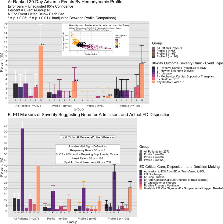
Comparison of 30-day events by profile are presented in Fig 3A and Table 2. Fig 3B shows rates for ED critical care interventions, unstable vital signs, dispositions, and loop diuretic administration all of which were similar between profiles (p>0.05). A 30-day fatal or non-fatal adverse event (secondary outcome) occurred in 16% of profile 3 patients, 13% profile 1, and 1% of profile 2 (p = 0.008). Median event severity/rank in profile 2 was lower compared to 1 or 3 (p = 0.003, Table 2). The likelihood of any 30-day adverse event (Table 2) was higher in Profile 3 vs. Profile 2 (OR = 12.8, 95%CI: 1.7–97.9) and Profile 1 vs. 2 (OR = 10.0, 95%CI: 1.2–81.2), but similar in Profile 3 vs. Profile 1 (OR = 1.28, 95%CI 0.5–3.0).

Fig 3. Comparison of cardiac index vs. vascular tone hemodynamic profiles by 30-day adverse events, emergency department (ED) characteristics, and ED disposition.

Fig 3

(A) Hemodynamic profiles 1–3 in the validation cohort (inset) are compared by individual components of the composite 30-day secondary outcome. Compared to profile 2 (gold in inset), profile 3 (black) and profile 1 (purple) had greater rates of any outcome in the composite. (B) The were no statistically significant differences between profiles in actual ED disposition decisions (ICU, or discharge from ED), ED treatments administered, or the presence of unstable vital signs or need for supplemental oxygen.

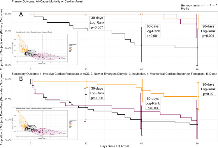
Fig 4 presents Kaplan-Meier survival for mortality (4A) and the secondary outcome(4B) stratified by profile and compared at each of 30, 60, and 90 days (all log-rank p<0.05, both outcomes). Time-to-death was worst in profile 3 (90-day hazard ratio {HR} = 4.83, 95%CI 1.36–17.1), while time to any event (90-day HR = 0.36, 95%CI: 0.15–0.85) was best in profile 2.

Fig 4. Survival to primary and secondary study outcomes by hemodynamic profile.

Fig 4

Kaplan Meier curves for three hemodynamic profiles by cardiac index and vascular tone (see inset) in the validation cohort, through 90-day follow-up. (A) Primary outcome: All-cause mortality or cardiac arrest. Profile 3 (black) has significantly worse survival compared to profiles 1 or 2 (purple and gold, respectively) through each of 30, 60 and 90 days. (B) Secondary outcome: A composite of invasive cardiac procedure, new or emergent dialysis, intubation, mechanical cardiac support or transplant, and death or cardiac arrest. Profile 2 has significantly better event-free survival compared to profiles 1 or 3 through each of 30, 60 and 90 days.

Sensitivity analyses

On principle components analysis of the VC hemodynamic profiles, using all clinical variables collected (Table 1) other than cardiac index and vascular tone, we found that ≥7 principal components were required to explain ≥75% of between-profile variance.

116/257 (45.1%) patients met our specified criteria (Supplement S1 File—Sensitivity analysis 2) for a “clear indication” for hospital admission in the ED. After excluding these patients, just 12.1% of those remaining (17/141) were discharged from the ED, but 56.7% (80/141) were hemodynamic Profile 1 or 2 (low risk for 90-day mortality) and 30.0% (42/141) were Profile 2 (low risk for any 30-day adverse event). If Profile 2 vs. Profile 1 or 3 was used as a discharge criterion in these 141 patients without a clear indication for hospital admission, significantly more patients (p<0.001) would have been discharged from the ED compared to the actual discharge rate, without any missed deaths (100% negative predictive value for Profile 2).

CC2 (patients with non-cardiac dyspnea) had p = 0.001 difference in profiling compared to AHF-adjudicated patients in the VC by PERMANOVA.

Discussion

In this multicenter prospective observational study, we report 3 main objectives and their findings for what is to our knowledge the first time: 1. We externally validated non-invasive hemodynamic profiling of ED AHF patients by cardiac index and vascular with a finger-cuff monitor, 2. We show that this profiling was specific to the diagnosis of AHF among patients in the ED, as compared to one control group (CC) of septic patients and another (CC2) of patients with diagnostically-adjudicated non-cardiac dyspnea. 3. We show that these profiles have significant association with mortality and other clinically-important and patient-centered outcomes, without this association being clearly explained by other clinical variables. Overall, these results build upon the prior study [18] to suggest that these previously described hemodynamic profiles by finger-cuff have external validity, specificity to AHF, and potential clinical utility as novel markers for ED risk-stratification.

Hemodynamic profiling has long been used to subgroup AHF patients by clinically-important differences in physiology, particularly in the relationship between cardiac index and vascular tone. Despite being recommended as part of routine AHF assessment in guidelines [1,22,23], invasive measures are so specialized and uncommon [19] that ED use is virtually unheard of, while current non-invasive methods [20,21] based on physical exam are subjective and lack sufficient interrater reliability [12,18,24,41] in the ED. Finger-cuff monitors are a non-invasive approach which provides objective data, and the replication of the exploratory cluster analysis results in the prior study’s DC [18] adds external validity to the observed profiles as a novel marker in ED AHF patients.

In the prior study [18] patients had marked heterogeneity by cardiac index and vascular tone and clustered into three novel two-dimensional profiles. We were able to replicate these profiles de novo in a prospectively enrolled and diagnostically adjudicated external cohort, whereas the DC was a retrospective analysis without diagnostic adjudication. Additionally, we used a specialized cluster analysis procedure which was less reliant on analyst assumptions and theoretically more robust to data overfitting than what was used in the DC’s profiling. Our findings nevertheless replicated the heterogeneity and distribution of cardiac index and vascular tone first noted in the DC study [18], while clearly differing from the septic CC. Replication achieved by these methods add rigor and bolster the case that these hemodynamic profiles represent a true feature of AHF patient is the ED, rather than artifact and overfitting of the prior study’s single retrospective sample.

Both 90-day mortality and a composite of 30-day adverse events differed significantly between profiles in the VC. Assessing these differences was a novel and primary goal of the current study, since the DC study was neither powered for nor designed [18] to test differences in clinical outcomes between profiles. The between-profile differences in adverse events observed were clinically significant: no patients died within the first 60 days outside the high-risk profile 3, and death remained five times more likely in profile 3 at 90 days. Additionally, the low-risk profile 2 had roughly 12 times lower likelihood of any adverse 30-day event compared to profiles 1 or 3.

Novel risk markers for AHF are needed in the ED [1], and particularly markers of low risk [1,2,810]. Over 80% of the 1 million AHF patients presenting to US EDs annually are admitted, including over 90% of the current sample, many of whom are at low risk for short term adverse events [1]. The burden on patients and healthcare resources is correspondingly high. Among the 141 patients in the VC who lacked one or more clear criteria for ED-to-hospital admission at our institutions (sensitivity analysis 2), the low-risk profile 2 was present in more than double the actual number of patients discharged. Similarly, over 57% of those without clear admission criteria were in profile 1 or 2, among which no patient died within 60 days. Physicians were blinded to monitoring of cardiac index and vascular tone in this study, and it is possible that knowledge of a patient’s hemodynamic profile would have improved risk-stratification and facilitated ED discharges.

As in the DC study [18], hemodynamic profile was not clearly explained by other clinical variables. Two validated CDRs related to the primary outcome, and one for the secondary outcome, did not differ significantly between the high vs. low-risk profiles observed. Among a long list of common clinical variables, only BUN and history of diabetes differed between profiles. While the absence of diabetes and a lower BUN would be expected to correlate with better clinical outcomes, these variables were paradoxically the lowest in the highest risk hemodynamic profile (profile 3). Principle components analysis (sensitivity analysis 1) failed to yield any simple combination of clinical variables which would explain the variance in patients’ hemodynamic profile. It is unlikely, based on the results in the VC and the prior description of the DC [18], that these hemodynamic profiles are simple functions of more common and available clinical measures. Instead, these results suggest that hemodynamic profiling by finger-cuff monitor cardiac index and vascular tone add novel information not already captured in the standard of care. Further research is needed to assess if the information added, particularly regarding association of these profiles and clinical outcomes as a risk-measure, is incremental with other established AHD risk measures. A pre-planned analysis of the VC to assess for incremental value in risk-stratification and prognosis is currently underway.

Limitations

This study had several limitations. First, finger-cuff hemodynamic monitors have a roughly +/- 30% error in cardiac index compared to invasive monitoring [18,35], which is below the level of construct validity to completely replace invasive catheter based methods in the cardiac ICU [18,35], though relatively low as an absolute amount (mean difference in cardiac index on finger-cuff vs. invasive standard = 0.07 L/min/m2 {95%CI: 0.01–0.13} [42]). The specific formula by which cardiac index, mean arterial pressure (MAP) and SVRI are calculated from the finger cuff monitor are proprietary details we are not privy to, with the only specific measures known to be incorporated being the time in systole vs. diastole, heart rate, and the velocity time integral of the pulse waveform. It is possible the device includes the calculated cardiac index and MAP in the derivation of SVRI, which would in turn extend the error rate for cardiac index to vascular tone as well. The device is highly accurate compared to invasive monitoring of MAP (R2 = 0.96 [34], mean difference in MAP from finger cuff vs. invasive 4.2 mmHg {95%CI: 2.8–5.6 mmHg}), so if calculated in this way the error rate in SVRI would be unlikely to vary much from the error rate of cardiac index. Regardless, invasive monitoring is not feasible in the ED and physical exam based non-invasive alternatives are far more unreliable and inaccurate [12,18,24,41], making the error rate in finger-cuff monitors likely the best achievable in this patient population and setting at present. Moreover, the primary goals of this study were 1. to show that clustering of ED patients by finger-cuff monitor hemodynamics were repeatable in an external sample and unique from patients without AHF and 2. to show that these profiles were associated with clinically important outcomes in ED AHF patients such as mortality and the ranked composite outcome. Both goals (and their subsequent findings) are novel compared to what has been examined in the prior literature [18] in examining extremal validity/reliability (goal 1) and predictive validity for patient-oriented outcomes (goal 2). Concurrent validity (i.e. comparison of numerical cardiac index and SVRI to a right-heart catheterization gold standard) was explicitly not a goal of this study, and may be the subject of future investigations. With this said, such a study is unlikely to be feasible or ethical in this population (ED patients with AHF and without cardiogenic shock, some of whom are discharged without hospitalization) since pulmonary artery catheterization is outside the current scope of practice of emergency medicine providers [43] and only performed in less than 1% of inpatient AHF encounters [19]. Thus, while this study was not designed to provide evidence of concurrent validity of finger cuff monitors compared to invasive monitoring, it is unlikely that such an investigation is possible in this population and this in turn underlines the potential utility of finger cuff monitoring for hemodynamic profiling of AHF. Namely, the lack of ability to employ invasive standards or useful non-invasive alternatives of hemodynamic profiling indicate a need for a novel non-invasive method feasible for the ED which is reliable between studies (i.e. external validity and reliability), consistent in differentiating profiling of AHF patients from non-AHF patients (i.e. face validity), and informative about patient-oriented and clinically-important outcomes (i.e. predictive validity) as we show here for the first time.

Second, in our approach to replication and validation using PERMANOVA means we failed to reject the null hypothesis that the VC did not replicate the DC profiles, which is not the same as accepting the hypothesis that they were the same. We used a more conservative alpha threshold of 0.3 to test this hypothesis to decrease the chance of type II error, but we cannot guarantee that additional replication studies or a larger statistical power would have failed to reject the null hypothesis. Moreover, the clear differences of the VC and CC profiles enhance our confidence in the results, given that the CC patients were enrolled at the same times and hospitals as the DC but with a different underlying ED diagnosis (sepsis, rather than AHF).

Third, our study was performed at 5 high-volume academic EDs, and results may not generalize to dissimilar settings or AHF patient populations significantly different than our sample (Table 1).

Fourth, unaccounted for lost-to-follow-up is possible for the secondary outcome, such as if a patient had an adverse event at an outside hospital. We confirmed 100% of patient follow-up for the primary outcome at 90 days between the use of telephone follow-up, HIEs including the largest hospital systems near the study sites, and dual-review with adjudication for outcome record review.

Fifth, AHF is a clinical diagnosis without a gold standard. We used double-blinded diagnostic adjudication by experienced AHF researchers and clinicians to limit the analysis of patients who could have met the inclusion criteria with signs, symptoms, and lab/imaging findings due to a diagnosis other than AHF (e.g. non-cardiac causes of dyspnea, such as chronic obstructive pulmonary disease, etc.). Moreover, a sensitivity analysis showed that profiling of patients with vs. without adjudicated AHF in the current study (i.e. included patients vs. patients excluded after adjudication as “Not AHF”) was significantly different between the two groups (p<0.001). In particular, patients initially included in the current study who were adjudicated as “not-AHF” had higher cardiac index than those adjudicated as AHF (4.04 L/min vs. 3.71, 2.08 vs. 1.71, 2.88 vs. 2.4 for profiles 1–3 respectively, 2.72 vs. 2.4 overall). This adds internal validity to the diagnostic adjudication and face validity overall, by reinforcing that profiling of patients with AHF in the current study were distinguishable from those adjudicated as “not-AHF” (i.e. but otherwise meeting inclusion criteria). Diagnostic adjudication in our cohort, by standard methods for the field, is an improvement in scientific rigor over the prior study [18]. Nevertheless, the lack of a gold-standard for diagnosis of AHF (i.e. being a clinical syndrome) is a limitation to our study and all AHF literature.

Finally, while no clear combination of individual variables or CDRs appear to explain the difference in profiles, this does not imply that adding the hemodynamic profiles would add incremental prognostic value to existing AHF risk measures. Rather, incremental prognostic utility is a separate question, to be addressed in a pre-planned future analysis of the VC.

Conclusion

In this prospective observational cohort study, we validate 3 distinct hemodynamic profiles of ED AHF patients by cardiac index and vascular tone, as measured on a non-invasive finger cuff monitor and described in prior work [18]. Mortality and a composite of adverse short-term events differed markedly between these profiles, suggesting a potential for use in the ED as a marker for risk-stratification.

Supporting information

S1 File. Supplemental methods.

I—Further methodological detail on consensus clustering, and contrast to k-means cluster analysis. II—Consensus Clustering Dendrograms. III—Delta-area under the cumulative distribution function (CDF) for each additional level K in consensus clustering. IV—Sensitivity Analyses—Methods, Goals, and Rationale.

(DOCX)

S2 File. Code for analysis and minimal dataset.

Code for the R statistical programming language to reproduce results with the provided minimal datasets S3S5 Files.

(R)

S3 File. Minimal dataset for the validation cohort.

(CSV)

S4 File. Minimal dataset “VC_and_DC”.

(CSV)

S5 File. Minimal dataset “VC_and_CC”.

(CSV)

Data Availability

All relevant data are within the manuscript and its Supporting Information files. The datasets are attached as CSV files, and code for reproduction of the primary statistical analyses in R are attached in the submission.

Funding Statement

The ClearSight monitor’s manufacturer, Edwards Lifesciences Corporation, provided funding for this study in the form of the Edwards Lifesciences Investigator-Initiated Grant. The funding was used for research assistant time and the monitors used in the study. Edwards Lifesciences Corporation also provided salary support for PL. The specific roles of these authors are articulated in the ‘author contributions’ section. The funders had no role in study design, data collection and analysis, decision to publish, or preparation of the manuscript.

References

  • 1.Collins SP, Storrow AB, Levy PD, et al. Early management of patients with acute heart failure: state of the art and future directions—a consensus document from the SAEM/HFSA acute heart failure working group. Acad Emerg Med. 2015;22(1):94–112. doi: 10.1111/acem.12538 [DOI] [PMC free article] [PubMed] [Google Scholar]
  • 2.Benjamin EJ, Muntner P, Alonso A, et al. Heart Disease and Stroke Statistics—2019 Update: A Report From the American Heart Association. Circulation. 2019;139(10):e56–e528. doi: 10.1161/CIR.0000000000000659 [DOI] [PubMed] [Google Scholar]
  • 3.Pandey A, Patel KV, Liang L, et al. Association of Hospital Performance Based on 30-Day Risk-Standardized Mortality Rate With Long-term Survival After Heart Failure Hospitalization: An Analysis of the Get With The Guidelines-Heart Failure Registry. JAMA Cardiol. 2018;3(6):489–497. doi: 10.1001/jamacardio.2018.0579 [DOI] [PMC free article] [PubMed] [Google Scholar]
  • 4.McCausland JB, Machi MS, Yealy DM. Emergency physicians’ risk attitudes in acute decompensated heart failure patients. Acad Emerg Med. 2010;17(1):108–110. doi: 10.1111/j.1553-2712.2009.00623.x [DOI] [PubMed] [Google Scholar]
  • 5.Lee DS, Lee JS, Schull MJ, et al. Prospective Validation of the Emergency Heart Failure Mortality Risk Grade for Acute Heart Failure. Circulation. 2019;139(9):1146–1156. doi: 10.1161/CIRCULATIONAHA.118.035509 [DOI] [PubMed] [Google Scholar]
  • 6.Lee DS, Schull MJ, Alter DA, et al. Early deaths in patients with heart failure discharged from the emergency department: a population-based analysis. Circ Heart Fail. 2010;3(2):228–235. doi: 10.1161/CIRCHEARTFAILURE.109.885285 [DOI] [PubMed] [Google Scholar]
  • 7.Collins SP, Pang PS. ACUTE Heart Failure Risk Stratification. Circulation. 2019;139(9):1157–1161. doi: 10.1161/CIRCULATIONAHA.118.038472 [DOI] [PMC free article] [PubMed] [Google Scholar]
  • 8.Chang PP, Wruck LM, Shahar E, et al. Trends in Hospitalizations and Survival of Acute Decompensated Heart Failure in Four US Communities (2005–2014). Circulation. 2018;138(1):12–24. doi: 10.1161/CIRCULATIONAHA.117.027551 [DOI] [PMC free article] [PubMed] [Google Scholar]
  • 9.Collins SP, Lindsell CJ, Naftilan AJ, et al. Low-risk acute heart failure patients: external validation of the Society of Chest Pain Center’s recommendations. Crit Pathw Cardiol. 2009;8(3):99–103. doi: 10.1097/HPC.0b013e3181b5a534 [DOI] [PMC free article] [PubMed] [Google Scholar]
  • 10.Butler J, Hanumanthu S, Chomsky D, Wilson JR. Frequency of low-risk hospital admissions for heart failure. The American journal of cardiology. 1998;81(1):41–44. doi: 10.1016/s0002-9149(97)00851-5 [DOI] [PubMed] [Google Scholar]
  • 11.Harrison N, Favot M, Levy P. The Role of Troponin for Acute Heart Failure. Curr Heart Fail Rep. 2019;16(1):21–31. doi: 10.1007/s11897-019-0420-5 [DOI] [PubMed] [Google Scholar]
  • 12.Ahmad T, Desai N, Wilson F, et al. Clinical Implications of Cluster Analysis-Based Classification of Acute Decompensated Heart Failure and Correlation with Bedside Hemodynamic Profiles. PLoS One. 2016;11(2):e0145881. doi: 10.1371/journal.pone.0145881 [DOI] [PMC free article] [PubMed] [Google Scholar]
  • 13.Collins SP, Levy PD, Fermann GJ, et al. What’s Next for Acute Heart Failure Research? Acad Emerg Med. 2018;25(1):85–93. doi: 10.1111/acem.13331 [DOI] [PubMed] [Google Scholar]
  • 14.Harrison N, Pang P, Collins S, Levy P. Blood Pressure Reduction in Hypertensive Acute Heart Failure. Curr Hypertens Rep. 2021;23(2):11. doi: 10.1007/s11906-021-01127-8 [DOI] [PubMed] [Google Scholar]
  • 15.Viau DM, Sala-Mercado JA, Spranger MD, Leary DS, Levy PD. The pathophysiology of hypertensive acute heart failure. Heart (British Cardiac Society). 2015;101(23):1861. doi: 10.1136/heartjnl-2015-307461 [DOI] [PubMed] [Google Scholar]
  • 16.Fudim M, Hernandez AF, Felker GM. Role of Volume Redistribution in the Congestion of Heart Failure. J Am Heart Assoc. 2017;6(8):e006817. doi: 10.1161/JAHA.117.006817 [DOI] [PMC free article] [PubMed] [Google Scholar]
  • 17.Bilchick KC, Mejia-Lopez E, McCullough P, et al. Clinical Impact of Changes in Hemodynamic Indices of Contractile Function During Treatment of Acute Decompensated Heart Failure. Journal of cardiac failure. 2018;24(1):43–50. doi: 10.1016/j.cardfail.2017.07.001 [DOI] [PubMed] [Google Scholar]
  • 18.Nowak RM, Reed BP, DiSomma S, et al. Presenting phenotypes of acute heart failure patients in the ED: Identification and implications. Am J Emerg Med. 2017;35(4):536–542. doi: 10.1016/j.ajem.2016.12.003 [DOI] [PubMed] [Google Scholar]
  • 19.Hernandez GA, Lemor A, Blumer V, et al. Trends in Utilization and Outcomes of Pulmonary Artery Catheterization in Heart Failure With and Without Cardiogenic Shock. Journal of cardiac failure. 2019;25(5):364–371. doi: 10.1016/j.cardfail.2019.03.004 [DOI] [PubMed] [Google Scholar]
  • 20.Forrester JS, Diamond G, Chatterjee K, Swan HJ. Medical therapy of acute myocardial infarction by application of hemodynamic subsets (second of two parts). N Engl J Med. 1976;295(25):1404–1413. doi: 10.1056/NEJM197612162952505 [DOI] [PubMed] [Google Scholar]
  • 21.Nohria A, Tsang SW, Fang JC, et al. Clinical assessment identifies hemodynamic profiles that predict outcomes in patients admitted with heart failure. Journal of the American College of Cardiology. 2003;41(10):1797–1804. doi: 10.1016/s0735-1097(03)00309-7 [DOI] [PubMed] [Google Scholar]
  • 22.Hollenberg SM, Warner Stevenson L, Ahmad T, et al. 2019 ACC Expert Consensus Decision Pathway on Risk Assessment, Management, and Clinical Trajectory of Patients Hospitalized With Heart Failure. Journal of the American College of Cardiology. 2019;74(15):1966. doi: 10.1016/j.jacc.2019.08.001 [DOI] [PubMed] [Google Scholar]
  • 23.Ponikowski P, Voors AA, Anker SD, et al. 2016 ESC Guidelines for the diagnosis and treatment of acute and chronic heart failure: The Task Force for the diagnosis and treatment of acute and chronic heart failure of the European Society of Cardiology (ESC)Developed with the special contribution of the Heart Failure Association (HFA) of the ESC. Eur Heart J. 2016;37(27):2129–2200. doi: 10.1093/eurheartj/ehw128 [DOI] [PubMed] [Google Scholar]
  • 24.Chaudhry A, Singer AJ, Chohan J, Russo V, Lee C. Interrater reliability of hemodynamic profiling of patients with heart failure in the ED. The American Journal of Emergency Medicine. 2008;26(2):196–201. doi: 10.1016/j.ajem.2007.04.029 [DOI] [PubMed] [Google Scholar]
  • 25.Weil BR, Techiryan G, Suzuki G, Konecny F, Canty JM Jr. Adaptive Reductions in Left Ventricular Diastolic Compliance Protect the Heart From Stretch-Induced Stunning. JACC Basic Transl Sci. 2019;4(4):527–541. doi: 10.1016/j.jacbts.2019.04.002 [DOI] [PMC free article] [PubMed] [Google Scholar]
  • 26.Obokata M, Nagata Y, Kado Y, Kurabayashi M, Otsuji Y, Takeuchi M. Ventricular-Arterial Coupling and Exercise-Induced Pulmonary Hypertension During Low-Level Exercise in Heart Failure With Preserved or Reduced Ejection Fraction. Journal of cardiac failure. 2017;23(3):216–220. doi: 10.1016/j.cardfail.2016.10.001 [DOI] [PubMed] [Google Scholar]
  • 27.Harjola V-P, Mullens W, Banaszewski M, et al. Organ dysfunction, injury and failure in acute heart failure: from pathophysiology to diagnosis and management. A review on behalf of the Acute Heart Failure Committee of the Heart Failure Association (HFA) of the European Society of Cardiology (ESC). Eur J Heart Fail. 2017;19(7):821–836. doi: 10.1002/ejhf.872 [DOI] [PMC free article] [PubMed] [Google Scholar]
  • 28.Weil BR, Suzuki G, Young RF, Iyer V, Canty JM Jr. Troponin Release and Reversible Left Ventricular Dysfunction After Transient Pressure Overload. J Am Coll Cardiol. 2018;71(25):2906–2916. doi: 10.1016/j.jacc.2018.04.029 [DOI] [PMC free article] [PubMed] [Google Scholar]
  • 29.Favot M, Ehrman R, Gowland L, et al. Changes in speckle-tracking-derived mechanical dispersion index are associated with 30-day readmissions in acute heart failure. Ultrasound J. 2019;11(1):9. doi: 10.1186/s13089-019-0125-8 [DOI] [PMC free article] [PubMed] [Google Scholar]
  • 30.Reddy YNV, Andersen MJ, Obokata M, et al. Arterial Stiffening With Exercise in Patients With Heart Failure and Preserved Ejection Fraction. J Am Coll Cardiol. 2017;70(2):136–148. doi: 10.1016/j.jacc.2017.05.029 [DOI] [PMC free article] [PubMed] [Google Scholar]
  • 31.Collins SP, Jenkins CA, Harrell FE Jr., et al. Identification of Emergency Department Patients With Acute Heart Failure at Low Risk for 30-Day Adverse Events: The STRATIFY Decision Tool. JACC Heart Fail. 2015;3(10):737–747. doi: 10.1016/j.jchf.2015.05.007 [DOI] [PMC free article] [PubMed] [Google Scholar]
  • 32.Sax DR, Mark DG, Huang J, et al. Use of Machine Learning to Develop a Risk-Stratification Tool for Emergency Department Patients With Acute Heart Failure. Ann Emerg Med. 2021;77(2):237–248. doi: 10.1016/j.annemergmed.2020.09.436 [DOI] [PubMed] [Google Scholar]
  • 33.von Elm E, Altman DG, Egger M, Pocock SJ, Gøtzsche PC, Vandenbroucke JP. The Strengthening the Reporting of Observational Studies in Epidemiology (STROBE) statement: guidelines for reporting observational studies. PLoS Med. 2007;4(10):e296. doi: 10.1371/journal.pmed.0040296 [DOI] [PMC free article] [PubMed] [Google Scholar]
  • 34.Martina JR, Westerhof BE, van Goudoever J, et al. Noninvasive continuous arterial blood pressure monitoring with Nexfin(R). Anesthesiology. 2012;116(5):1092–1103. doi: 10.1097/ALN.0b013e31824f94ed [DOI] [PubMed] [Google Scholar]
  • 35.Ameloot K, Palmers PJ, Malbrain ML. The accuracy of noninvasive cardiac output and pressure measurements with finger cuff: a concise review. Curr Opin Crit Care. 2015;21(3):232–239. doi: 10.1097/MCC.0000000000000198 [DOI] [PubMed] [Google Scholar]
  • 36.Peterson PN, Rumsfeld JS, Liang L, et al. A validated risk score for in-hospital mortality in patients with heart failure from the American Heart Association get with the guidelines program. Circ Cardiovasc Qual Outcomes. 2010;3(1):25–32. doi: 10.1161/CIRCOUTCOMES.109.854877 [DOI] [PubMed] [Google Scholar]
  • 37.Garg N, Pekmezaris R, Stevens G, et al. Performance of Emergency Heart Failure Mortality Risk Grade in the Emergency Department. West J Emerg Med. 2021;22(3):672–677. doi: 10.5811/westjem.2021.1.48978 [DOI] [PMC free article] [PubMed] [Google Scholar]
  • 38.Shiraishi Y, Kohsaka S, Abe T, et al. Validation of the Get With The Guideline–Heart Failure risk score in Japanese patients and the potential improvement of its discrimination ability by the inclusion of B-type natriuretic peptide level. American heart journal. 2016;171(1):33–39. doi: 10.1016/j.ahj.2015.10.008 [DOI] [PubMed] [Google Scholar]
  • 39.Suzuki S, Yoshihisa A, Sato Y, et al. Clinical Significance of Get With the Guidelines-Heart Failure Risk Score in Patients With Chronic Heart Failure After Hospitalization. J Am Heart Assoc. 2018;7(17):e008316. doi: 10.1161/JAHA.117.008316 [DOI] [PMC free article] [PubMed] [Google Scholar]
  • 40.Wilkerson MD, Hayes DN. ConsensusClusterPlus: a class discovery tool with confidence assessments and item tracking. Bioinformatics. 2010;26(12):1572–1573. doi: 10.1093/bioinformatics/btq170 [DOI] [PMC free article] [PubMed] [Google Scholar]
  • 41.Almeida Junior GLG, Clausell N, Garcia MI, et al. Natriuretic Peptide and Clinical Evaluation in the Diagnosis of Heart Failure Hemodynamic Profile: Comparison with Tissue Doppler Echocardiography. Arquivos brasileiros de cardiologia. 2018;110(3):270–277. doi: 10.5935/abc.20180046 [DOI] [PMC free article] [PubMed] [Google Scholar]
  • 42.Saugel B, Hoppe P, Nicklas JY, et al. Continuous noninvasive pulse wave analysis using finger cuff technologies for arterial blood pressure and cardiac output monitoring in perioperative and intensive care medicine: a systematic review and meta-analysis. British Journal of Anaesthesia. 2020;125(1):25–37. doi: 10.1016/j.bja.2020.03.013 [DOI] [PubMed] [Google Scholar]
  • 43.Beeson MS, Ankel F, Bhat R, et al. The 2019 Model of the Clinical Practice of Emergency Medicine. Journal of Emergency Medicine. 2020;59(1):96–120. doi: 10.1016/j.jemermed.2020.03.018 [DOI] [PubMed] [Google Scholar]

Decision Letter 0

Gianluigi Savarese

7 Jan 2022

PONE-D-21-36949Hemodynamic profiles by non-invasive monitoring of cardiac index and vascular tone in acute heart failure patients in the emergency department: external validation and clinical outcomesPLOS ONE

Dear Dr. Harrison,

Thank you for submitting your manuscript to PLOS ONE. After careful consideration, we feel that it has merit but does not fully meet PLOS ONE’s publication criteria as it currently stands. Therefore, we invite you to submit a revised version of the manuscript that addresses the points raised during the review process.

Please submit your revised manuscript by Feb 21 2022 11:59PM. If you will need more time than this to complete your revisions, please reply to this message or contact the journal office at plosone@plos.org. When you're ready to submit your revision, log on to https://www.editorialmanager.com/pone/ and select the 'Submissions Needing Revision' folder to locate your manuscript file.

Please include the following items when submitting your revised manuscript:

  • A rebuttal letter that responds to each point raised by the academic editor and reviewer(s). You should upload this letter as a separate file labeled 'Response to Reviewers'.

  • A marked-up copy of your manuscript that highlights changes made to the original version. You should upload this as a separate file labeled 'Revised Manuscript with Track Changes'.

  • An unmarked version of your revised paper without tracked changes. You should upload this as a separate file labeled 'Manuscript'.

If you would like to make changes to your financial disclosure, please include your updated statement in your cover letter. Guidelines for resubmitting your figure files are available below the reviewer comments at the end of this letter.

If applicable, we recommend that you deposit your laboratory protocols in protocols.io to enhance the reproducibility of your results. Protocols.io assigns your protocol its own identifier (DOI) so that it can be cited independently in the future. For instructions see: https://journals.plos.org/plosone/s/submission-guidelines#loc-laboratory-protocols. Additionally, PLOS ONE offers an option for publishing peer-reviewed Lab Protocol articles, which describe protocols hosted on protocols.io. Read more information on sharing protocols at https://plos.org/protocols?utm_medium=editorial-email&utm_source=authorletters&utm_campaign=protocols.

We look forward to receiving your revised manuscript.

Kind regards,

Gianluigi Savarese

Academic Editor

PLOS ONE

Journal Requirements:

When submitting your revision, we need you to address these additional requirements.

1. Please ensure that your manuscript meets PLOS ONE's style requirements, including those for file naming. The PLOS ONE style templates can be found at

https://journals.plos.org/plosone/s/file?id=wjVg/PLOSOne_formatting_sample_main_body.pdf and

https://journals.plos.org/plosone/s/file?id=ba62/PLOSOne_formatting_sample_title_authors_affiliations.pdf

[Note: HTML markup is below. Please do not edit.]

Reviewers' comments:

Reviewer's Responses to Questions

Comments to the Author

1. Is the manuscript technically sound, and do the data support the conclusions?

The manuscript must describe a technically sound piece of scientific research with data that supports the conclusions. Experiments must have been conducted rigorously, with appropriate controls, replication, and sample sizes. The conclusions must be drawn appropriately based on the data presented.

Reviewer #1: Yes

Reviewer #2: Partly

**********

2. Has the statistical analysis been performed appropriately and rigorously?

Reviewer #1: Yes

Reviewer #2: Yes

**********

3. Have the authors made all data underlying the findings in their manuscript fully available?

The PLOS Data policy requires authors to make all data underlying the findings described in their manuscript fully available without restriction, with rare exception (please refer to the Data Availability Statement in the manuscript PDF file). The data should be provided as part of the manuscript or its supporting information, or deposited to a public repository. For example, in addition to summary statistics, the data points behind means, medians and variance measures should be available. If there are restrictions on publicly sharing data—e.g. participant privacy or use of data from a third party—those must be specified.

Reviewer #1: Yes

Reviewer #2: No

**********

4. Is the manuscript presented in an intelligible fashion and written in standard English?

PLOS ONE does not copyedit accepted manuscripts, so the language in submitted articles must be clear, correct, and unambiguous. Any typographical or grammatical errors should be corrected at revision, so please note any specific errors here.

Reviewer #1: Yes

Reviewer #2: Yes

**********

5. Review Comments to the Author

Please use the space provided to explain your answers to the questions above. You may also include additional comments for the author, including concerns about dual publication, research ethics, or publication ethics. (Please upload your review as an attachment if it exceeds 20,000 characters)

Reviewer #1: This manuscript by Professor Levy et al. addresses the use of a non-invasive finger-cuff monitor measuring CI and SVRI in AHF patients in an ED setting. The research group is well established in the field. The manuscript is well written, and a lot of effort has gone into the work.

In summary, my main criticism of this work relates to the limitations of finger-cuff monitors. 1) The authors do discuss this as a limitation, with a roughly +/- 30% error in CI compared to invasive monitoring. However, the calculations of SVRI are also based on CI, resulting in a higher risk of error, which is not addressed. I also wonder if the results to a large extent are associated with blood pressure. What is the clinically additional value of CI and SVRI? Figure 2 shows correlations which are well known: The higher the SVRI (afterload), the lower the CI (CO).

2) Secondly, I wonder why the authors have chosen prespecified clusters instead of clustering all patients de novo. In my opinion, this would have been preferable. I understand the rationale behind prespecified clusters but suggest a reanalysis.

3) Thirdly, in table 1, I don´t see that multiple hypothesis tests have been performed. Were there any significant differences?

4) Lastly, although validation of previous published data could be valuable, the additional new information from this study is limited.

Reviewer #2: The authors presented a validation study of a novel non-invasive monitoring technique for the hemodynamic profiling of patients with AHF. The study has a good rationale and novel techniques are strongly warranted in the management of patients with AHF. However, I have several concerns related to the structure of the study:

- although this is a validation study, the definition of hemodynamic profile in AHF should require, at least for a subgroup of patients, direct invasive hemodynamic data for comparison

- the control cohort of patients with sepsis might not be optimal and an additional comparison with healthy individuals would be appreciated

- the diagnosis of AHF is clinical, the authors should reinforce (or acknwoledge in limitation) the criteria for inclusion as the risk of including patients with non-cardiac dyspnea is not minimal

- the text is too long, in particular the introduction and methodological section, and sohuld be shortened and made clearer for clinical readers

**********

6. PLOS authors have the option to publish the peer review history of their article (what does this mean?). If published, this will include your full peer review and any attached files.

If you choose “no”, your identity will remain anonymous but your review may still be made public.

Do you want your identity to be public for this peer review? For information about this choice, including consent withdrawal, please see our Privacy Policy.

Reviewer #1: No

Reviewer #2: No

[NOTE: If reviewer comments were submitted as an attachment file, they will be attached to this email and accessible via the submission site. Please log into your account, locate the manuscript record, and check for the action link "View Attachments". If this link does not appear, there are no attachment files.]

While revising your submission, please upload your figure files to the Preflight Analysis and Conversion Engine (PACE) digital diagnostic tool, https://pacev2.apexcovantage.com/. PACE helps ensure that figures meet PLOS requirements. To use PACE, you must first register as a user. Registration is free. Then, login and navigate to the UPLOAD tab, where you will find detailed instructions on how to use the tool. If you encounter any issues or have any questions when using PACE, please email PLOS at figures@plos.org. Please note that Supporting Information files do not need this step.

PLoS One. 2022 Mar 31;17(3):e0265895. doi: 10.1371/journal.pone.0265895.r002

Author response to Decision Letter 0


8 Feb 2022

January 9, 2022

RE: PONE-D-21-36949, “Hemodynamic profiles by non-invasive monitoring of cardiac index and vascular tone in acute heart failure patients in the emergency department: external validation and clinical outcomes”

Dear Dr. Savarese, our peer reviewers, and PLOS ONE staff,

On behalf of myself and my co-authors, thank you for your time and effort in considering our manuscript the opportunity to revise it for publication. In our "Response to Reviewers" document included in the resubmission, we have offered our responses to each of the reviewer comments including line numbers referencing the edits made in the manuscript. We have further included the revised manuscript in two versions, with and without changes tracked, in the resubmission.

Sincerely,

Nicholas Eric Harrison, MD, MSc

Assistant Professor of Emergency Medicine

Indiana University School of Medicine

REVIEWERS' COMMENTS:

Reviewer's Responses to Questions

Comments to the Author

1. Is the manuscript technically sound, and do the data support the conclusions?

The manuscript must describe a technically sound piece of scientific research with data that supports the conclusions. Experiments must have been conducted rigorously, with appropriate controls, replication, and sample sizes. The conclusions must be drawn appropriately based on the data presented.

Reviewer #1: Yes

Reviewer #2: Partly

- Thank you for this feedback. Please see our responses to your comments below and the corresponding edits, and please let us know if there are any further concerns.

2. Has the statistical analysis been performed appropriately and rigorously?

Reviewer #1: Yes

Reviewer #2: Yes

3. Have the authors made all data underlying the findings in their manuscript fully available?

The PLOS Data policy requires authors to make all data underlying the findings described in their manuscript fully available without restriction, with rare exception (please refer to the Data Availability Statement in the manuscript PDF file). The data should be provided as part of the manuscript or its supporting information, or deposited to a public repository. For example, in addition to summary statistics, the data points behind means, medians and variance measures should be available. If there are restrictions on publicly sharing data—e.g. participant privacy or use of data from a third party—those must be specified.

Reviewer #1: Yes

Reviewer #2: No

- Thank you for this feedback. The full dataset and the R code to reproduce the statistical analyses were included in the original submission. Please let us know if there is something missing from the uploaded dataset and the R code we may have missed, so that we may provide anything further that is needed. Our hope has been to ensure we meet the highest standards of data transparency possible for this publication.

4. Is the manuscript presented in an intelligible fashion and written in standard English?

PLOS ONE does not copyedit accepted manuscripts, so the language in submitted articles must be clear, correct, and unambiguous. Any typographical or grammatical errors should be corrected at revision, so please note any specific errors here.

Reviewer #1: Yes

Reviewer #2: Yes

5. Review Comments to the Author

Please use the space provided to explain your answers to the questions above. You may also include additional comments for the author, including concerns about dual publication, research ethics, or publication ethics. (Please upload your review as an attachment if it exceeds 20,000 characters)

Reviewer #1: This manuscript by Professor Levy et al. addresses the use of a non-invasive finger-cuff monitor measuring CI and SVRI in AHF patients in an ED setting. The research group is well established in the field. The manuscript is well written, and a lot of effort has gone into the work. In summary, my main criticism of this work relates to the limitations of finger-cuff monitors.

1) The authors do discuss this as a limitation, with a roughly +/- 30% error in CI compared to invasive monitoring. However, the calculations of SVRI are also based on CI, resulting in a higher risk of error, which is not addressed.

Thank you for this feedback. We agree this is also a limitation, and like most of the comments here, much was left out of this paper to that effect because it was already quite long (as reviewer 2 points out). However, we understand the concerns raised and have tried to add back an extensive amount to the limitations section to address this. In response to reviewer 2’s feedback, we have moved a great deal to the supplement, while also adding text to meet the requested changes by yourself and reviewer 2. We have tried our best overall to strike a balance between parsimony and being thorough, so please let us know (for the edits in response to this comment as well as all the others) whether more detail in the text is required because we can always add more. In many cases we left language out we would have liked to include simply in an effort to meet the request for more brevity.

With regards to the specific point raised here - we actually cannot be completely certain if this is or is not how the machine calculates SVRI (i.e. by first calculating CO and MAP, and then deriving it by SVR = MAP/CO). The manufacturer keeps this information proprietary, and we are unfortunately not privy to the specific details. The details that are public include that CO is calculated from the systolic pressure time integral of the arterial pulse waveform and heart rate, plus a proprietary model adjusting for physiologic variables including age, gender, and of course height and weight. Please see https://education.edwards.com/clearsighttm-system-technology-overview/258045#. We have added text at line 601-611 to clarify that the specifics of this formula are unknown besides the basics (i.e. that the volume clamp method is used to construct a continuous arterial pulse waveform, integrated to derive a stroke volume, and adjusted by a proprietary formula).

However, to your point, we can at least speculate that the device likely calculates CO and MAP first, and thereafter calculates SVR after this from these two values (and SVRI by including the body surface area adjustment). We have added language from lines 601-680 to help clarify to the reader what the implications would be if this is indeed a correct assumption on how SVRI is calculated, and what would be expected as far as error in SVRI. Overall, the contribution of error from MAP is likely nominal because the device has R2 = 0.96 agreement (interclass correlation) in blood pressure compared to invasive monitoring (i.e. arterial catheter) as referenced in the added text. A metaanalysis (Saugel et al 2020, referenced in the added text) found that the absolute mean difference between the finger-cuff device and invasive monitoring for MAP was +4.2 mmHg (95%CI: 2.8-5.6 mmHg). Thus, since this error is quite small in a clinical sense, the error in SVR (and therefore SVRI) likely comes predominantly from the known error in CO (and is thus likely to be of a similar magnitude, i.e. +/- 30%).

We have also added text at line 601-602 to clarify the error of the CO in absolute terms. While the percent error is around ~30% as previously referenced in the text, the absolute value of error between finger-cuff and invasive cardiac index is about 0.07 L/min/m2 {95%CI: 0.01-0.13} (Saugel 2020 metaanalysis, added as a reference in lines 601-602). From the standpoint of clinical significance, and in comparison to the overall spread of measured cardiac index in the sample, this is quite small. For instance, the IQR of cardiac index in the sample and for each profile were as below -

All Patients {n=257} 2.4 L/min/m2 ; IQR: 2.00-3.08

Hemodynamic Profile 1 {n=68} 3.71 L/min/m2 ; IQR: 3.35-4.12

Hemodynamic Profile 2 {n=69} 1.81 L/min/m2 ; IQR: 1.64-1.97

Hemodynamic Profile 3 {n=120} 2.4 L/min/m2 ; IQR: 2.18-2.65

On an absolute basis, an error rate of 0.07 L/min/m2 compared to invasive monitoring, as cited in the added text, is relatively small compared to the spread of data overall and the differences between each profile.

Finally, one of the reasons we left out some of this context from the limitations section in the original submission (in addition to a long text, as reviewer 2 points out) is that ultimately, we were explicitly not trying in this study to validate the finger cuff monitor for SVRI or cardiac index measurement (i.e. as compared to pulmonary artery catheterization). Please see responses to reviewer 2 comments about comparison to invasive monitoring for more detail here. In brief - we have added text to be more explicit that: 1. A comparison to an invasive standard was not a goal or a finding of this study 2. While such a comparison could be a goal of future study, it is highly unlikely such a study could be approved in this population (ED patients before disposition, including those who may be discharged without admission) since invasive SVRI and cardiac index monitoring (with a pulmonary artery catheter) is outside the scope of practice for emergency physicians and performed less than 1% of the time even among AHF patients after admission, 3. The goals of this study were to show that A. Hemodynamic profiling by the finger cuff device could externally validate the prior study (Nowak 2017 as cited in text), B. This profiling, regardless of its error rate in SVRI and cardiac index compared to invasive monitoring, nevertheless predicts patient-oriented outcomes which could be useful to emergency physicians as a novel tool for risk stratification (see further discussion and edits regarding your question about novelty and impact).

2) I also wonder if the results to a large extent are associated with blood pressure.

Thank you for this comment. This is certainly a possibility to consider which we had a lack of clarity on as currently written, and have added further language at lines 467-472 to add clarity. As mentioned, there was no significant difference between profiles (see table 1) for blood pressure.

Overall, differences in SBP and DBP were neither statistically significant nor relatively different in a clinically-significant manor. DBP was virtually identical across profiles. The profile (1) with the highest SBP had the lowest SVRI, and the profile (2) with the next highest SBP had the highest SVRI. At the same lines we have added clarification from a recent reference, where finger cuff monitoring was used in the same population, and a Bayesian multivariate analysis showed that the finger cuff SVRI was only weakly correlated with MAP (r = 0.479, 95% interval 0.379-0.568) and SBP (r = 0.324, 95% interval 0.206-0.426). SVRI on the finger cuff was moderately correlated with DBP (r = 0.587, 0.498-0.660), but again the profiling had virtually no discernable difference by DBP (let alone a statistically significant one).

For background, the point of the above prior analysis whose reference we have added at the lines indicated, was because we wondered exactly the same questions as you have presented. Namely, if SVRI (or cardiac index) were strongly associated with blood pressure in a highly predictable way it would lessen the utility of the finger-cuff monitor. As we found out, SBP was just as strongly correlated with on the finger cuff monitor in this population with cardiac index (r=0.295, 0.181-0.404) as it was SVRI.

3) What is the clinically additional value of CI and SVRI? Figure 2 shows correlations which are well known: The higher the SVRI (afterload), the lower the CI (CO).

Thank you for pointing out that this was unclear. Similar to comments elsewhere, we have added text to better clarify what was and was not a goal of the study. Showing the simple relationship between cardiac index and SVRI was not a goal of this study - this has already been done on the finger-cuff monitors in this population in the original Nowak et. al 2017 study (Am J Emerg Med. 2017;35(4):536-542) and as you correctly point out is simply an expected physiologic relationship regardless (i.e. MAP= CO x SVR). Figure 2 was included to give graphical/visual comparison for the external validation (and replication) aim of the study - i.e. showing in panel 2B that the clusters of observations look similar between the VC and the DC, but the clusters of observations of the VC and non-AHF patients in the CC look different. In other words, showing that in this study we validated the clustering (i.e. 2-dimensional/multivariate distribution on cardiac index and SVRI) of AHF patients in a new/external population compared to the original Nowak study, this in turn shows that the clusters first described/derived in Nowak 2017 are able to be replicated. The rationale here is this: if the finger-cuff is to at all have any clinical utility (in the ED, in AHF patients) the profiling must be able to be reproduced (externally-validated) between samples. An inability to replicate profiling among AHF patients in a new sample would suggest that the profiling in Nowak 2017 was simply random statistical noise (i.e. rather than a generalizable feature of AHF patients in the ED compared to non-AHF patients, see response to Reviewer 2 inquiry regarding comparison to non-cardiac dyspnea), and therefore any clinical use of the finger cuff monitor would be effectively moot. An important point to understand here (and which hopefully we have helped clarify with the edits to the text) is that again we assume nothing here about the accuracy compared to an invasive monitor - i.e. concurrent validity - beyond what is reported in the prior literature (all outside the ED setting, for reasons discussed in the response to reviewer 2). Rather, we only were interested in between-study reliability of the clustering by the monitor (i.e. external validity). Showing reliability/external validity makes it plausible that the finger-cuff monitor could be used in the ED to obtain numbers that are similar regardless of site/center/local population of AHF patients, and adds rigor to the very exploratory and descriptive results shown in the prior study (Nowak 2017). See later answers to your question about novelty/clinical impact for more detail on this.

To your original question, given the above - Figure 2 is therefore simply an effort to give the reader a sense of how closely things were able to be replicated in the VC compared to the DC (Nowak 2017 AHF patient original clustering), and the CC (septic controls enrolled at the same time and from the same places as the Nowak 2017 AHF patients). Put simply: in an external sample, AHF patients had similar clustering on the finger-cuff as the prior study of AHF patients (Figure 2B), and clustering was visibly different from patients with sepsis (which would be expected since sepsis has very different hemodynamics than AHF, figure 2C). Note in response to a comment from reviewer 2 we also performed an additional analysis comparing AHF patients in the VC to those patients enrolled at the same time and adjudicated to have non-cardiac dyspnea (see response to Reviewer 2 below), to give an additional control.

The visual comparison in figure 2 is simply qualitative, and of course we showed (as described in the text) quantitatively using PERMANOVA that clustering in the VC was similar to the DC and different in a statistically-significant way from the CC (as well as the non-cardiac dyspnea cohort we added in response to reviewer 2). The point of figure 2 beyond the quantitative analysis reported in the text was twofold: 1. We assumed many readers would find a visual comparison helpful, since it is hard to understand intuitively the multivariate (i.e. a 2-dimensional response variable) statistical comparison we made to perform the validation 2. For someone reading the paper to be able to read an output from the finger-cuff monitor and directly pinpoint which profile their patient was in based on the cardiac index and SVRI numbers (by just looking where in figure 2A their patient’s numbers corresponded to). For similar reasons as #1 (difficulty of simply “thinking through” a reference range in multi(2)-dimensional space), it would be less difficult for a busy clinician to figure out which profile their patient fits into without a visual aid.

2) Secondly, I wonder why the authors have chosen prespecified clusters instead of clustering all patients de novo. In my opinion, this would have been preferable. I understand the rationale behind prespecified clusters but suggest a reanalysis.

Thank you for this comment. All patients were, in fact, clustered de novo. We have edited for clarity at line 305. The supplement contains the detailed results of the de novo clustering (placed here because it was felt the technical detail of consensus clustering was a bit much for the typical reader within an already quite long paper). Please let us know if further clarity is needed, if more detail is needed, and/or if a specific reanalysis of the (de novo) consensus clustering we performed is still needed following this clarification (i.e. something perhaps which we have not considered or misunderstood from the comment here). We appreciate you pointing out that this was unclear before.

4) Thirdly, in table 1, I don´t see that multiple hypothesis tests have been performed. Were there any significant differences?

Thank you for this comment. We certainly could apply an adjustment of significance for multiple comparisons to table 1. However, we should note that all standard statistical methods which adjust for multiple comparisons do so with the result (and the goal of) reducing the significance of differences (i.e. all result in it being less likely to reject the null hypothesis, rather than more, to limit spurious discovery of a significant difference). Please let us know if there is a specific technique you mean and would like us to apply (i.e. perhaps one of which we are unaware) which adjust for multiple comparisons and result in the non-significant differences throughout table 1 potentially becoming significant.

5) Lastly, although validation of previous published data could be valuable, the additional new information from this study is limited.

Thank you for this feedback. We have added an extensive amount of text from lines 156-174, 526-597 and 616-640 and 657-680 to clarify the goals of the study and why the results from these goals are relevant. To be honest, however, there is far more detail yet we could go into towards this end (as evidenced by the unusual length of this response letter, for which we apologize) and once again should be clear that we wanted to balance addressing these suggestions with the desire for more brevity in the paper from reviewer 2. Please let us know if more detail is needed in considering our response as we are happy to add more and/or revise further.

To your specific point about clinical impact -

First, it certainly is true that even if the finger cuff monitor produces reliable/consistent clustering in separate studies in an ED AHF population (i.e. external validation of the profiling with this machine in this population, our first objective), this says nothing about the clinical utility of this tool. Namely, a reliable tool (even one reliable across studies/populations) is only useful if it also has validity for some clinically useful goal. In other words, even if profiling as described first by Nowak et al is externally validated as we showed, this is only clinically impactful if that reliable information can be used towards a clinically useful goal. In light of this, we could have defined a “clinically useful goal” in either of two ways: 1. concurrent validity (i.e. the finger-cuff monitor has strong accuracy compared to invasive monitoring on a pulmonary artery catheter), or 2. the profiling as described first in Nowak 2017 and externally validated here is associated with patient-oriented outcomes (i.e. predictive validity). We explicitly decided that #1 (concurrent validity) could not be a goal of this study due to the reasons discussed in the response to Reviewer 2’s comment about comparison to an invasive standard - e.g. invasive hemodynamic monitoring is outside the scope of practice of emergency physicians and therefore never performed in AHF patients prior to ED disposition, and it is rarely performed even after hospital admission (~1% of contemporary AHF admissions {Hernandez 2019 Journal of cardiac failure. 2019;25(5):364-371}) and generally reserved for the sickest or most clinically complex patients. The latter introduces spectrum bias, compared to the other 99% of admitted AHF patients, and certainly in comparison to those never admitted (i.e. those treated and evaluated in the ED only). Put simply, it would be nice to show that cardiac index and SVRI correlate strongly with a pulmonary artery catheter, but such a study is likely infeasible and unethical in ED patients before disposition (since emergency physicians do not have invasive hemodynamic monitoring in their scope of practice, and invasive monitoring with a pulmonary artery catheter does have greater than minimal risks). Moreover, in our view, the large amount of literature out there comparing finger-cuff monitors to invasive monitoring (all in settings where the clinicians do have pulmonary artery catheterization in their scope of practice, namely the OR and ICU) would actually make a comparison to invasive monitoring in an ED setting less novel and of less clinical benefit, since invasive monitoring isn’t even an option in the ED to begin with, and accuracy compared to invasive SVRI or cardiac index is certainly not a patient-centered outcome. All of which brings us to goal 2 - i.e. do these profiles on the finger cuff monitor, first described in the ED population of AHF patients by Nowak et. al and able to be externally validated here, have predictive validity for patient-oriented clinically important outcomes? As we describe in our introduction and elsewhere (but which was pared back since, as reviewer 2 points out, the paper is already quite long) risk stratification of AHF patients by emergency physicians before disposition is one of the greatest unmet challenges in ED care of AHF. The HFSA and SAEM guidelines on early management of AHF (Collins 2015, as referenced in manuscript) singled out the identification of novel tools for ED risk stratification as a major unmet need. Finger-cuff monitoring in the ED, and particularly using the profiling first described in Nowak 2017 and validated here, is certainly a novel method: finger cuff monitors are used in ED patients prior to disposition as often as pulmonary artery catheterization, which is to say never. Therefore, if this novel method was reliable and externally valid (our objective 1) and it corresponded to clinically important outcomes (like mortality and the other adverse events we examined) this could in turn aid ED risk-stratification (predictive validity, our objective 2). We would argue that this would be a clinically impactful result regardless of how closely it could be compared to an invasive standard which cannot be used in the ED anyway (i.e. concurrent validity, which was not a goal of the study). Certainly, it would be more patient-oriented, at least.

Second, note that once again because of the already great length of this paper and the number/complexity of the statistical analyses already therein, a separate analysis which further highlights the clinical impact is to be published separately. We allude to this in the limitations section: even though the profiling was associated with clinically important outcomes and no other clinical variables clearly distinguished the profiles, this only suggests in a basic way that the prognostic (risk-stratification) value of the profiling is new information when added to standard clinical variables obtained in the ED. Showing incremental predictive value - i.e. a formal multivariable hypothesis test to see if this profiling adds predictive value for mortality and other adverse events after adjusting for the predictive value of current clinical standards in ED-based AHF risk-stratification - is a separate aim involving several additional analyses which we performed but nevertheless felt were too much to include in 1 paper (pursuant to Reviewer 2’s comment that the paper is already too long without any of that). In that analysis (planned to be published separately) we found that the prediction for mortality and other adverse events by profile remained significant after adjusting for multiple validated AHF clinical risk scores (e.g. Get With the Guidelines Heart Failure risk score, Emergency Heart Failure Mortality Grade, STRATIFY by Collins et al.). The profiling appropriately reclassified a significant number of low-risk patients who were erroneously classified as high-risk by these risk scores/clinical decision instruments. In other words, several patients whom the validated decision instruments would have classified as high risk prior to ED disposition (and therefore whom the ED clinician would certainly would have admitted to the hospital), but who in actuality had no adverse events, would have been reclassified as low-risk by use of the finger-cuff profiling. In turn, these patients would have been able to have been identified as appropriate for observation unit or even possibly direct discharge from the ED thanks to the added information of the hemodynamic profiles we evaluated.

To your point about the clinical impact of the current paper, we do think this separate analysis regarding incremental prognostic value (as briefly described above) is the more clinically-impactful finding. However, as mentioned, putting all of what we report in the current paper in a single manuscript with this additional analysis was felt simply to be too much (both for size of the manuscript, and ease of interpretability). We also considered dropping from the manuscript the reporting on externally validation of the Nowak 2017 clustering (objective 1), and/or the unadjusted associations with clinical outcomes between profiles (objective 2) which make up the current manuscript in favor of just reporting the analysis of incremental prognostic utility (“objective 3” as described above, and planned as of now as a separate manuscript). However, we felt the current approach to be better for a few reasons: 1. Incremental prognostic value and clinical utility for risk stratification (objective 3, forthcoming manuscript) means nothing if the original profiling done by Nowak in 2017 turned out to be spurious (i.e. without external validity, unable to be replicated) 2. Similarly, we felt it was important to compare the AHF profiling to a control cohort (the septic CC in the original draft, and in response to reviewer 2 the additional control cohort of patients with non-cardiac dyspnea). Incremental prognostic utility of the profiling (and the profiling overall) would have less face validity if it was not able to be shown that the profiling of AHF patients was different from non-AHF patients 3. The current manuscript directly responds to/addresses what Nowak et. al identified as the primary limitations of their original study, and thereby is a natural next step in evaluating this paradigm in a way that improves upon the scientific rigor of what came before. Because of each of these reasons, we felt publishing the results in the current manuscript a necessary step to establish the rigor and validity underlying the more clinically relevant analysis about incremental value in risk-stratification.

To the latter point, prior to the original study [Nowak et. al, 2017, Am J Emerg Med. 2017;35(4):536-542] quantitative cardiac index and SVRI monitoring had not been described previously in an ED population of AHF patients, and while the results were interesting as a descriptive/exploratory analysis, there were legitimate methodological concerns which the authors identified as needing addressed in future study (i.e. this one) to strengthen the methodological rigor and certainty of inference. First, Nowak et al identified that their analysis was limited by not having any diagnostic adjudication. AHF is a clinical diagnosis and uncertainty in diagnosis (and therefore study inclusion) is one of the greatest limitations of all AHF literature. As reviewer 2 points out, even our current diagnostically-adjudicated study (e.g. versus non-cardiac dyspnea) has this limitation, and it is certainly true that the completely unadjudicated cohort in Nowak 2017 was even more limited. Thus our work here is an intentional improvement in methodological rigor over the prior study. Second, Nowak et. al noted that their study was underpowered to detect any between-profile differences in patient-oriented clinically-significant outcomes. Because of this, our study was designed to be able to detect such differences (i.e. objective 2), with more rigorous methods of outcome collection in addition to a larger sample size/more statistical power, as an explicit response to the Nowak 2017 study.

With all that said, we felt publication of the current results (i.e. because of how they establish rigor for the more clinically-important analysis, by improving upon the primary limitations in the previous study) was necessary. Moreover, due to this being reported separately from the more clinically-focused analysis, we felt this manuscript appropriate for a journal like PLOS ONE where clinical impact is not among the criteria considered for publication. Rather, it is our understanding of the journal’s mission and publication criteria that the focus is on rigor of the underlying methods and interpretation of results, without regard to clinical impact. However, if it is felt necessary and not too burdensome to an already long manuscript, we can certainly add the further analysis regarding incremental value in risk-stratification in to what is here currently. Please let us know if this is desired.

Thank you for your time in peer reviewing and your thoughtful comments.

Reviewer #2: The authors presented a validation study of a novel non-invasive monitoring technique for the hemodynamic profiling of patients with AHF. The study has a good rationale and novel techniques are strongly warranted in the management of patients with AHF. However, I have several concerns related to the structure of the study:

- although this is a validation study, the definition of hemodynamic profile in AHF should require, at least for a subgroup of patients, direct invasive hemodynamic data for comparison

Thank you for the feedback. We thought a lot about this too, much of which was ultimately removed from the manuscript because (as you point out later) this manuscript is already quite long. We have added back a large amount of text from lines 601 -680 discussing this point in the limitations section, and to hopefully reinforce a lack of comparison to an invasive standard as a limitation of the study.

From line 601-680 we have added text to help clarify that finger-cuff monitors have been compared with an invasive gold standard in numerous past studies for cardiac output/cardiac index (i.e. pulmonary artery catherization) and blood pressure (i.e. invasive blood pressure monitoring) in the ICU and perioperative/OR setting . We added a reference to a 2020 metaanalysis (Saugel 2020) summarizing these studies comparing finger cuff monitors to a gold standard. We added a clarification on the error rate for cardiac index on the finger-cuff monitor (based on the several studies in the metaanalysis with comparison to invasive methods) in terms of the absolute difference between the finger-cuff and pulmonary artery catheter thermodilution (+0.07 L/min/m2, 95%CI: 0.01-0.13) which likely better gives context to the degree of error than percent difference. Additionally, we clarify in the added text the accuracy of the finger cuff for MAP, which as previously cited in our introduction represents an extremely high intraclass correlation for invasive vs. non-invasive (R2=0.96). Please see the comments in response to reviewer 1 for more discussion on the above.

Despite the numerous prior studies comparing the finger-cuff in the ICU and OR settings as outlined in the newly cited metaanalysis and clarified text, we agree that in an ideal world a comparison to pulmonary artery catheter monitoring would be helpful, since despite the aforementioned evidence there are no studies known to us comparing finger-cuff monitors to invasive monitoring in the ED setting specifically. As mentioned above, we considered this, but ultimately a comparison compared to an invasive standard was deemed infeasible and likely unethical in the ED setting, since pulmonary artery catheterization is not performed in the emergency department. We have added a citation in these lines to help clarify that pulmonary artery catheterization is not within the scope of practice of emergency physicians. Further we have added text in the same lines to clarify that as rarely as invasive hemodynamic monitoring is performed in inpatients (1% of contemporary AHF hospitalizations, as referenced in the text), it is never performed in the ED among patients prior to disposition (i.e. those who are not yet admitted to the hospital and may in fact even be discharged without hospitalization, our study’s population) because of the lack of training and expertise among emergency physicians to perform this procedure and the risks of the procedure even when performed by specialists who do receive training to perform it (e.g. cardiologists, intensivists, surgeons, and some anesthesiologists). In the lines mentioned for the added text, we briefly clarify the goals of the study and the reason why, explicitly, a comparison to an invasive standard was not a goal and how this exists as limitation.

In short, recognizing that a comparison to invasive monitoring (i.e. concurrent validity) in the ED prior to disposition was unlikely feasible or ethical, we sought in this study to test whether the finger-cuff hemodynamic profiles provided predictive validity (i.e. ability to predict clinically relevant and patient-centered outcomes like mortality). As discussed in the responses to reviewer 1, a lack of statistical power and methodological rigor to test differences in clinical outcomes between the finger-cuff profiles was named as a primary limitation of the original study (Nowak 2017, as cited in text). The naming of this as a limitation and unanswered question of the Nowak 2017 study was one of two primary justifications for the current study - i.e. the current study was designed explicitly to be able to better assess for clinical outcome differences between profiles as we showed. This goes along with the other primary goal of this study (to externally validate the finger-cuff based hemodynamic profiling first described in Nowak 2017) for the overall goal of determining whether the profiles first described in Nowak 2017 have clinical utility for risk-stratification of AHF patients prior to ED disposition decision to admit or discharge the patient (i.e. one of the greatest current needs in ED evaluation and management of AHF, as cited/described in our introduction, though only briefly because as you mention later the introduction is already quite long). Please see responses to reviewer 1 for more details.

Ultimately our rationale for seeking to test predictive validity (do profiles predict clinically-important patient outcomes?) and not concurrent validity (does the finger cuff monitor produce the same SVRI and cardiac index measurements in ED patients as a pulmonary artery catheter) is perhaps best explained first through an analogy: An ECG obviously is not as accurate at predicting the presence or absence of acute myocardial infarction (AMI) in ED patients with chest pain as it would be for the physician to perform a cardiac catheterization in all those presenting to the ED with chest pain. However, there is no such thing as an emergency physician performed cardiac catheterization (let alone one performed at the point-of-care/bedside, because it would be unsafe and outside the scope of practice of emergency physicians just like pulmonary artery catheterization). For this reason, no study has ever compared ECG versus the gold standard of cardiac catheterization at the bedside, and in the ED. Even if a study comparing ED-performed cardiac catheterization to ECG were feasible by limiting the comparison to only the highest risk population (e.g. only those with a dynamic troponin elevation, wall motion abnormalities on echocardiography, etc.), it is questionable how useful this would be since restricting the comparison would result in spectrum bias - i.e. the patient population would fairly closely represent patients admitted to the hospital or ICU with an already high suspicion (pretest probability) of AMI. Since comparison of ED ECG to subsequent performance of left heart catheterization after hospital admission and with a high pretest probability has already been extensively studied, this hypothetical ED-based study involving only the highest-risk patients would provide little new information to what is known. Furthermore, we know quite clearly that ECG abnormalities in the ED do not perfectly predict intervention-amenable coronary occlusion being detected on those patients who the emergency physician decides to admit to the hospital, and whom the cardiologist ultimately decides to take for cardiac catheterization. Many ECG findings which can relate to AMI are non-specific and/or insensitive. Yet despite this known lack of accuracy and no past studies directly comparing to a gold standard in the ED setting for a broad population of ED chest pain patients across the risk spectrum, the ECG is nevertheless still a critical tool in the initial ED evaluation and risk-stratification (including decision to admit patient to hospital, consult a cardiologist, get further testing, or simply discharge directly from the emergency department). This is because it does have some predictive value for AMI on later cardiac catheterization, it is able to be obtained by a nurse at the bedside quickly and non-invasively so as to be feasible in all patients presenting to the ED with chest pain, and obtaining the gold standard of cardiac catheterization in an ED patient is not feasible for emergency physicians to use. Nor would it be ethical to test cardiac catheterization in comparison to a screening test like ECG in this population (i.e. a broad swath of ED patients with chest pain across spectrums of risk) since the vast majority of ED chest pain patients do not have AMI and do not need invasive management or even hospital admission.

This is analogous to the issue with the finger cuff monitor: 1. We know it is not perfectly accurate for cardiac index and SVRI (error rate of +0.07 L/min/m2 {95%CI: 0.01-0.13} for cardiac index, Saugel 2020 as referenced in edited copy) in the ICU and OR setting, where the gold standard is within the clinical scope of practice (e.g. by the intensivist or cardiologist or other specialist who receives training in this procedure, unlike the emergency physician) 2. We know that the gold standard (again cardiac catheterization, though in this case a right-heart cath) is not in the scope of practice of ED clinicians (save those few with concomitant ICU fellowship training) and likely not feasible or ethical as a screening procedure in all AHF patients regardless (since the literature would suggest that as many as half of the 1 million AHF patients who present to US EDs annually have preventable admissions and low-risk phenotypes, and who potentially should be discharged rather than admitted) 3. Our results suggest that the profiling first described by Nowak et al in AHF patients in the ED is repeatable and consistent in an external population from the original derivation 4. Our results suggest that these profiles are significantly associated with relevant clinical outcomes like mortality. Taking point 1 and 2 together, it is unlikely a study comparing invasive monitoring to the finger-cuff in an ED population before disposition could currently meet the threshold of clinical equipoise needed to make such a study ethical, especially given an already large body of literature in other settings like the ICU and OR. On the other hand, taking points 3 and 4 together, whether the invasive to non-invasive correlation in cardiac index is different in the ED compared to the ICU or OR is effectively a moot point, since invasive options are not even possible in this population and the non-invasive option does appear to be reliable and have clinical utility for patient-oriented outcomes. Put another way, the goal of the study was not to show that the finger-cuff could be used to detect clinically-significant profiles that were the same as profiling on an invasive monitor, but rather just to show that the profiles the finger-cuff does detect may have some clinical utility (i.e. by predicting mortality and other outcomes) and be reliable (i.e. through external validation).

We apologize if this was not entirely clear and hope some of what we have added in the edits have helped clarify this, and highlight both the limitations and strengths of the study in an appropriate way. However, we are also highly cognizant of how long and methodologically complex the manuscript already is. As mentioned in the comments to reviewer 1, we originally intended to include even more detailed analyses on the prognostic value of the finger-cuff profiles in this manuscript but (as you point out later) the manuscript already is long enough in the methods and introduction to be somewhat difficult to read without add more.

To the question of performing a comparison to an invasive standard in only a select few patients - It is theoretically possible that a future study could overcome the ethical and feasibility barriers of an ED-performed comparison of the finger cuff to right-heart catheterization by limiting it to only the sickest/highest risk AHF patients. However, like the ECG example, this would in effect defeat the purpose: restricting the sample to the sickest or some other subset would introduce spectrum bias, and the sample would end up looking more similar to the populations in which we already have copious data (ICU, OR/perioperative) than it would to the population of interest (ED patients with AHF and an undifferentiated broad spectrum of risk for serious outcomes). As a result, the information gained from a limited comparison like this would not be particularly novel compared to what we already cite from the ICU and OR literature, which in turn would make the ethics of performing such a comparison even less in favor of doing so (i.e. the information gained is less, but the risks to those patients receiving right heart catheterizations by emergency physicians, not trained to perform this procedure, would remain just as high). Moreover, doing so would effectively be besides the point: restricting an ED-based comparison to only those patients in whom invasive hemodynamic monitoring is indicated (i.e. the sickest patients) would directly ignore the patients for whom improved risk-stratification may be a benefit (i.e. those ED patients with AHF who may actually be low risk, but who will be admitted to the hospital anyway, because ED clinicians lack reliable tools for risk-stratification and tend to be risk-averse as cited in the introduction). Again, this would also hinge on the premise that right heart catheterizations were able to be performed by ED clinicians, which currently it is not (as part of their training or their clinical scope of practice, as cited). Thus, like the ECG, the point is less about purity of measure compared to an invasive gold standard, and more about reliability of that measure and its ability to be used as a screening tool to help risk-stratify patients at risk for patient-oriented outcomes.

Once again, given the complexity of the above (and the length of our response here, for which we apologize), we could not capture everything in our edits to the manuscript. Please let us know if further edits are needed, and outweigh the reviewer’s request for more brevity, and if so we are obviously happy to oblige.

- the control cohort of patients with sepsis might not be optimal and an additional comparison with healthy individuals would be appreciated

Thank you for this feedback. We chose the septic cohort (CC) as a control for three primary reasons: 1. These patients were enrolled at the same time, same sites, and the same parent study, as the AHF patients in the derivation cohort (DC) 2. Sepsis is fairly well defined to have different hemodynamic derangements, particularly with cardiac index and SVRI, compared to AHF. 3. Like AHF (as you point out in the next comment) Sepsis is also a clinical diagnosis (i.e. it is a clinical syndrome). Given these, it was felt that the CC represented a reasonable external control for which there should be minimal differences at the level of study/site/time, and for which the primary discernable difference should be hemodynamics and clinical diagnosis

However, we certainly agree that perhaps another control cohort may add value. Pursuant to your comment below (about AHF as a clinical diagnosis, and the inherent risk of conflating non-cardiac dyspnea with AHF even in an adjudicated study such as ours) and in light of the comment here, we performed an additional sensitivity analysis with a second control group perhaps more optimal than the septic patients. This new, second control cohort (“CC2” in the amended text, now in Supplement S4 to accommodate the reviewer request for more brevity in the methods), includes all patients enrolled in the current study (meeting all inclusion and exclusion criteria) but who were ultimately excluded after adjudication as “Not-AHF”. Meeting the inclusion criteria (emergency physician suspicion of AHF and at least 1 of: 1. Dyspnea OR 2. BNP>300 OR 3. Edema on chest x-ray) but being adjudicated as “Not-AHF” means (with the same limitations/uncertainty to the diagnostic adjudication as included below and in all AHF research) these patients should mostly be those who had non-cardiac dyspnea and/or chronic heart failure that was not acutely decompensated. Given this, one would expect these patients would have different hemodynamics as well.

Lines 171-172, 521-522, 529-531 and 656-679 include added text to address the changes requested and described above. In short, there was significant difference (p<0.001 by PERMANOVA) in cardiac index/SVRI profiling (i.e. difference in hemodynamic profile) between the VC and this CC2 (non-cardiac dyspnea and/or heart failure without compensation), similar to the difference between the VC and the septic CC.

- the diagnosis of AHF is clinical, the authors should reinforce (or acknwoledge in limitation) the criteria for inclusion as the risk of including patients with non-cardiac dyspnea is not minimal

Thank you for pointing this out. As discussed in the responses to reviewer 1, having a diagnostically-adjudicated sample was an explicit methodological choice directly meant to improve upon what the prior study’s authors (Nowak et al 2017) identified as one of their biggest limitations. However, we completely agree that even this improvement in rigor compared to the Nowak study is not without bias, as all AHF studies ultimately suffer from the limitation of AHF being a clinical diagnosis. We have added text at lines 656-679 to further clarify this limitation. For more on differentiation from non-cardiac dyspnea, see our response to your feedback above regarding an additional control cohort beyond the sepsis patients.

- the text is too long, in particular the introduction and methodological section, and sohuld be shortened and made clearer for clinical readers

Thank you for your feedback. We have moved the methods for the 3 sensitivity analyses to the Supplemental material (S4). Text has been deleted and/or amended for brevity and clarity in the introduction.

We agree, it is a quite long and complex manuscript. We have tried to balance the desire for brevity and clarity with the need to accommodate the other requested edits and maintain methodological transparency, as discussed throughout the other responses to reviewer comments. Please let us know if further brevity is required, and we can move more text to the Supplement as needed and/or provide further edits.

Attachment

Submitted filename: Response to Reviewers.docx

Decision Letter 1

Gianluigi Savarese

10 Mar 2022

Hemodynamic profiles by non-invasive monitoring of cardiac index and vascular tone in acute heart failure patients in the emergency department: external validation and clinical outcomes

PONE-D-21-36949R1

Dear Dr. Harrison,

We’re pleased to inform you that your manuscript has been judged scientifically suitable for publication and will be formally accepted for publication once it meets all outstanding technical requirements.

Within one week, you’ll receive an e-mail detailing the required amendments. When these have been addressed, you’ll receive a formal acceptance letter and your manuscript will be scheduled for publication.

An invoice for payment will follow shortly after the formal acceptance. To ensure an efficient process, please log into Editorial Manager at http://www.editorialmanager.com/pone/, click the 'Update My Information' link at the top of the page, and double check that your user information is up-to-date. If you have any billing related questions, please contact our Author Billing department directly at authorbilling@plos.org.

If your institution or institutions have a press office, please notify them about your upcoming paper to help maximize its impact. If they’ll be preparing press materials, please inform our press team as soon as possible -- no later than 48 hours after receiving the formal acceptance. Your manuscript will remain under strict press embargo until 2 pm Eastern Time on the date of publication. For more information, please contact onepress@plos.org.

Kind regards,

Gianluigi Savarese

Academic Editor

PLOS ONE

Additional Editor Comments (optional):

Reviewers' comments:

Reviewer's Responses to Questions

Comments to the Author

1. If the authors have adequately addressed your comments raised in a previous round of review and you feel that this manuscript is now acceptable for publication, you may indicate that here to bypass the “Comments to the Author” section, enter your conflict of interest statement in the “Confidential to Editor” section, and submit your "Accept" recommendation.

Reviewer #1: All comments have been addressed

Reviewer #2: All comments have been addressed

**********

2. Is the manuscript technically sound, and do the data support the conclusions?

The manuscript must describe a technically sound piece of scientific research with data that supports the conclusions. Experiments must have been conducted rigorously, with appropriate controls, replication, and sample sizes. The conclusions must be drawn appropriately based on the data presented.

Reviewer #1: Yes

Reviewer #2: Yes

**********

3. Has the statistical analysis been performed appropriately and rigorously?

Reviewer #1: Yes

Reviewer #2: Yes

**********

4. Have the authors made all data underlying the findings in their manuscript fully available?

The PLOS Data policy requires authors to make all data underlying the findings described in their manuscript fully available without restriction, with rare exception (please refer to the Data Availability Statement in the manuscript PDF file). The data should be provided as part of the manuscript or its supporting information, or deposited to a public repository. For example, in addition to summary statistics, the data points behind means, medians and variance measures should be available. If there are restrictions on publicly sharing data—e.g. participant privacy or use of data from a third party—those must be specified.

Reviewer #1: Yes

Reviewer #2: Yes

**********

5. Is the manuscript presented in an intelligible fashion and written in standard English?

PLOS ONE does not copyedit accepted manuscripts, so the language in submitted articles must be clear, correct, and unambiguous. Any typographical or grammatical errors should be corrected at revision, so please note any specific errors here.

Reviewer #1: Yes

Reviewer #2: Yes

**********

6. Review Comments to the Author

Please use the space provided to explain your answers to the questions above. You may also include additional comments for the author, including concerns about dual publication, research ethics, or publication ethics. (Please upload your review as an attachment if it exceeds 20,000 characters)

Reviewer #1: Thank you for your answers and clarification. I think that the manuscript has improved and in my opinion can be accepted for pulblication.

Reviewer #2: All the comments have been addressed by the authors. However, the lack of a validation cohort with invasive hemodynamic assessment and the lack of a control healthy cohort remain strong limitations that limit the routine applicability of the technique in clinical practice

**********

7. PLOS authors have the option to publish the peer review history of their article (what does this mean?). If published, this will include your full peer review and any attached files.

If you choose “no”, your identity will remain anonymous but your review may still be made public.

Do you want your identity to be public for this peer review? For information about this choice, including consent withdrawal, please see our Privacy Policy.

Reviewer #1: No

Reviewer #2: No

Acceptance letter

Gianluigi Savarese

22 Mar 2022

PONE-D-21-36949R1

Hemodynamic profiles by non-invasive monitoring of cardiac index and vascular tone in acute heart failure patients in the emergency department: external validation and clinical outcomes

Dear Dr. Harrison:

I'm pleased to inform you that your manuscript has been deemed suitable for publication in PLOS ONE. Congratulations! Your manuscript is now with our production department.

If your institution or institutions have a press office, please let them know about your upcoming paper now to help maximize its impact. If they'll be preparing press materials, please inform our press team within the next 48 hours. Your manuscript will remain under strict press embargo until 2 pm Eastern Time on the date of publication. For more information please contact onepress@plos.org.

If we can help with anything else, please email us at plosone@plos.org.

Thank you for submitting your work to PLOS ONE and supporting open access.

Kind regards,

PLOS ONE Editorial Office Staff

on behalf of

Dr. Gianluigi Savarese

Academic Editor

PLOS ONE

Associated Data

    This section collects any data citations, data availability statements, or supplementary materials included in this article.

    Supplementary Materials

    S1 File. Supplemental methods.

    I—Further methodological detail on consensus clustering, and contrast to k-means cluster analysis. II—Consensus Clustering Dendrograms. III—Delta-area under the cumulative distribution function (CDF) for each additional level K in consensus clustering. IV—Sensitivity Analyses—Methods, Goals, and Rationale.

    (DOCX)

    S2 File. Code for analysis and minimal dataset.

    Code for the R statistical programming language to reproduce results with the provided minimal datasets S3S5 Files.

    (R)

    S3 File. Minimal dataset for the validation cohort.

    (CSV)

    S4 File. Minimal dataset “VC_and_DC”.

    (CSV)

    S5 File. Minimal dataset “VC_and_CC”.

    (CSV)

    Attachment

    Submitted filename: Response to Reviewers.docx

    Data Availability Statement

    All relevant data are within the manuscript and its Supporting Information files. The datasets are attached as CSV files, and code for reproduction of the primary statistical analyses in R are attached in the submission.


    Articles from PLoS ONE are provided here courtesy of PLOS

    RESOURCES