Abstract
The cause of pleural empyema is bacterial pneumonia and three stages has been described in the evolution of this disease: exudative, fibrino-purulent and organizational phases. The first therapeutic intervention is the antibiotic therapy; where pharmacological therapy alone is not sufficient to eradicate the infection, it is also necessary a surgical treatment. Since the province of Piacenza having been in the epicenter area during the first Sars-Cov 2 pandemic wave in March 2020 and the number of patients with Covid-related pneumonia requiring invasive and non-invasive respiratory support dramatically increased, had a considerable organizational impact on pulmonology and respiratory unit, hindering an optimal treatment of the bacterial pneumonia both in community as well as in the hospital. Among the many “collateral” damages of the epidemiological wave of the infection with Sars Cov-2, we have been able to observe in our Hospital, also an increase of pulmonary empyemas diagnosed at an advanced stage for what we believe to be organizational and social causes directly related to the pandemic: in order to cope with the emergency the Unit of Pneumology has been since March 2020 nearly uninterruptedly dedicated to the exclusive treatment of Covid patients so the pneumologist has been removed due to the need from outpatient and residential management of general pneumology. (www.actabiomedica.it)
Keywords: pleural empyema, Covid-19, hospital organization
The most common cause of pleural empyema is bacterial pneumonia: indeed almost half of the patients with acute bacterial pneumonia develop a pleural effusion (parapneumonic effusion); besides pneumonia, trauma and bacteremia can cause infectious pleural effusions (1). Although pleural effusions appear more frequently in patients with hospital-acquired pneumonia, community-acquired pneumonia is the most common underlying disease of pleural empyema with nearly two thirds of the cases (1, 2).
Despite widespread use of antibiotics and the availability of vaccines against pneumococcus, empyema remains the most common pneumonia complication and an important cause of worldwide morbidity and mortality (3). Three stages have been classically described in the evolution of empyema: the exudative, fibrino-purulent and organizational phases. Clinically they are classified as simple, complicated and empyema franco (4).
The first therapeutic intervention is the antibiotic therapy that must be empirically reasoned considering the clinical patient, local epidemiology of bacterial infections, the possibility of an onset of resistances and the treatment type and duration (oral or intravenous, home or hospital); where pharmacological therapy alone is not sufficient to eradicate the infection, it is also necessary to place a pleural evacuation drainage of the infected matter and pulmonary re-exportation, associated with possible endocavitary fibrinolysis: this maneuver is usually carried out by the specialist pneumologist to whom one refers to in the first instance for the treatment of community - acquired pneumonia complicated by parapneumonic effusion. In cases where pharmacological therapy and pleural drainage cannot reach clinical-radiological recovery, pleural decortication is recommended or medical thoracoscopy in the fibrinopurulent stages, or surgery, in advanced or organizational fibrinopurulent phases, that is currently recommended through videothoracoscopy (5–6); it has been shown that when performed later in the course of the disease, surgery is associated with a higher conversion rate to thoracotomy and more complications than when it is performed earlier in the disease process (5).
Because the province of Piacenza, northern Emilia Romagna, was in the epicenter area during the Sars-Cov 2 pandemic wave, in March 2020 the number of patients with Covid-related pneumonia in the period from February 21 to December 31, 2020 who required invasive respiratory support and not had a frightening increase, with significant organizational impact on both territorial health care and our hospital network. From March 2020 the majority of medical wards and non-medical wards had been converted into Covid wards for about 2 months and successively only some wards have returned to carrying out normal clinical practices of the pre-Covid era.
With regards to the treatment of pulmonary diseases in order to cope with the emergency the Unit of Pneumology has been since March nearly uninterruptedly dedicated to the exclusive treatment of Covid patients and a new respiratory intensive care unit (UTIR) equipped with 8 stations was created for the intensive care treatment and ventilation of patients affected by Covid-related pneumonia. Like this, the pneumology specialist has been removed due to the need from outpatient and residential management of general pneumology.
According to literature the patients with community-acquired pneumonia tend to be infected with streptococcus species and anaerobes (e.g., bacteroides and peptostreptococcus), whereas patients with hospital-acquired infection are more likely to have methicillin-resistant staphylococcus and gram-negative bacteria (e.g., enterobacter). (7). The local epidemiology of the pathogens responsible for parapneumonic empyema is similar to that described in the literature with a prevalence of infections by methicillin-resistant staphylococcus spp arising during hospitalization or in patients with severe comorbidities such as diabetes, COPD, immunodepression, advanced stage liver cirrhosis as indicated in Figure 1.
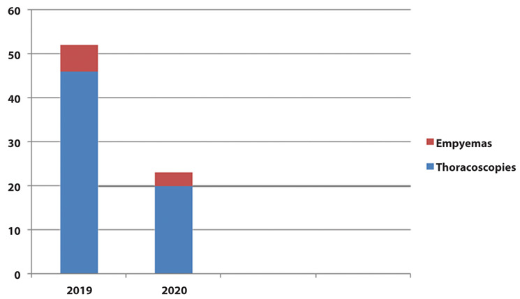
Figure 1.

Total number of medical thoracoscopies carried out by the unit of interventional pulmonology in 2019 versus those of 2020; empyemas treated through medical thoracoscopies in 2019 were 6 out of a total of 52, in 2020 2 out of a total of 23.
In clinical records of Thoracic surgery for the year 2020 an increase of 100% of surgical operations for the treatment of pleural empyema is highlighted; going from an extremely stabile figure of 6–8 interventions per year during the previous 5 years to 16 interventions in 2020.
Considering the average age and the comorbidities, being stabile enough throughout the years, the only factor which can possibly explain this figure is the Covid pandemic. It is interesting to actually ask in which way the pandemic has influenced this figure. Objectively the number of patients operated with current or previous positivity to Sars-cov2 is very limited, therefore the pandemic has probably influenced this, hence hindering an optimal treatment of the bacterial pneumonia both in community acquired pneumonia as well as in the hospital.
Firstly, the Sars-Cov 2 pandemic has made general medical practices impossible as an objective wave of fear has influenced hospitalization, which has very likely increased the number of non-covid pneumonia being treated at home exclusively with oral antibiotic therapy, therefore the number of patients accessing the emergency room has increased at the hospital with already diagnosed pleural empyema franco.
Secondly, the new organization has conditioned the treatment of non-Covid related pulmonary pathologies and carrying out procedures of interventional pulmonology: during the period from 21st February to 31st December 2020 pleural empyemas treated through medical thoracoscopies by the Pulmonologist have gone from a total of 6 in 2019 to a total of 2 in the same period of 2020.
Therefore in our opinion, such a net increase of pleural empyema cases treated surgically in the pandemic area presents a dual origin: on one side the reduction of practices both hospitalization as well as interventional pulmonology and on the other side the delayed diagnosis of patients coming from their domicile.
In our case studies, in fact, we have seen double cases of empyema treated in VATS surgery as can be seen from Table 1: among sixteen cases only one needed thoracotomy conversion for the presence of important pleural adhesion synekies. Mortality at 30 days in patients was 0%.
Table 1.
Epidemiology and number of patients affected by empyemas at an advanced stage treated surgically from the period 21st February-31st December 2019 compared to the same period in 2020: the increase is 100%
| Surgically treated pulmonary empyemas | 2019 | 2020 |
|---|---|---|
| N(=) | 8 | 16 |
| Patient sex | M=5; F= 3 | M=11; F=5 |
| Patients median age (y) | 62.4 y | 64.9 y |
| Patients co-morbidities | COPD (=1) Diabetes (=1) Tuberculosis (=1) Lung cancer (=1) No comorbidities (=4) |
COPD (=2) Diabetes (=2) Chronic kidney disease (=1) Lymphoma (=1) Child C liver disease (=1) No comorbidities (=10) |
| Surgery | VATS (=8) Open (=0) |
VATS (=15) Open (=1) |
| Covid status (swab test) | Pos (=0) Neg (=8) |
Pos (=3) Neg (=13) |
| Follow up (30-days) | Alive (=7) Death (=1) |
Alive (16) Death (=0) |
| Stage | II (=8) III (=0) |
II (=9) III (=7) |
| Main isolated pathogenes | Pneumococcus (=1) Staphyloccoccus spp (=4) |
VATS: video-assisted thoracic surgery. COPD: chronic obstructive pulmonary disease.
In conclusion among the many “collateral” damages of the epidemiological wave of the infection with Sars Cov-2, we have been able to observe in our Hospital, for the reasons set out above, also an increase of pulmonary empyemas diagnosed at an advanced stage, that is in a purulent and organized fibrino-phase that have requested surgical treatment as a first therapeutical practice.
Conflicts of interest:
Each author declares that he or she has no commercial associations (e.g. consultancies, stock ownership, equity interest, patent/licensing arrangement etc.) that might pose a conflict of interest in connection with the submitted article.
References
- Dean NC, Griffith PP, Sorensen J, McCauley L, Jones BE, Lee YC. Pleural effusions at first emergency department encounter predict worse clinical outcomes in pneumonia patients. Chest. 2016;149(6):1509–15. doi: 10.1016/j.chest.2015.12.027. [DOI] [PMC free article] [PubMed] [Google Scholar]
- Hamm H, Light RW. Parapneumonic effusion and empyema. Eur Respir J. 2016;10(5):1150–1156. doi: 10.1183/09031936.97.10051150. [DOI] [PubMed] [Google Scholar]
- Shen K.R, Bribriesco A, Crabtree T, et al. The American Association for Thoracic Surgery consensus guidelines for the management of empyema. The Journal of Thoracic and Cardiovascular Surgery. 2017;153(6):129–146. doi: 10.1016/j.jtcvs.2017.01.030. [DOI] [PubMed] [Google Scholar]
- Ala Eldin H. Ahmed and Tariq E. Yacoub. Empyema Thoracis. Clin Med Insights Circ Respir Pulm Med. 2010;4:1–8. doi: 10.4137/ccrpm.s5066. [DOI] [PMC free article] [PubMed] [Google Scholar]
- Wait MA, Sharma S, Hohn J, Dal Nogare A. A randomized trial of empyema therapy. Chest. 1997;111:1548–1551. doi: 10.1378/chest.111.6.1548. [DOI] [PubMed] [Google Scholar]
- Porcel JM, Valencia H, Bielsa S. Factors influencing pleural drainage in parapneumonic effusions. Rev Clin Esp. 2016;216:361–366. doi: 10.1016/j.rce.2016.04.004. [DOI] [PubMed] [Google Scholar]
- Maskell NA, Batt S, Hedley EL, Davies CW, Gillespie SH, Davies RJ. The bacteriology of pleural infection by genetic and standard methods and its mortality significance. Am J Respir Crit Care Med. 2006;174:817–823. doi: 10.1164/rccm.200601-074OC. [DOI] [PubMed] [Google Scholar]


