

Abstract
Foot ulceration is one of the biggest complications experienced by type 2 diabetes patients. The severity and prevention of new wounds can be overcome through early detection interventions. This systematic review aims to explain and provide a comparison of various interventions that have been developed to prevent the occurrence of Diabetes Foot Ulcers (DFU). We searched Scopus, Science Direct, PubMed, CINAHL, SAGE, and ProQuest for English, experimental studies, published between 2016-2021 that tested early detection for preventing diabetic foot ulcers in diabetic patients. The Joanna Briggs Institute guidelines were used to assess eligibility, and PRISMA quality and a checklist to guide this review. 25 studies were obtained that matched the specified inclusion criteria. The entire article has an experimental study design. Majority of respondents were type 2 diabetes patients who have not experienced ulceration. Based on the results of the review, there were 3 main types of interventions used in the early detection of DFU. The types of intervention used are 1) conventional intervention/physical assessment, 2) 3D thermal camera assessment system, and 3) DFU screening instrument. The three types of interventions have advantages and disadvantages, so their use needs to be adjusted to the conditions and needs of the patient. the development of DFU risk early detection intervention needs to be developed. Integration with modern technology can also be done to increase the accuracy of the results and the ease of examination procedures.
Significance for public health.
This systematic review aims to explain various digital and conventional-based early detection interventions along with their advantages and disadvantages that can be used to assess risk factors for DFU in DM patients. It is because several existing studies only discuss one model of early detection of DFU in DM patients, however, studies that describe various interventions that can be carried out for early detection in DM patients have not been found. By knowing several DFU prevention interventions, it is expected to increase the independence of patients and families in preventing complications such as diabetic foot.
Key words: Foot ulceration, foot disorder, application, early detection, diabetes type 2
Introduction
One of the serious complications that can occur in people with type 2 diabetes mellitus (DM) is diabetic peripheral neuropathy or often known as diabetic foot ulcer (DFU). The frequency of occurrence of DFU in patients with type 2 diabetes is quite high, especially in individuals with type 2 diabetes for more than 10 years, and 60% experience disability even to the point of leg amputation.1 Complications of DFU can result in increased treatment costs, increased disability rates, decreased quality of life, and also increased risk of death. DFU can be prevented through 5 main elements, namely 1) identification of the feet at risk, 2) regular examinations, 3) education of patients, families and health workers, 4) ensuring the use of appropriate footwear and 5) treating risk factors.2 The risk of DFU can be prevented if it is detected early, but often does not get enough attention from care givers.
Assessment of risk factors for DFU needs to be done as an early prevention of complications. Some of the problems that occur are that early detection of risk for DFU can only be done by health workers, while patients and families do not yet have the ability to independently assess these risk factors.3 This is because the media or instruments used for early detection are only limited to use by medical personnel. In addition, treatment so far has focused more on other body systems that are considered more important, such as the heart, kidneys, brain and eyes. This is reinforced by previous studies which revealed that complications of DFU occur due to delays in early detection and poor case management. 4
DFU can be avoided or delayed if treated adequately at an early stage. DFU risk factor assessment carried out by health workers, especially doctors through analysis of blood circulation, plantar foot pressure, and foot neuropathy. In addition, specialists usually assess the vascular status of the lower extremities using Doppler ultrasound. This is considered to result in an accurate analysis of the conditions and risk factors for DFU. However, patients are forced to visit the doctor often for diabetic foot examinations, which are considered to be disruptive to activities and expensive costs. In addition, patients do not have the ability to carry out independent examinations due to low patient knowledge of the disease, and the absence of medical equipment, therefore currently the development of technology-based DFU early detection instruments that can be carried out independently by patients is starting to be developed.5 The Health Belief Model (HBM) is the most widely used theory to explain health behavior. In relation to the behavior of early detection of diabetic foot in type 2 DM patients, a person will take health actions such as early detection if he has confidence in vulnerability, seriousness of a disease, benefits, obstacles in taking action and taking into account self-efficacy or self-confidence in carrying out an action plus the existence of cues to action (cues to action) both from within and or outside the individual.6,7
Nowadays, technological advances have developed rapidly, accompanied by the increasing use of the internet and smartphones as a means of communication in everyday life. In addition, the current use of smartphones is mainly based on Android, which is also used as a means of accessing health information in the form of telemedicine or telenursing. The use of technology in the health sector also plays a role in several interventions for early detection of DFU in diabetic patients. The combination of technology in the form of cameras and computers to detect the presence of neuropathic disorders in DM patients has been widely developed. In addition, direct intervention using conventional tools in the form of footwear equipped with sensors,8 and pin-prick tests are also used to perform early detection of DFU.9 Several existing studies only discuss one model of early detection of DFU in DM patients, however, studies that describe various interventions that can be carried out for early detection in DM patients have not been found. Therefore, this systematic review aims to explain various digital and conventional-based early detection interventions along with their advantages and disadvantages that can be used to assess risk factors for DFU in DM patients.
Design and methods
Information sources and search strategy
The literature search was carried out in November 2020- January 2021. The data used in this study were secondary data obtained from the results of research conducted by previous researchers and not from direct observation. Sources of secondary data obtained in the form of journal articles of national and international repute with a predetermined theme. The literature used was obtained from the Scopus database, ProQuest CINAHL, Science Direct, Nature, PubMed, and SAGE. Keyword search using the term MeSH. The specific keywords used to search the article were foot disorder OR foot ulceration, AND early detection, AND Diabetes type 2.
Study eligibility and selection criteria
In this section, reviewers select the articles that have been obtained based on predetermined keywords. Previously, reviewers made the PICOS format as an indicator for the assessment of the suitability of the article. The PICOS criteria that are made can be seen in Table 1. Based on the results of the literature search, 1,076 articles that match keywords with article details were obtained from the Scopus database (n=166), Pro Quest (n=204), Science Direct (n=183), PubMed (n=170), SAGE (n=165) and CINAHL (n=188). From a total of 1076 articles found, article duplication checks were then carried out and 783 of the same articles were found so that they were excluded from the search results and 293 articles were left. Reviewers then conducted screening based on title (n=293), abstract (n=94) and full text (n=25) adjusted to the theme. Based on the eligibility screening carried out against the inclusion and exclusion criteria, 25 articles were found that could be used in this review. The results of the study article selection can be illustrated in the PRISMA (Figure 1).
Results
Study characteristic
Based on the results of the literature search that has been carried out, all of the articles are experimental studies. Several research designs used were quasi-experimental, pilot study and mixed method study. The research came from various countries, and most of them came from the European continent. This systematic review aims to determine several interventions and procedures for early detection of DFU in diabetic patients. Based on the search results, three intervention themes were found that were mostly carried out, namely 1) conventional intervention/direct physical examination, 2) 3D thermal camera assessment system and 3) development of DFU risk factor examination instruments.
Respondents’ characteristic
Respondents who participated in the study were patients with diabetes mellitus, with the majority being type 2 diabetes. The age of the respondents varied, with the youngest age being 18 years old to the oldest being 80 years old. The gender of the respondents also varies, but in some studies the majority of the respondents are male. The duration of suffering from diabetes is also one of the characteristics of the respondents required. DM patients who participated had a history of suffering from DM for more than 10 years, up to those who had just been diagnosed for less than 6 months but already had symptoms, especially neuropathic disorders. Some respondents in the study had experienced diabetic ulcers and some had not yet experienced ulceration but began to feel the signs of DFU symptoms. Several studies also included respondents who were healthy and did not have a history of DM or other neuropathic symptoms as a comparison group, to determine the effectiveness of the interventions carried out on research subjects.
Table 1.
PICOS criteria.
| Criteria | Inclusion | Exclusion |
|---|---|---|
| Population | Studies comprised DM patient with or without DFU | Neuropathy patient but not diabetes |
| Intervention | Early detection/intervention for DFU | Not discuss DFU intervention |
| Comparison | No comparator | - |
| Outcome | Detection/intervention for DFU | Neuropathy detection in general |
| Study design and publication type | Quasi-experimental, randomized control and trial, mixed methods | Literature review, systematic review, narrative review |
| Publication year | Post 2016 | Pre 2016 |
| Language | English | Language other than English |
Diabetes foot ulcer (DFU) early detection intervention
Conventional intervention/physical assessment
The first intervention theme found from the results of the review conducted was conventional intervention or direct physical examination of the patient. In this case, the patient will be asked to use some of the tools that have been prepared. The first tool used in this conventional model is footwear or shoe soles that have been connected to a computer system. This tool will analyze risk factors for DFU based on recorded foot pressure. In this intervention, research subjects were asked to walk using footwear that was connected to the Data Acquisition System (DAQ). Furthermore, the measurement of the pressure and voltage obtained will be recorded and the results will be displayed on the LabView system panel. The panel is equipped with an LED that will light up when the pressure is more than 62kpa. In addition, this LabVIEW panel will also identify the metabolic status of the respondents, so they can find out their blood sugar levels, which will be compared at the end of intervention.8
Another conventional intervention used to examine risk factors for DFU is to utilize the vibrations generated by a tuning fork placed on the medial edge of the hallux. Analysis of the perception of vibration and noise generated by the device at a certain percentage limit will be categorized and can be used as an interpretation of the patient’s condition. Patients who are at risk of DFU will feel a tuning fork vibration that is relatively shorter than normal people can feel.10,11 In addition, examination of the limbs is also carried out by evaluating the function of the sudomotor or sweat glands in humans. Evaluation of sudomotor function was measured with the Sudoscan medical device, which consists of a set of two electrodes for the feet and hands connected to a computer. The average test duration was 3 min, in which 4 combinations of 15 different lowvoltage stimuli were applied. Patients do not require any preparation, and only need to place their palms and soles on the stainlesssteel electrodes and remain for the duration of the test. The device measures the conductance produced in response to an electrical stimulus, expressed in Siemen micros for the right and left sides. It is a method based on stimulation of sweat glands by low level voltage, which allows evidence of sweat dysfunction that is not detectable under physiological conditions. No subject preparation is required for this test. The performance and accuracy of this method have been evaluated in various clinical studies.5
The last type of conventional intervention found from the results of the review was an examination using a pinprick test. Pin pricks are inserted into pain receptors, namely the Meissner and Pacini nerves in the legs. Respondents entered one by one in the room, then asked to lie down in a supine position and without using footwear. After that the respondent will be explained the procedure to be carried out and introduced to the tools used. Then, a stimulus test was carried out using 2 objects that had a sharp and blunt surface to check whether the respondent had the appropriate pain stimulus. If the respondent feels a stab, the respondent is asked to provide a pain scale that is felt from a score of 0-10. Pinprick stimulation was repeated 2 times on 3 parts of the sole of the foot, with a stimulation interval of 5-10 sec. The area is the plantar arch and the skinfolds of the second and third toe innervated by the terminal branches of the tibial nerve. While the area of the sole of the foot that is thickened (metatarsal) is not stimulated. The stimulation course was run top down from hand to feet and was repeated about three minutes later in reversed order. All stimulations were performed by the same examiner.9
Figure 1.
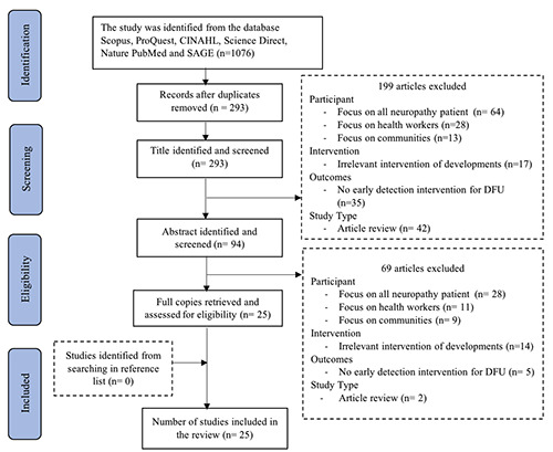
Preferred reporting items for systematic reviews and meta-analysis (PRISMA).
3D thermal camera assessment system
The second intervention theme that was obtained was the use of cameras equipped with a 3D thermal system which involved the storage and transfer of heat from the patient to the system.12 This thermal imaging system has a simulated temperature gradient of more than 2.2°C, which represents a temperature difference that could indicate a possible ulcer development.13 The obtained images are processed and segmented using basic image processing techniques. The analysis and interpretation were carried out using two techniques, namely the Otsu thresholding technique and the Point-to-Point mean difference technique.13,14 The procedure was carried out by means of the wound on the diabetic foot in the photo using a smartphone which was then connected to Wi-Fi and a router to the wound management system. on laptops. The majority of DFUs are found on the soles of the feet of DM patients, therefore this examination is also equipped with an acrylic box which is used to place the cellphone when taking pictures on the soles of the feet.15
In addition to using a camera on a smartphone, inspection using 3D thermal also uses a DSLR camera for clearer resolution and image quality.16 The procedure is almost the same, namely the legs are photographed using a Nikon D3300, and photographed in a close-up position at a distance of 30-40cm. The camera used to take pictures is the same camera and lens. The use of flash is not allowed, and only relies on light from the room. After that, the image will be interpreted by health workers, namely doctors based on the Region of Interest (ROI) of the ulcer. From this interpretation, several types of foot patches or models of wounds that occur on the feet of DM patients will be produced.17
DFU screening instrument
The last theme obtained from the results of the review is the development of the DFU examination instrument in the form of a questionnaire. The reason for developing this instrument was to complete components that did not exist in the previous instrument. In addition, several developments were also carried out to adapt the increasingly modern developments in technology and diagnostic methods of DFU.18 The International Working Group on the Diabetic Foot (IWGDF) publishes guidelines for the prevention and management of foot problems in diabetes. This guide aims at better disease management for diabetic patients, especially in multidisciplinary care settings. In addition, there is a need for a valid and appropriate instrument to assess the risk of DFU in clinical practice. The developed instrument has been tested for validity using classical and modern test theory among diabetic patients from various regions of China. This screening instrument also examines the risk factors for DFU and the novelties added are i) the existence of a process of developing and validating the instrument; ii) assessment dimensions; iii) the rationality of the assessment weights; iv) different items and measurement methods; and v) more developed use objectives (multidisciplinary, quantitative, predictive assessment).19,20
The development of quite interesting and modern instruments was also found from the results of the review. The development procedure carried out is to collect images of diabetic wounds from several open-source databases. Then the color segmentation is carried out using the Particle Swarm Optimization (PSO) technique. Region Of Interest (ROI) was extracted from the segmented image, different texture and color-based features were extracted and classified into three types of diabetic wound images using two classifiers namely Navie Bayes and Hoeffeding tree classifier. The imaging results can then be used as a guideline for the percentage of DFU in DM patients, so that risk factor assessment can be done more quickly and easily.2.
Subsequent development of the instrument is based on the theory that DFU can cause neurological disorders at the motor level (deformity), sensitivity level (burning, tingling, cramping and the like) and at the autonomic level (dryness, hyperkeratosis, cracking), and that damage to nerve level is usually accompanied by damage at the vascular level. Based on this, different techniques were chosen to identify risk factors for DFU which were eventually organized into 29 assessment items.22,23
Discussion
Conventional intervention/physical assessment
Early detection of foot ulceration in DM patients by conventional methods or direct physical examination is the most widely used in various clinical practices. The detection method that utilizes the use of footwear is considered good for identifying ulcerations, because there is a walking practice carried out by the patient. The gait owned by DM patients is not only able to measure the pressure in the legs, but can also indicate an abnormality which is predicted to be one of the signs of neuropathic disorders.8 Meanwhile, the use of the pinprick test as a method of early diagnosis of DFU is also considered good to do, because the procedure is simple and can identify the risk of DFU well.9 Overall, conventional interventions have several advantages and disadvantages. Based on the level of accuracy, this intervention has the highest level of accuracy when compared to other types of examinations. However, this conventional intervention also has several weaknesses. The first weakness is that there are several medical devices that must be prepared before carrying out the examination, this method cannot be carried out independently by the patient due to limited tools and also the inability to operate the examination procedure. 24
The second weakness is, the physical examination of the patient must be guaranteed safety, where the patient will not feel pain and discomfort when undergoing the examination procedure performed. If it is related to the current Covid-19 pandemic condition, this conventional examination procedure will be highly avoided because DM patients are one of the groups that are vulnerable to contracting the virus and are at high risk of getting worse if infected.25 On the other hand, many physical interventions are chosen because the results of the examination are more accurate and carried out by expert health workers, so that the risks that occur during the intervention can be minimized and the costs required for one examination are more affordable.
3D thermal camera assessment system
Measurement of foot ulcers with accurate instruments will help provide an assessment of the healing status of the wound. This is important to accelerate healing and to reduce the risk of lower extremity amputation for type 2 diabetes patients.16 Based on the second type of intervention, namely the use of a thermal camera, it was found that efficient image processing algorithms and costeffective imaging devices can meet the clinical needs of DFU identification. Wound assessment is implemented efficiently on a smartphone system, which allows patients to self-manage their foot ulcers and provides an opportunity for health workers, namely doctors, to carry out evaluations.13 The use of camera technology for early detection of the risk of ulceration on the feet of DM patients is starting to be widely used. This technology makes inspection easier, because it can be done in any place, as long as it is connected to internet and Wi-Fi facilities.14 The examination procedure is simple and judged not to hurt the patient is also a plus for this type of intervention. But in other cases, early detection by using a camera is considered to be quite expensive, because the type of camera used can only be installed on certain types of smartphones.
In addition, another weakness is the level of accuracy of the examination results is also not too high, it depends on the results of the photos obtained. The influence of room light, and skill in taking pictures can affect the results of the photos obtained. To overcome this, some examination tools are equipped with the use of shooting boxes, which function to ensure consistent lighting, image spacing and image quality, so that the wound assessment carried out will be effective.12 The results of the clinical assessment of the healing score algorithm is relevant to the assessment carried out by medical personnel. This shows that the use of thermal camera media is effective for detecting DFU in DM patients. More validation data is needed to evaluate the algorithm further. Inevitably, the physician’s determination of healing scores is influenced by experience and training, similar to what has been observed in the determination of wound area.15 The use of the same camera in the DFU examination is recommended, so that the results obtained are accurate and heterogeneous. In addition, the experiment took pictures on different skin textures which were classified into 3 classes of facial skin patches, namely normal, freckles and wrinkles. also need to be done before the tool is used to identify risk factors for DFU.17 Further testing and validation of the system must be carried out under a clinical environment, which is not possible at this stage due to the stringent regulations imposed. In addition, this intervention can also be developed into other possible applications such as wound healing and trauma monitoring.26
DFU screening instrument
The development of the DFU screening instrument was carried out to improve the assessment according to the standards carried out by nurses in clinical practice settings. All of the instruments developed have passed the validity and reliability requirements.19 In addition to improving the assessment according to the protocol, the development of the instrument also aims to reduce the subjectivity in interpreting the results of the diabetic foot assessment. One of the instruments developed, NeuDiaCan, allows the examination to be completed with an objective score that will help stratify the risk of diabetic foot and can be combined with standard nursing interventions. Thus, the use of this instrument can improve the skills of using technology in nurses and improve early detection of patients who are at risk of developing ulcers that can lead to amputation.22
Although it has been through various tests and has a good cutoff value, lower threshold limits should be used, especially for those with a history of ulceration. This was done for the purpose of protecting more patients, and minimizing the number of new and recurring DFUs. In addition, estimates and comparisons between medical costs used for preventive interventions, and costs for treatment when DFU occurs, must also be taken into account.20 The use of an instrument in the form of a questionnaire in the early detection of DFU is an effort that does not provide treatment to patients directly, therefore, sometimes the results of screening through this questionnaire are not as accurate as other types of interventions. Some of the main factors are the patient’s psychological condition that can affect the filling of answers, therefore it is necessary to ensure that the patient is ready to undergo screening, before the examination is carried out.23
In addition, the limited number of items in a dimension result in less-than-optimal internal consistency. Apart from the limitations, the development of the DFU early detection instrument also has its advantages. First, the developed instrument has been validated using classical test theory combined with modern test theory, so that item properties are ensured to have gone through careful examination. Furthermore, based on the estimated value obtained, the use of this screening instrument can also be integrated into the patient’s smartphone, so that patients and families can screen independently at home.18
Conclusions
Based on the systematic review that has been carried out, it is known that there are 3 main types of early detection interventions that are used to assess the risk of DFU in DM patients. The three interventions have strengths and weaknesses in several aspects. The use of the type of intervention chosen must be adjusted to several things, namely: 1) the condition of the patient to be examined, both physically and psychologically, 2) the availability of adequate resources, such as health workers, costs, time, and available tools, 3) desired level of effectiveness and severity of symptoms felt by the patient. This will help to increase patient comfort during the examination. In addition, further development to correct some of the shortcomings of existing instruments also needs to be done.
Acknowledgements
The authors thank the local authorities (Faculty of Nursing, Universitas Airlangga) for their support and contributes in this study.
References
- 1.Shabibi P, Abedzadeh Zavareh MS, Sayehmiri K, et al. Effect of educational intervention based on the Health Belief Model on promoting self-care behaviors of type-2 diabetes patients. Electron Physician 2017;9:5960–8. [DOI] [PMC free article] [PubMed] [Google Scholar]
- 2.Schaper NC, van Netten JJ, Apelqvist J, et al. Practical guidelines on the prevention and management of diabetic foot disease (IWGDF 2019 update). Diabetes Metab Res Rev 2020;36:1-10. [DOI] [PubMed] [Google Scholar]
- 3.Banik PC, Barua L, Moniruzzaman M, et al. Risk of diabetic foot ulcer and its associated factors among Bangladeshi subjects: A multicentric cross-sectional study. BMJ Open 2020;10:e034058. [DOI] [PMC free article] [PubMed] [Google Scholar]
- 4.Tolossa T, Mengist B, Mulisa D, et al. Prevalence and associated factors of foot ulcer among diabetic patients in Ethiopia: a systematic review and meta-analysis. BMC Public Health 2020;20:41. [DOI] [PMC free article] [PubMed] [Google Scholar]
- 5.Carbajal-Ramírez A, Hernández-Domínguez JA, Molina-Ayala MA, et al. Early identification of peripheral neuropathy based on sudomotor dysfunction in Mexican patients with type 2 diabetes. BMC Neurol 2019;19:1–6. [DOI] [PMC free article] [PubMed] [Google Scholar]
- 6.Hsieh YL, Lee FH, Chen CL, et al. Factors influencing intention to receive examination of diabetes complications. Asian Nurs Res 2016;10: 289–94. [DOI] [PubMed] [Google Scholar]
- 7.Ghobadi A, Sarbarzeh PA, Jalilian M, et al. Evaluation of factors affecting the severity of diabetic foot ulcer in patients with diabetes referred to a diabetes centre in Kermanshah. Diabetes Metab Syndr Obes Targets Ther 2020;13:693–703. [DOI] [PMC free article] [PubMed] [Google Scholar]
- 8.Damanhuri NS, Othman NA, Zaidi WFAW, Abdullah S. A development of plantar pressure sensor for foot ulcer detection in diabetic neuropathy individuals–A pilot study. J Phys Conf Ser 2020;1535:12019. [Google Scholar]
- 9.Chantelau E-A. A novel diagnostic test for end-stage sensory failure associated with diabetic foot ulceration: Proof-of-principle study. J Diabetes Sci Technol 2021;15:622-9. [DOI] [PMC free article] [PubMed] [Google Scholar]
- 10.Azzopardi K, Gatt A, Chockalingam N, Formosa C. Hidden dangers revealed by misdiagnosed diabetic neuropathy: a comparison of simple clinical tests for the screening of vibration perception threshold at primary care level. Prim Care Diabetes 2018;12:111–5. [DOI] [PubMed] [Google Scholar]
- 11.Zwaferink JBJ, Hijmans JM, Schrijver CM, et al. Mechanical noise improves the vibration perception threshold of the foot in people with diabetic neuropathy. J Diabetes Sci Technol 2018;14:16–21. [DOI] [PMC free article] [PubMed] [Google Scholar]
- 12.van Doremalen RFM, van Netten JJ, van Baal JG, et al. Infrared 3D thermography for inflammation detection in diabetic foot disease: A proof of concept. J Diabetes Sci Technol 2019;14:46–54. [DOI] [PMC free article] [PubMed] [Google Scholar]
- 13.Fraiwan L, AlKhodari M, Ninan J, et al. Diabetic foot ulcer mobile detection system using smart phone thermal camera: a feasibility study. Biomed Eng Online 2017;16:1-19. [DOI] [PMC free article] [PubMed] [Google Scholar]
- 14.Fraiwan L, Ninan J, Al-Khodari M. Mobile application for ulcer detection. Open Biomed Eng J 2018;12:16. [DOI] [PMC free article] [PubMed] [Google Scholar]
- 15.Wang L, Pedersen PC, Strong DM, et al. An automatic assessment system of diabetic foot ulcers based on wound area determination, color segmentation, and healing score evaluation. J Diabetes Sci Technol 2016;10:421-8. [DOI] [PMC free article] [PubMed] [Google Scholar]
- 16.Cassidy B, Reeves ND, Pappachan JM, et al. The DFUC 2020 dataset: Analysis towards diabetic foot ulcer detection. touchREV Endocrinol 2021;17:5-11. [DOI] [PMC free article] [PubMed] [Google Scholar]
- 17.Goyal M, Reeves ND, Davison AK, et al. Dfunet: Convolutional neural networks for diabetic foot ulcer classification. IEEE Trans Emerg Top Comput Intell 2018;4:728-39. [Google Scholar]
- 18.Zhou Q, Peng M, Zhou L, et al. Development and validation of a brief diabetic foot ulceration risk checklist among diabetic patients: A multicenter longitudinal study in China. Sci Rep 2018;8:1–9. [DOI] [PMC free article] [PubMed] [Google Scholar]
- 19.Baker N, Al-Muzaini A. A user’s guide to skin and nail conditions in diabetes. Diabet Foot 2018;4:31-9. [Google Scholar]
- 20.Akturan S, Kaya ÇA, Ünalan PC, Akman M. The effect of the BATHE interview technique on the empowerment of diabetic patients in primary care: a cluster randomised controlled study. Prim Care Diabetes 2017;11:154–61. [DOI] [PubMed] [Google Scholar]
- 21.Babu KS, Sabut S, Nithya DK. Efficient detection and classification of diabetic foot ulcer tissue using PSO technique. Int J Eng Technol 2018;7:1006-10. [Google Scholar]
- 22.Martínez-Alberto CE, Brito-Brito PR, Fernández-Gutiérrez DA, et al. Evaluation of the risk of diabetic peripheral neuropathy: Design and validation of the NeuDiaCan nursing screening procedure. Enfer Clin (English Ed) 2020;30:89-98. [DOI] [PubMed] [Google Scholar]
- 23.Binns-Hall O, Selvarajah D, Sanger D, et al. One-stop microvascular screening service: an effective model for the early detection of diabetic peripheral neuropathy and the high-risk foot. Diabet Med 2018;35:887–94. [DOI] [PMC free article] [PubMed] [Google Scholar]
- 24.Peterson M, Pingel R, Rolandsson O, Dahlin LB. Vibrotactile perception on the sole of the foot in an older group of people with normal glucose tolerance and type 2 diabetes. SAGE Open Med 2020;8:2050312120931640. [DOI] [PMC free article] [PubMed] [Google Scholar]
- 25.Santos TRM, Melo JV, Leite NC, Salles GF, Cardoso CRL. Usefulness of the vibration perception thresholds measurement as a diagnostic method for diabetic peripheral neuropathy: Results from the Rio de Janeiro type 2 diabetes cohort study. J Diabetes Complicat 2018;32:770–6. [DOI] [PubMed] [Google Scholar]
- 26.Goyal M, Reeves ND, Rajbhandari S, Yap MH. Robust methods for real-time diabetic foot ulcer detection and localization on mobile devices. IEEE J Biomed Health Inform 2018;23:1730–41. [DOI] [PubMed] [Google Scholar]