Abstract
Objectives:
To examine (1) the laterality of asymmetry in movements of the right and left corners of the mouth in space during voluntary smile and (2) the laterality of asymmetry in relation to the difference between the right and left hemiface size and the handedness.
Materials and Methods:
Participants were 155 volunteer Japanese female adults. They were categorized into the symmetric group (n = 120) and the right-side hemiface dominant group (n = 26) according to the hemiface size. In addition, the symmetric group was categorized into the right-handed group (n = 98) and the left-handed group (n = 22) according to the Edinburgh Handedness Inventory. Position vectors of the right and left corners of the mouth were obtained from the three-dimensional facial images for the rest, the maximal lip corner retraction, and the portrait smile. The displacements of the right and left corners of the mouth for each expression and the proportions of the subjects with the right- and left-sided laterality were compared.
Results:
The left corner of the mouth showed significantly greater displacement (P < .01) than the right in the symmetric group for the portrait smile. The left-sided laterality was found regardless of the handedness.
Conclusions:
Displacements of the right and left corners of the mouth during voluntary smile were asymmetric, and the left-sided laterality was found. Also, the laterality of the facedness differed in relation to the hemiface size, but was not related to the handedness.
Keywords: Facial expression, Smile, Asymmetry, Three-dimensional, Measurement, Human
INTRODUCTION
Facial expressions are classified as voluntary and spontaneous.1 Voluntary smile can be a learned greeting or a signal of appeasement2 and is known as an expression that people can easily adopt with high reproducibility.3,4 Dynamic analysis of the perioral area during voluntary smile has been applied as a means of objective diagnostic assessment of facial function in orthodontics.5–8
The term “facedness” indicates greater muscular control on one side of the face relative to the other.9 In terms of the movements of the corners of the mouth during voluntary smile, most previous studies7,10–13 have reported the left-sided facedness with the laterality (preference in using one side of the body over the other). But other studies8,14 found neither the facedness14 nor the laterality.8
Although the difference in size between the right and left hemifaces has been claimed as a cause of the facedness during voluntary smile,15 the study by Sackeim and Gur16 did not find any significant correlation between the hemiface width and the hemiface mobility or measures of the hemiface intensity. Moreover, the relationship between the facedness and the handedness is also controversial.9,10
The purpose of the present study was to investigate three-dimensionally if there is laterality of asymmetry in displacements of the right and left corners of the mouth during voluntary smile in humans, and if so, to investigate if the laterality of asymmetry observed is explained by the difference between the right and left hemiface size and the handedness.
MATERIALS AND METHODS
Participants were selected from 288 volunteers, including students and faculty of the university. One hundred fifty-five participants (mean age: 28 years 5 months; range: 18 years 0 months–53 years 6 months) met the following selection criteria: female, over 18 years old, no congenital facial deformities including cleft lip and palate, no facial paralysis, no history of noticeable scars or skin disease in the neck and dentofacial regions, no history of psychiatric disorder, no subjectively or objectively discernible jaw dysfunction, body mass index ranging from 18.5 to 25.0, Aesthetic Component of the Index of Orthodontic Treatment Need ranging from 1 to 4,17 overbite ranging between 1.0 mm and 5.0 mm; overjet ranging from 0.0 mm to 7.0 mm,18 and the soft tissue facial convexity angle ranging between −1.4 degrees and 21.6 degrees.19 The range of values for the overbite, overjet, and the facial convexity were determined on the basis of the normative mean ±2 standard deviations of the Japanese female adults.
Subjects were asked to sit in a semidark, air-conditioned room on a fixed chair, with a natural head position. They were then asked to perform tasks as provided in Table 1. The subjects were instructed vocally for each task and asked to maintain the expressions for about 5 seconds. After several rehearsals, each expression was recorded once with a three-dimensional image capturing device (®Danae100 NEC Engineering Co, Tokyo, Japan). Recordings of each type of expression were made with a resting interval of about 30 seconds between the expressions. The experimenter operated the system from a position out of the subject's view. The device contained two fixed CCD cameras and employed a shutter speed of 0.6 seconds for a photographic single frame and a focal length of 600 mm. The dimensional accuracy of the system was 0.12 mm.
Table 1.
Definitions of the Tasks

In a pilot study, five repeated data recordings were made with an interval of 30 seconds for the rest, the maximal lip corner retraction, and the portrait smile for 20 randomly selected subjects. The mean deviations of five measures for intrasubject intercommissure distances for aforementioned tasks were 0.16 mm, 0.39 mm, and 0.32 mm, respectively. The mean deviations for the two measures which were recorded with an interval of longer than 2 weeks for each of the three experimental tasks for the same subjects were 0.16 mm, 0.39 mm, and 0.32 mm, respectively. In addition, the movements of the corners of the mouth became smaller as the tasks were repeated because of fatigue. Thus, we considered the stability and reproducibility of the measurements were sufficient to detect statistical significances for intergroup comparisons, and we employed a single recording for each task.
Data Analysis
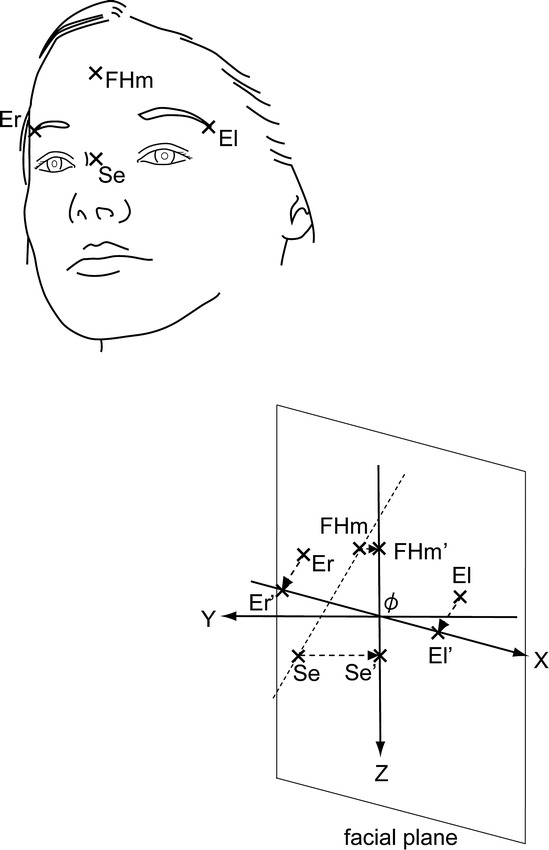
For each subject, the least squares mean plane was created as the facial plane from the positions of four reference feature points on the facial image at rest (Figure 1), and we developed a facial coordinate system (Figure 1). The definitions of the points used for the development of the coordinate system and the coordinate system are given in Table 2.
Figure 1.

Reference feature points and facial coordinate system employed in the present study. ϕ indicates the origin.
Table 2.
Definitions of the Measuring Points and a Facial Coordinate System

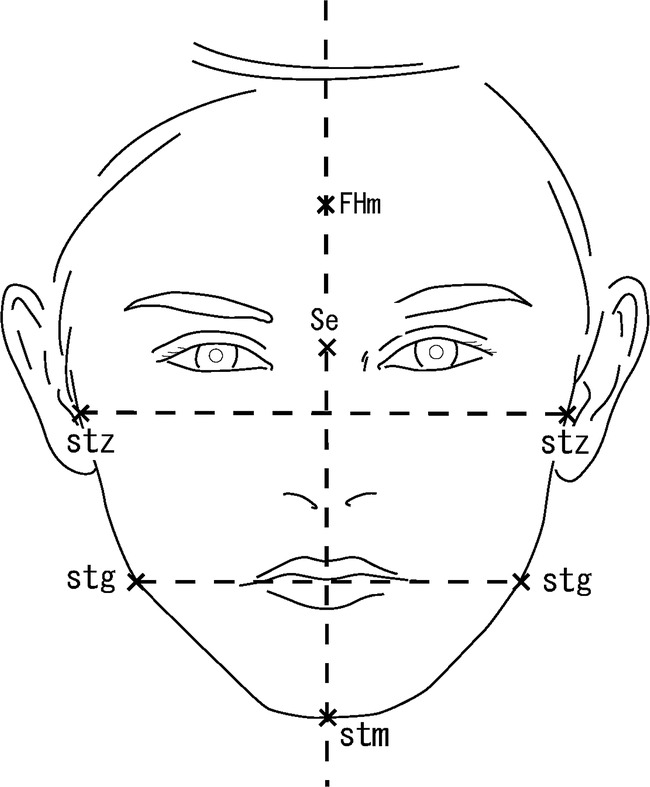
According to the right and left hemiface size at rest, the samples were categorized into three subgroups (Figure 2): the symmetric group (n = 120), in which the soft tissue menton deviated less than 4.0 mm from the facial midline (connecting the FHm and Se22) and differences in the distances between the right- and left-side soft tissue zygoma and soft tissue gonion from the facial midline were less than 2.0 mm23; the right-side hemiface dominant group (n = 26), in which the soft tissue menton deviated more than 4.0 mm toward the left from the facial midline, or differences in the distances between the right- and left-side soft tissue zygomas and soft tissue gonions from the facial midline exceeded 2.0 mm toward the right side; and the left-side hemiface dominant group (n = 3), in which the hemifaces were judged to be left-side dominant according to the similar criteria adopted for the right-side hemiface dominant group. The left-side hemiface dominant group was excluded because the sample size was too small for statistical analysis. Additionally, we divided the symmetric group into the right-handed group (n = 98) and the left-handed group (n = 22) according to the laterality index of the Edinburgh Handedness Inventory.24
Figure 2.

Reference feature points used to categorize subjects into three subgroups according to the right and left hemiface size. Stm, stz, and stg indicate the soft tissue menton, soft tissue zygoma, and soft tissue gonion, respectively.
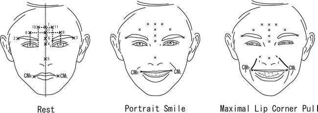
We defined the right and left corners of the mouth as CMR and CML, respectively, and identified them on the facial images of the rest, the maximal lip corner retraction, and the portrait smile (Figure 3), respectively. In addition, 11 anatomic and geometric feature points were identified on the facial images for each expression (Figure 3). Conversion coefficients that minimized the difference between the position vectors for each corresponding feature point for the rest and the maximal lip corner retraction, and for the rest and the portrait smile were obtained for each subject for each task according to the least square method. The position vectors of CMR and CML for the maximal lip corner retraction and for the portrait smile were transformed into position vectors in the facial coordinate system, created on the facial image at rest, using the obtained conversion coefficient to eliminate possible effects of spontaneous head movement during the recording session.
Figure 3.

The measurement points were the right and left corners of the mouth (CMR and CML). In addition, 11 feature points were used to minimize the effects of spontaneous head movement during the recording session in each subject. (1) FHm. (2) Er. (3) El. (4) Se. (5) Pronasale. (6) The internal point 2/3 of the distance from FHm to Se. (7) The internal point 1/3 of the distance from FHm to Se. (8) The point vertically aligned with point 7 and horizontally aligned with the right pupil. (9) The point vertically aligned with point 7 and horizontally aligned with the left pupil. (10) The point vertically aligned with FHm and horizontally aligned with the right endocanthion. (11) The point vertically aligned with FHm and horizontally placed in tandem with the left endocanthion.
Position vector lengths from CMR and CML for the rest to CMR and CML for the maximal lip corner retraction and for the portrait smile were defined as |ΔCMR| and |ΔCML|. Absolute values of the position vector components from CMR and CML for the rest to CMR and CML for the maximal lip corner retraction and for the portrait smile were defined as |Δx|, |Δy|, and |Δz|, respectively. According to the difference between |ΔCMR| and |ΔCML|, the results were categorized as follows: the no-laterality, in which the difference between |ΔCMR| and |ΔCML| was less than 2 mm; the right-laterality, in which |ΔCMR| was at least 2 mm greater than |ΔCML|; the left-laterality, in which |ΔCML| was at least 2 mm greater than |ΔCMR|. Measurements were made using a three-dimensional registration software (3D-Rugle, Medic Engineering Co, Kyoto, Japan) in the present study. All feature points were visually identified on the monitor (Diamondcrysta RDTI74LM-H, Mitsubishi Electronic Corp, Tokyo, Japan) by the same examiner.
Statistical Analyses
Student t-test was employed for comparisons between CMR and CML for the symmetric group and the right-side hemiface dominant group for |Δx|, |Δy|, and |Δz|. Fisher's exact test was employed for comparisons between proportions of the right-laterality and the left-laterality for each symmetric group and the right-side hemiface dominant group. The proportions of the right-laterality and the left-laterality were also compared for the right-handed group and the left-handed group. Statistical analyses were made using a software program.25 P values less than 0.05 were assigned as significant. For multiple comparisons, the significance levels were adjusted to P < .017 with Bonferroni correction for three comparisons.
RESULTS
For the maximal lip corner retraction, significant difference was not seen between CMR and CML for any of |Δx|, |Δy|, and |Δz| in the symmetric group or the right-side hemiface dominant group (Table 3). For the portrait smile, CML exhibited significantly greater |Δx| and |Δy| than CMR in the symmetric group (P = .0040 and P = .0008, respectively), meanwhile, significant difference was not seen for |Δz| [Table 4]).
Table 3.
Comparisons of Displacements Between CMR and CML for the Symmetric Group and the Right-Side Hemiface Dominant Group Computed for the Maximal Lip Corner Retraction

Table 4.
Comparisons of Displacements Between CMR and CML for the Symmetric Group and the Right-Side Hemiface Dominant Group Computed for the Portrait Smile

For the portrait smile, the proportion of the left-laterality was significantly higher than that with the right-laterality (84.1% vs 15.9%, P = .0001) in the symmetric group (Table 5). Significant difference was not seen in the proportion of the right-laterality and the left-laterality in the right-side hemiface dominant group (right-laterality and left-laterality were both 50.0%; Table 5).
Table 5.
Proportions of the Right-Laterality and the Left-Laterality Within the Symmetric Group (n = 44) and the Right-Side Hemiface Dominant Group (n = 6)

In addition, for the portrait smile, the proportion of the subjects with the left-laterality was significantly greater than the proportion of the subjects with the right-laterality both for the right-handed and the left-handed, respectively (82.4% vs 17.6%, P = .0023, and 90.0% vs 10.0%, P = .0485, respectively; Table 6).
Table 6.
Proportions of the Right-Laterality and the Left-Laterality for the Right-Handed Group (n = 34) and the Left-Handed Group (n = 10)

DISCUSSION
Numerous studies26–28 have reported that the right-hemiface tends to be wider than the left. The size difference between the right and left hemiface is suggested as one possible cause of the asymmetric displacements of the right and left corners of the mouth during voluntary smile.
A previous study15 that examined the effect of the hemiface size on the hemiface mobility has reported that if the two hemifaces differed in size, the expression on the wider side could appear diluted and be perceived as less expressive. The other study by Sackeim and Gur16 however, did not find any significant correlation between the hemiface size and the hemiface mobility or measures of the hemiface intensity of expression. In the present study, as for the portrait smile, the left corner of the mouth demonstrated greater displacement than the right in subjects whose faces were judged to be symmetric. Meanwhile, the right and left corners of the mouth did not differ in displacement in subjects whose faces were judged to be right dominant. This suggests that the difference in the hemiface size is likely to cause the difference in displacement of the right and left corners of the mouth. We need to consider the difference between the right and left hemiface size when evaluating the displacements of the corners of the mouth during voluntary smile.
In contrast to the portrait smile, the task of retracting the lip corners with a maximum effort did not show any asymmetry in the displacement distances in space between the right and left corners of the mouth. A previous study29 has reported the different association between the electromyographic (EMG) activities recorded from the zygomatic and orbicularis oculi muscles and the regional cerebral blood flow for the natural smile and the voluntary smile. The observed difference may be explained by possible difference in neuronal controls between the portrait smile, which is assumed as a conditioned voluntary smile to simulate the imaginary “natural” smile, and the maximal lip corner retraction, which simulated a border retracting movements of the lip corners with maximum voluntary efforts.
The results of the present study, suggesting the left-sided laterality in the displacements of the corners of the mouth for the portrait smile, ie, the artificial smile the subjects were asked to perform in a manner they felt natural, were congruent with those of previous studies11–13,30 that employed conventional two-dimensional frontal facial photographs. Regarding voluntary smile, motor command is thought to arise from the primary motor area and end in the contralateral motor facial nucleus in the lower third of the face.31 The ipsilateral motor cortex receives an inhibitory signal from the other side of the motor cortex and asymmetric recruitment of neuronal activity at the cortical level through the uncrossed corticospinal tract has also been reported.32 The asymmetric response of the motor cortex during ipsilateral movement probably represents the asymmetry of the summation of the ipsilateral innervation and transcallosal inhibitory control.32,33 It can, therefore, be speculated that the asymmetries of neuronal activity in the motor cortices might be a possible explanation of the asymmetry and laterality in the facial motor output to the lower one third of the face determined in the present study.
The handedness is known to be correlated with footedness.34 The basal ganglia motor circuit, the supplementary motor area that projects to the putamen, is somatotopically organized with arm and leg representation.35 Thus, the arm and leg have similar neuronal pathways independent of the circuit of voluntary smile. The present finding, that the laterality of the displacements of the corners of the mouth for the portrait smile is independent from subjects' handedness, may be explained by the aforementioned difference in neuronal pathways.
In summary, though we must be cautious about the interpretation of the information obtained from the present study, which recorded the displacements of the corners of the mouth, the present findings suggest that facial expressions in orthodontic diagnosis should be made cautiously in light of the knowledge on lateralization of the facial motor output, especially when diagnosing those having facial asymmetry.
CONCLUSIONS
Displacements of the right and left corners of the mouth during voluntary smile were asymmetric, and the left-sided laterality was found.
The laterality of the facedness differed in relation to the hemiface size, but was not related to the handedness.
REFERENCES
- 1.Myers R. E. Comparative neurology of vocalization and speech: proof of a dichotomy. Ann N Y Acad Sci. 1976;280:745–760. doi: 10.1111/j.1749-6632.1976.tb25537.x. [DOI] [PubMed] [Google Scholar]
- 2.Ackerman J. L, Ackerman M. B, Brensinger C. M, Landis J. L. A morphometric analysis of the posed smile. Clin Orthod Res. 1998;1:2–11. doi: 10.1111/ocr.1998.1.1.2. [DOI] [PubMed] [Google Scholar]
- 3.Trotman C. A, Faraway J. J, Essick G. K. Three-dimensional nasolabial displacement during movement in repaired cleft lip and palate patients. Plast Reconstr Surg. 2000;105:1273–1283. doi: 10.1097/00006534-200004040-00003. [DOI] [PubMed] [Google Scholar]
- 4.Johnston D. J, Millett D. T, Ayoub A. F, Bock M. Are facial expressions reproducible? Cleft Palate Craniofac J. 2003;3:291–296. doi: 10.1597/1545-1569_2003_040_0291_afer_2.0.co_2. [DOI] [PubMed] [Google Scholar]
- 5.Trotman C. A, Faraway J. J, Phillips C. Visual and statistical modeling of facial movement in patients with cleft lip and palate. Cleft Palate Craniofac J. 2005;42:245–254. doi: 10.1597/04-010.1. [DOI] [PMC free article] [PubMed] [Google Scholar]
- 6.Proffit W. R, White R. P, Jr, Sarver D. M. Contemporary Treatment of Dentofacial Deformity. St Louis, Mo: Mosby; 2003. pp. 92–126. [Google Scholar]
- 7.Coulson S. E, Croxson G. R, Gilleard W. L. Three-dimensional quantification of the symmetry of normal facial movement. Otol Neurotol. 2002;23:999–1002. doi: 10.1097/00129492-200211000-00032. [DOI] [PubMed] [Google Scholar]
- 8.Giovanoli P, Tzou C. H, Ploner M, Frey M. Three-dimensional video-analysis of facial movements in healthy volunteers. Br J Plast Surg. 2003;56:644–652. doi: 10.1016/s0007-1226(03)00277-7. [DOI] [PubMed] [Google Scholar]
- 9.Borod J. C, Caron H. S, Koff E. Asymmetry of facial expression related to handedness, footedness, and eyedness: a quantitative study. Cortex. 1981;18:381–390. doi: 10.1016/s0010-9452(81)80025-1. [DOI] [PubMed] [Google Scholar]
- 10.Chaurasia B. D, Goswami H. K. Functional asymmetry in the face. Acta Anat (Basel) 1975;91:154–160. doi: 10.1159/000144379. [DOI] [PubMed] [Google Scholar]
- 11.Campbell R. Asymmetries in interpreting and expressing a posed facial expression. Cortex. 1978;14:327–342. doi: 10.1016/s0010-9452(78)80061-6. [DOI] [PubMed] [Google Scholar]
- 12.Sackeim H. A, Gur R. C, Saucy M. C. Emotions are expressed more intensely on the left side of the face. Science. 1978a;202:434–436. doi: 10.1126/science.705335. [DOI] [PubMed] [Google Scholar]
- 13.Borod J. C, Haywood C. S, Koff E. Neuropsychological aspects of facial asymmetry during emotional expression: a review of the normal adult literature. Neuropsychol Rev. 1997;7:41–60. doi: 10.1007/BF02876972. [DOI] [PubMed] [Google Scholar]
- 14.Strauss E, Kaplan E. Lateralized asymmetries in self-perception. Cortex. 1980;16:289–293. doi: 10.1016/s0010-9452(80)80064-5. [DOI] [PubMed] [Google Scholar]
- 15.Nelson C. A, Horowitz F. D. Asymmetry in facial expression. Science. 1980;209:834. doi: 10.1126/science.7403852. [DOI] [PubMed] [Google Scholar]
- 16.Sackeim H. A, Gur R. C. Asymmetry in facial expression. Science. 1980;209:834–836. doi: 10.1126/science.7403854. [DOI] [PubMed] [Google Scholar]
- 17.Evans R, Shaw W. Preliminary evaluation of an illustrated scale for rating dental attractiveness. Eur J Orthod. 1987;9:314–318. doi: 10.1093/ejo/9.4.314. [DOI] [PubMed] [Google Scholar]
- 18.Wada K. A study on the individual growth of maxillofacial skeleton by means of lateral cephalometric roentgenograms. J Osaka Univ Dent Soc. 1977;22:239–269. [Google Scholar]
- 19.Alcalde R. E, Jinno T, Orsini M. G, Sasaki A, Sugiyama R. M, Matsumura T. Soft tissue cephalometric norms in Japanese adults. Am J Orthod Dentofacial Orthop. 2000;118:84–89. doi: 10.1067/mod.2000.104411. [DOI] [PubMed] [Google Scholar]
- 20.Takada K, Lowe A. A, Yoshida K, Sakuda M. Tongue posture at rest: an electromyographic and cephalometric appraisal. J Osaka Univ Dent Sch. 1985;25:139–151. [PubMed] [Google Scholar]
- 21.Richardson C. K, Bowers D, Bauer R. M, Heilman K. M, Leonard C. M. Digitizing the moving face during dynamic displays of emotion. Neuropsychologia. 2000;38:1028–1039. doi: 10.1016/s0028-3932(99)00151-7. [DOI] [PubMed] [Google Scholar]
- 22.Haraguchi S, Takada K, Yasuda Y. Facial asymmetry in subjects with skeletal Class III deformity. Angle Orthod. 2002;72:28–35. doi: 10.1043/0003-3219(2002)072<0028:FAISWS>2.0.CO;2. [DOI] [PubMed] [Google Scholar]
- 23.Severt T. R, Proffit W. R. The prevalence of facial asymmetry in the dentofacial deformities population at the University of North Carolina. Int J Adult Orthodon Orthognath Surg. 1997;12:171–176. [PubMed] [Google Scholar]
- 24.Oldfield R. C. The assessment and analysis of handedness: the Edinburgh inventory. Neuropsychologia. 1971;9:97–113. doi: 10.1016/0028-3932(71)90067-4. [DOI] [PubMed] [Google Scholar]
- 25.R Development Core Team R A Language and Environment for Statistical Computing. Vienna: R Foundation for Statistical Computing; 2006. [Google Scholar]
- 26.Weeden J. C, Trotman C. A, Faraway J. J. Three dimensional analysis of facial movement in normal adults: influence of sex and facial shape. Angle Orthod. 2001;71:132–140. doi: 10.1043/0003-3219(2001)071<0132:TDAOFM>2.0.CO;2. [DOI] [PubMed] [Google Scholar]
- 27.Koff E, Borod J, Strauss E. Development of hemiface size asymmetry. Cortex. 1985;21:153–156. doi: 10.1016/s0010-9452(85)80023-x. [DOI] [PubMed] [Google Scholar]
- 28.Haraguchi S, Iguchi Y, Takada K. Asymmetry of the face in orthodontic patients. Angle Orthod. 2008;78:421–426. doi: 10.2319/022107-85.1. [DOI] [PubMed] [Google Scholar]
- 29.Iwase M, Ouchi Y, Okada H, et al. Neural substrates of human facial expression of pleasant emotion induced by comic films: a PET study. NeuroImage. 2002;17:758–768. [PubMed] [Google Scholar]
- 30.Borod J. C, Caron H. S. Facedness and emotion related to lateral dominance, sex and expression type. Neuropsychologia. 1980;18:237–241. doi: 10.1016/0028-3932(80)90070-6. [DOI] [PubMed] [Google Scholar]
- 31.Urban P. P, Wicht S, Vucorevic G, et al. The course of corticofacial projections in the human brainstem. Brain. 2001;124:1866–1876. doi: 10.1093/brain/124.9.1866. [DOI] [PubMed] [Google Scholar]
- 32.Hayashi M. J, Saito D. N, Aramaki Y, Asai T, Fujibayashi Y, Sadato M. Hemispheric asymmetry of frequency-dependent finger movement: a functional magnetic resonance imaging study. Cereb Cortex. 2008;18:2932–2940. doi: 10.1093/cercor/bhn053. [DOI] [PMC free article] [PubMed] [Google Scholar]
- 33.Spraker M. B, Yu H, Corcos D. M, Vaillancourt D. E. Role of individual basal ganglia nuclei in force amplitude generation. J Neurophysiol. 2007;98:821–834. doi: 10.1152/jn.00239.2007. [DOI] [PMC free article] [PubMed] [Google Scholar]
- 34.Kang Y, Harris L. J. Handedness and footedness in Korean college students. Brain Cogn. 2000;43:268–274. [PubMed] [Google Scholar]
- 35.Alexander G. E, Crutcher M. D. Functional architecture of basal ganglia circuits: neural substrates of parallel processing. Trends Neurosci. 1990;13:266–271. doi: 10.1016/0166-2236(90)90107-l. [DOI] [PubMed] [Google Scholar]


