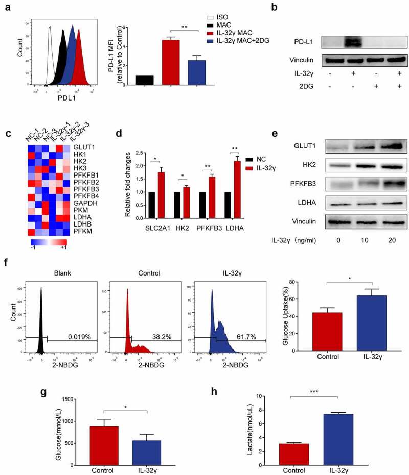
Figure 6.

Enhanced glycolysis mediates IL-32γ-induced PD-L1 expression in macrophages. (a, b) Flow cytometry and western blot analysis of PD-L1 expression in macrophages pretreated with 2DG (5 mM) for 2 h before stimulation with IL-32γ (40 ng/ml) for 24 h. (c) Microarray analysis of RNA transcripts of IL-32γ (40 ng/ml) treated macrophages and control, with heatmap display of key glycolytic genes up-regulated in IL-32γ treated macrophages. (d) Macrophages were left untreated or treated with IL-32γ (40 ng/ml) for 24 h, the levels of glycolysis-related gene expression were determined by qRT-PCR. (e) Western blot analysis of glycolysis-related gene expression in macrophages treated with IL-32γ (0, 10, 20 ng/ml) for 24 h. (f, g) Glucose uptake rate (f) and glucose content (g) in macrophages treated with IL-32γ (20 ng/ml) for 24 h were determined with Glucose Uptake Assay Kit and Glucose Assay Kit, respectively. (h) The levels of lactate production were determined with a lactate assay kit. Data are presented as the mean ± SD of at least three independent experiments; *p < .05, **p < .01, ***p < .001, #, not significant.
