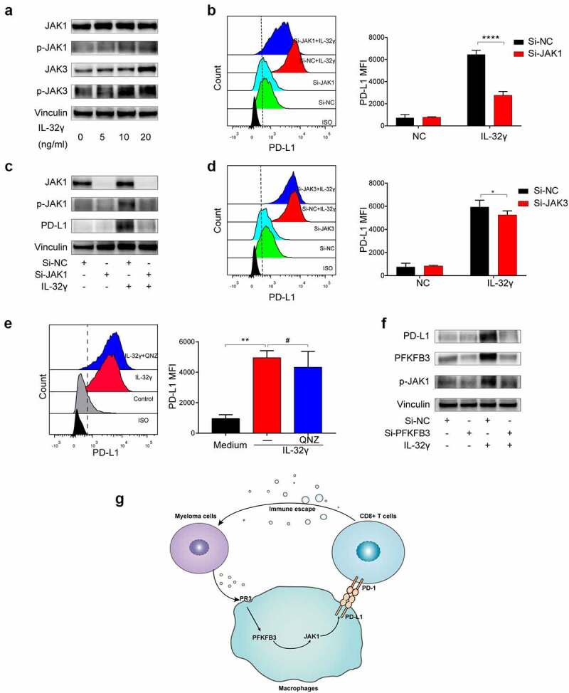
Figure 8.

PFKFB3-JAK1 axis induces PD-L1 expression in IL-32γ treated macrophages. (a) Macrophages were treated with IL-32γ (24 h) for the indicated concentrations. Cell extracts were analyzed by western blotting using indicated antibodies. (b) Macrophages were transfected with control siRNA (Si-NC) or JAK1 siRNA (Si-JAK1) and then treated with or without IL-32γ (20 ng/ml) for 24 h. The PD-L1 expression was subsequently examined by flow cytometry. (c) The JAK1, p-JAK1, and PD-L1 expressions were analyzed by western blotting. (d) Macrophages were transfected with control siRNA (Si-NC) or JAK3 siRNA (Si-JAK3) and then treated with IL-32γ (20 ng/ml) for 24 h. The PD-L1 expression was examined by flow cytometry. (e) Macrophages were pretreated with QNZ (20uM) or not for 2 h, after which IL-32γ (20 ng/ml) was added into cell culture for 24 h, and PD-L1 expression was subsequently examined by flow cytometry. (f) Macrophages were transfected with control siRNA (Si-NC) or PFKFB3 siRNA (Si-PFKFB3) and then treated with or without IL-32γ (20 ng/ml) for 24 h. Cell extracts were analyzed by western blotting using indicated antibodies. (g) Schematic illustration of the molecular mechanism of IL-32γ induced PFKFB3 dependent PD-L1 expression in macrophages. Data are presented as the mean ± SD of at least three independent experiments; *p < .05, **p < .01, ***p < .001.
