Abstract
Vacuolar protein sorting 33B (VPS33B) is important for intracellular vesicular trafficking process and protein interactions, which is closely associated with the arthrogryposis, renal dysfunction, and cholestasis syndrome. Our previous study has shown a crucial role of Vps33b in regulating metabolisms of bile acids and lipids in hepatic Vps33b deficiency mice with normal chow, but it remains unknown whether VPS33B could contribute to cholestatic liver injury. In this study we investigated the effects of hepatic Vps33b deficiency on bile acid metabolism and liver function in intrahepatic cholestatic mice. Cholestasis was induced in Vps33b hepatic knockout and wild-type male mice by feeding 1% CA chow diet for 5 consecutive days. We showed that compared with the wild-type mice, hepatic Vps33b deficiency greatly exacerbated CA-induced cholestatic liver injury as shown in markedly increased serum ALT, AST, and ALP activities, serum levels of total bilirubin, and total bile acid, as well as severe hepatocytes necrosis and inflammatory infiltration. Target metabolomics analysis revealed that hepatic Vps33b deficiency caused abnormal profiles of bile acids in cholestasis mice, evidenced by the upregulation of conjugated bile acids in serum, liver, and bile. We further demonstrated that the metabolomics alternation was accompanied by gene expression changes in bile acid metabolizing enzymes and transporters including Cyp3a11, Ugt1a1, Ntcp, Oatp1b1, Bsep, and Mrp2. Overall, these results suggest a crucial role of hepatic Vps33b deficiency in exacerbating cholestasis and liver injury, which is associated with the altered metabolism of bile acids.
Keywords: cholestasis, liver injury, Vps33b, bile acids, metabonomics
Introduction
Cholestatic liver disease is a disorder of bile acid (BA) metabolism, pathologically manifested as biliary tract and hepatocyte injury [1]. Various factors such as pregnancy status, hereditary BA transporter gene deficiency, drug injury, and cholangiolithiasis may cause abnormity of BA metabolism and further liver injury [2–4]. Till now, only ursodesoxycholic acid and recently FDA-approved obeticholic acid were identified in treating cholestatic liver diseases such as primary biliary cirrhosis (PBC) and primary sclerosing cholangitis [3, 5, 6]. Therefore, exploring potential novel targets or candidates in regulating BA homeostasis and cholestasis pathological mechanism is crucial in the development of therapeutic drug for the treatment of cholestasis.
Vacuolar protein sorting 33B (VPS33B), a member of the Sec1/Munc18 family of class C vacuolar protein sorting proteins, is involved in the vesicular intracellular trafficking process and protein interactions [7]. VPS33B plays important roles in hepatic polarity that directly maintains the structure and/or function of the liver [8]. Mutation of VPS33B can cause arthrogryposis renal dysfunction and cholestasis syndrome (ARC), a severe autosomal recessive multisystem disorder typically characterized by congenital joint contractures, renal tubular acidosis, and neonatal jaundice leading to death in infancy [9, 10]. Patients surviving into adulthood have an elevated BA but normal bilirubin levels in serum [10]. Clinical therapy for ARC simply relieves patient discomfort, such as ursodeoxycholate therapy, a promising drug for cholestasis [9, 11]. VPS33B has also been reported to play roles in liver related diseases [8, 12]. Downregulation of VPS33B expression is a critical step for inflammation-driven hepatocellular carcinoma, and VPS33B serves as an important tumor suppressor in hepatocarcinogenesis [12]. Vps33b was demonstrated a critical role in establishing structural and functional aspects of hepatocyte polarity and may point toward gene transfer mediated the treatment of ARC liver disease [8].
Our recent study demonstrated that mice with hepatic Vps33b deficiency displayed BA accumulation and slight liver damage [13], which was similar with the clinical characteristics of ARC patients. Targeted metabolomics analysis of BAs revealed that taurine-conjugated BAs were increased in the serum of hepatic Vps33b-depleted mice, while unconjugated BAs were prone to decrease. These data suggested that Vps33b played a crucial role in BA metabolism under normal physiological condition. However, whether Vps33b is involved in the progression of cholestatic liver disease was still unclear. Therefore, in the present study, the effect of hepatic Vps33b deficiency on BA metabolism and liver function were further investigated in intrahepatic cholestatic mice induced by cholic acid (CA) overload, to fully elucidate the role of Vps33b in cholestatic liver injury.
Materials and methods
Animals and treatments
Vps33b hepatic knockout male mice (Vps33bflox/flox, alb-cre) and wild-type mice (Vps33bflox/flox) with a C57BL/6 genetic background were obtained from Jun-ling Liu’s Laboratory (Shanghai Jiao Tong University School of Medicine, China). Mice were generated and maintained in a specific-pathogen-free environment under a standard 12-h light/12-h dark cycle. Food and water were freely assessed. Both mice (male, 8–12 weeks, n = 6) were fed with normal chow diet or 1% CA (Sigma-Aldrich, Germany) diet for 5 consecutive days respectively. Mice chow diets were produced by Guangdong Medical Laboratory Animal Center (Guangzhou, China). At day 6, all mice were sacrificed for further analysis. All of the animal experiments were approved by the Institutional Animal Care and Use Committee at Sun Yat-sen University (Guangzhou, China). All procedures were conducted in accordance with the guidelines established by the Institutional Animal Care and Use Committee of Sun Yat-sen University (Guangzhou, China).
Histological and biochemical assessments
Mice liver was sectioned from center of the largest lobular and then fixed in 4% formalin immediately. After dehydration, waxing, and sectioning, hematoxylin and eosin staining was performed to assess liver injury. Whole blood collected from the orbit of mice was kept at room temperature for 30 min, serum was obtained after centrifugation of the whole blood at 1000× g for 15 min at 4 °C. Serum levels of alanine transaminase (ALT), aspartate transaminase (AST), alkaline phosphatase (ALP), total bilirubin (TBILI), and total BA (TBA), as well as liver TBA were measured by commercially available kits (Kehua Bio-Engineering Co., Ltd, Shanghai, China) on automatic biochemistry analyzer (Hitachi-7020, Japan).
qRT-PCR analysis
The expression of BA disposition-related genes in mice liver was measured by qRT-PCR analysis as previously described [13]. The sequences of gene-specific primers (Supplementary Table 1) were obtained from a PrimerBank [14, 15] and synthesized by Thermo Fisher Scientific.
Liquid chromatography/mass spectrometry and metabolomics analysis
Samples for metabolomic analysis were prepared according to our previously described method [13]. Briefly, serum, bile collected from gall bladder, liver, and intestinal tissue samples were prepared or extracted in 67% or 50% aqueous acetonitrile to obtain supernatant of extraction. The obtained extraction was injected into Thermo Scientific Q Exactive™ benchtop Orbitrap high-resolution mass spectrometer (Thermo Fisher Scientifics, San Jose, CA, USA) and in combination with Thermo Scientific Dionex Ultimate 3000 UHPLC system (Diones Corporation, Sunnyvale, CA, USA). The chromatography separation was performed by using ACQUITY UPLC BEH C18 column 1.7 μm (2.1 mm × 50 mm, Waters Corporation, MA, USA). Electrospray negative ionization mode was used for BAs targeted analysis. Multivariate data were analyzed via SIMCA 13.0 software (Umetrics, Kinnelon, NJ, USA). BAs were identified from authentic standards as described before [13].
Statistical analysis
All values are expressed as the mean ± SD. Statistical analysis was performed by unpaired Students’ t-test or Man–Whitney U test with SPSS statistical software and GraphPad Prism 7 (GraphPad Software Inc., San Diego, CA, USA). Only the comparisons indicated above the bars are being made, and a P value of less than 0.05 was considered with significance.
Results
Hepatic Vps33b deficiency aggravates CA-induced cholestatic liver injury
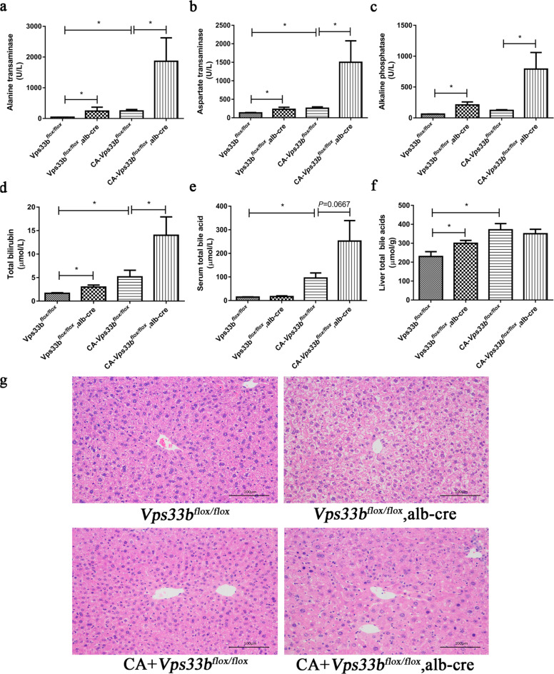
The effect of hepatic Vps33b deficiency on CA-induced intrahepatic cholestatic mice was investigated. Compared to Vps33bflox/flox mice, Vps33bflox/flox, alb-cre mice exhibited a characteristic of minor cholestasis and liver injury under normal chow condition (Fig. 1), as demonstrated by elevated trends in serum enzyme activities including ALT, AST, and ALP, as well as serum TBILI and TBA levels and liver TBA level. Among them, significant differences were observed in serum ALP activities and TBILI level (P < 0.05). When mice were treated with chow containing 1% CA, serum ALT, AST, ALP activities, and TBILI level were significantly increased in Vps33bflox/flox, alb-cre mice compared to those in Vps33bflox/flox mice. But only mild increase was found in serum TBA (P = 0.067). In addition, the hematoxylin and eosin staining showed that hepatocyte degeneration, necrosis around the portal area were more serious in the CA feeding Vps33bflox/flox, alb-cre mice than that in Vps33bflox/flox mice. These data suggested that hepatic Vps33b deficiency aggravated CA-induced cholestatic liver injury in mice.
Fig. 1. Hepatic Vps33b deficiency aggravates CA-induced cholestasis in mice.
Both Vps33bflox/flox, alb-cre and Vps33bflox/flox mice were fed with standard or cholic acid supplemented chow for 5 days. Activities of serum ALT, AST, and ALP (a–c), as well as levels of serum total BA and total bilirubin and live total BA (d–f) were measured. Hematoxylin and eosin staining (magnification of ×200) was performed to assess liver injury (g). Data are expressed as the mean ± SD (n = 5). *P < 0.05.
Hepatic Vps33b deficiency alters bile acid homeostasis in cholestasis mice
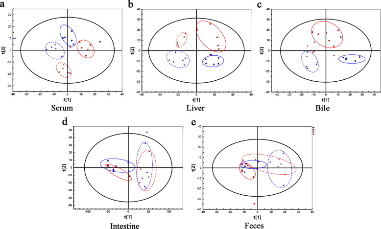
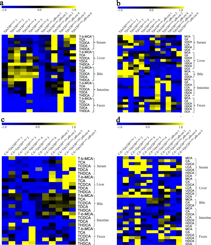
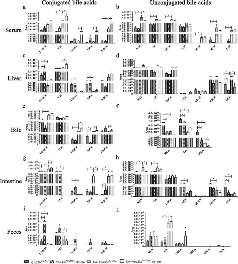
Target metabolomics analysis was performed to examine the changes in the BA homeostasis. Serum, liver, bile, intestine, and feces samples were detected, respectively. PCA analysis revealed a distribution pattern in four groups of mice. Clear separations of scatter plot were shown among four groups in samples of serum, liver, and bile instead of intestine and feces, indicating significant differences in the endogenous metabolome in serum, liver, and bile (Fig. 2). Target metabolomics analysis on BAs was further performed. BAs consist of conjugated and unconjugated types in vivo, and the majority of conjugated BAs are in the form of taurine-conjugation in mice. The heat map (Fig. 3) and corresponding relative quantitative analysis using log intensity of peak area showed that in serum samples (Fig. 4a, b), CA treatment caused apparent elevation of taurocholic acid (TCA), taurohyodeoxycholic acid (THDCA), CA, muricholic acid (MCA), ursodeoxycholic acid (UDCA), hyodeoxycholic acid (HDCA), and deoxycholic acid (DCA) levels in Vps33bflox/flox mice. Hepatic Vps33b deficiency significantly resulted in the increase of conjugated BAs including TCA, taurochenodeoxycholic acid (TCDCA), and taurodeoxycholic acid (TDCA) in the CA feeding mice, but the main unconjugated BA MCA was significantly reduced. Under normal chow condition, hepatic Vps33b deficiency was prone to cause accumulation of the main conjugated BAs such as TCA and tauro-β-muricholic acid (T-β-MCA) and decrease of unconjugated BAs in the serum, although no significant differences were observed between the two groups.
Fig. 2. Principal component analysis score plot of Vps33bflox/flox mice (Blue) and Vps33bflox/flox, alb-cre mice (Red) fed with normal (solid line) or 1% CA chow (dotted line).
Serum, liver, bile, intestine, and feces samples were analyzed, respectively (a–e).
Fig. 3. Heat maps of BA profiles in serum, liver, bile, intestine, and feces samples.
Conjugated (a, c) and unconjugated (b, d). BAs were mapped in both Vps33bflox/flox and Vps33bflox/flox, alb-cre mice.
Fig. 4. BA component quantitative analysis.
Amounts of individual BAs in serum (a, b), liver (c, d), bile (e, f), intestine (g, h) and feces (i, j) were quantified and displayed as log intensity of peak area. Data are expressed as the mean ± SD (n = 5). *P < 0.05.
In liver (Fig. 4c, d), most of the conjugated BAs including T-β-MCA, TCDCA, and TDCA levels were downregulated after CA feeding in Vps33bflox/flox mice, while most of the unconjugated BAs remained unchanged except DCA. Hepatic Vps33b deficiency resulted in significant increase of conjugated BA TCA and decrease of unconjugated BA DCA in the liver of the CA feeding mice. In normal chow mice, hepatic Vps33b deficiency resulted in significant increase of conjugated BA T-β-MCA and decrease of unconjugated BA DCA in the liver.
For bile samples (Fig. 4e, f), significant elevations of T-β-MCA and TCDCA levels were observed after CA feeding in Vps33bflox/flox mice, but CA amount was significantly reduced. Hepatic Vps33b deficiency led to the increase of conjugated BAs including TCDCA, TDCA, and THDCA, and decrease of unconjugated BA CA in the CA-induced cholestatic mice. In normal chow mice, hepatic Vps33b deficiency led to decrease of unconjugated BAs including MCA, CA, and CDCA, but had little effect on conjugated BAs, except the downregulation of TCDCA.
For intestine and feces samples (Fig. 4g–j), only significant increase of T-β-MCA was observed in the CA feeding Vps33bflox/flox mice, and this trend was further enhanced by hepatic Vps33b deficiency.
Generally, the above data demonstrated that hepatic Vps33b deficiency changed the BA homeostasis in CA-induced cholestasis mice, as evidenced by the upregulation of conjugated BAs in serum, liver, and bile; conversely, these conjugated BAs were downregulated in intestine and feces.
Hepatic Vps33b deficiency causes alternations of bile acid disposition-related gene expression in cholestatic mice
The mRNA levels of hepatic genes involved in BA homeostasis were further measured. As shown in Fig. 5, the BA synthesis rate-limiting enzyme Cyp (cytochrome p450) 7a1 was found to decrease to an almost undetected level after CA feeding with or without hepatic Vps33b deficiency. Besides, CA treatment also caused upregulation of the two Phase I metabolizing enzymes Cyp2b10 and Cyp3a11, but the upregulation was not further impacted by hepatic Vps33b deficiency. The Phase II metabolizing enzyme Ugt1a1 has shown significant decrease by hepatic Vps33b deficiency in the CA feeding mice. For BA transporters, CA feeding caused the decrease in BA uptake transporter Ntcp, but an increase in the expression of output transporters Bsep and Mrp4 in Vps33bflox/flox mice, while hepatic Vps33b deficiency led to significant decrease in both uptake transporters Ntcp and Oatp1b1, as well as the output transporter Mrp2. In the CA feeding mice, hepatic Vps33b deficiency further reduced the Ntcp and Oatp1b1 expression, and reversed the upregulation of Bsep. Collectively, these data demonstrated that hepatic Vps33b deficiency led to significant change in the mRNA expression of hepatic genes involved in BA homeostasis such as Cyp3a11, Ugt1a1, Ntcp, Oatp1b1, Bsep, and Mrp2.
Fig. 5. Hepatic Vps33b deficiency alters the expression of BA disposition-related gene in mice.
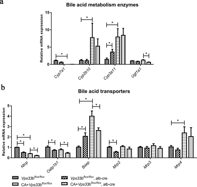
The RNAs were isolated from live tissues and measured by qRT-PCR analysis. a BA metabolizing enzymes; b BA transporters. Data are expressed as the mean ± SD (n = 5). *P < 0.05.
Discussion
The current study demonstrated that hepatic Vps33b deficiency aggravated CA-induced cholestatic liver injury in mice, and targeted metabolomics revealed that hepatic Vps33b deficiency changed the BA homeostasis in cholestasis mice, as evidenced by the upregulation of conjugated BAs in serum, liver, and bile. Furthermore, hepatic Vps33b deficiency caused significant changes in the mRNA expression of BA metabolizing enzymes and transporters.
Cholestasis is one of the three main symptoms in ARC patients with VPS33B mutations, and the patients are normally characterized with high plasma ALP activity and BA level, and slightly increased ALT and AST activities [10, 16, 17]. The disease is also associated with conjugated hyperbilirubinemia, especially appearing in ARC patients with severe cholestasis [10, 16]. Consistently, the Vps33bflox/flox, alb-cre mice showed significant increases in plasma ALP activity and TBILI level, as well as mild upregulation of ALT and AST activities and TBA level compared to the control mice (Fig. 1). Moreover, the above phenotypes were exacerbated after CA treatment, indicating the function deficiency in liver Vps33b is an important factor contributing to cholestasis and liver injury.
Upregulation of TBA level is well defined in ARC patients, which lead to cholestasis. But the changes of individual BAs have not been revealed. In the current study, individual BAs were measured using targeted metabolomics analysis on BAs (Fig. 4). Under normal chow condition, the main conjugated BAs such as TCA and T-β-MCA were accumulated in the serum of Vps33bflox/flox, alb-cre mice, while unconjugated BAs were prone to decrease. It has been reported that the PBC patients were associated with decreased conversion of conjugated to unconjugated BAs, leading to higher levels of conjugated BAs in the serum [18], which is consistent with our finding. This phenomenon may be due to a higher rate of conjugation reaction and conversion to hydrophilic biliary components as a feedback mechanism to relieve BA toxicity [19, 20]. CA is a primary BA in both humans and rodents, and is substantially elevated in patients with cholestatic diseases [21, 22]. Meanwhile, feeding of a diet supplemented with excessive CA was proved a well-established cholestasis model [23, 24], and thus the CA-induced cholestatic mice were used in this study. CA treatment also caused the increase of conjugated BAs in serum, and the increase was enhanced when accompanied by liver Vps33b depletion, which was in line with the biochemical result that liver Vps33b depletion aggravated cholestasis (Fig. 1). The change of individual BA in liver and bile was not as obvious as that in serum. Only increased TCA level in liver and decreased CA level in bile were found in the CA feeding Vps33bflox/flox, alb-cre mice as compared to those in the CA feeding Vps33bflox/flox mice. Overall, hepatic Vps33b deficiency caused abnormal profiles of BAs in cholestasis mice, as evidenced by the upregulation of conjugated BAs in serum, liver, and bile, which suggests that BA profile in ARC patients with VPS33B mutation might mainly represent as the elevation of conjugated BAs in blood.
BA homeostasis is under the control of complex signaling networks [25]. Briefly, BA is synthesized in mice liver via a rate-limiting enzyme CYP7A1, and further undergoes hydroxylation by CYP3A4 (mouse CYP3A11) and CYP2B6 (mouse CYP2B10) and subsequent conjugation by enzymes such as UGT1A1 to reduce toxicity and facilitate BA output from hepatocyte to bile duct. The transporters responsible for BA transport mainly include uptake transporters such as NTCP and OATPs, and output transporters such as BSEP and MRPs. Hepatic depletion of Vps33b induced significant increase in Cyp3a11 expression in normal chow diet mice, but not in the CA feeding mice (Fig. 5a), which might explain that the inductive effect of Vps33b loss in Cyp3a11 would be overwhelmed by negative feedback in gene regulation when BA accumulated. Actually, this phenomenon has been proved elsewhere [26–28]. Ntcp and Oatp1b1 were downregulated in both normal chow and CA feeding mice when liver Vps33b was depleted (Fig. 5b), suggesting that the uptake of BAs was reduced in cholestasis status. Meanwhile, Bsep, the critical transporter mediating BA output, was significantly upregulated in the normal chow diet mice by Vpss33b depletion. However, in the CA feeding mice, Vps33b depletion caused no further Bsep induction (Fig. 5b), which was consistent with the elevation of BA found in serum. Among Mrps, only Mrp2 was reduced by Vpss33b depletion in mice (Fig. 5b). MRP2 is responsible for mediating bilirubin excretion, and the function impairment of MRP2 will lead to hyperbilirubinemia and jaundice [29, 30], which were also characterized in ARC patients. Thus, the Mrp2 reduction in Vpss33b depletion mice supports the finding of high bilirubin level in serum. Nuclear receptors such as farnesoid X receptor (FXR) and pregnane X receptor (PXR) play critical role in regulating BA disposition, the genetic alterations of these enzymes and transporters caused by Vps33b deficiency are probably associated with the modulation of nuclear receptors. The downregulation of Ntcp and Oatp1b1 by Vps33b deficiency may be due to the activation of FXR-SHP signaling, and the upregulation of Bsep and Cyp3a11 could be caused by direct FXR or PXR activation. Overall, the above alterations of BA-related genes may be due to FXR- or PXR-mediated signaling responses. For further mechanistic study, it is necessary to design both in vivo and in in vitro experiments to investigate whether nuclear receptors mediate the effects of Vps33b on BA disposition.
The BA disposition is under the control of complex signaling networks involving dozens of nuclear receptors and corresponding metabolizing enzymes and transporters; although we tried to measure the key enzymes and transporters involved in BA disposition, it still needs to further detect more genes to fully recognize the role of Vps33b in cholestasis. Another limitation of our present study is that only the mRNA expression of these enzymes and transporters were measured, it is necessary to further detect their corresponding protein expression to confirm the results.
In conclusion, liver-specific depletion of Vps33b aggravated cholestatic liver injury in mice and caused abnormal BA profile, as characterized by high levels of conjugated BAs and the alternations of BA metabolizing enzymes and transporters. These findings confirm the important role of VPS33B in cholestasis and provide new insights to understand the pathogenesis of ARC symptoms and also provide potential diagnostic markers for ARC in clinical practice.
Supplementary information
Acknowledgements
The work was supported by the National Natural Science Foundation of China (Grants: 82025034, 81973392, 81320108027), the National Key Research and Development Program (Grant: 2017YFE0109900), the Shenzhen Science and Technology Program (KQTD20190929174023858), the Key Laboratory Foundation of Guangdong Province (Grant: 2017B030314030), the Local Innovative and Research Teams Project of Guangdong Pearl River Talents Program (2017BT01Y093), and the National Engineering and Technology Research Center for New Drug Druggability Evaluation (Seed Program of Guangdong Province, 2017B090903004).
Author contributions
HCB and MH conceived and designed the study. KLF, PC, YYZ, YMJ, and YG performed the experiments. KLF, PC, HZZ, and LHG analyzed the data. CHW and JLL provide Vps33bflox/flox mice and Vps33bflox/flox, alb-cre mice (C57/BL6 strain). PC and KLF wrote the manuscript. HCB revised the manuscript.
Competing interests
The authors declare no competing interests.
Footnotes
These authors contributed equally: Kai-li Fu, Pan Chen
Supplementary information
The online version contains supplementary material available at 10.1038/s41401-021-00723-3.
References
- 1.European Association for the Study of the Liver. EASL Clinical Practice Guidelines: management of cholestatic liver diseases. J Hepatol. 2009;51:237–67. doi: 10.1016/j.jhep.2009.04.009. [DOI] [PubMed] [Google Scholar]
- 2.Jansen PL, Ghallab A, Vartak N, Reif R, Schaap FG, Hampe J, et al. The ascending pathophysiology of cholestatic liver disease. Hepatology. 2017;65:722–38. doi: 10.1002/hep.28965. [DOI] [PubMed] [Google Scholar]
- 3.Trauner M, Fuchs CD, Halilbasic E, Paumgartner G. New therapeutic concepts in bile acid transport and signaling for management of cholestasis. Hepatology. 2017;65:1393–404. doi: 10.1002/hep.28991. [DOI] [PubMed] [Google Scholar]
- 4.Chatterjee S, Annaert P. Drug-induced cholestasis: mechanisms, models, and markers. Curr Drug Metab. 2018;19:808–18. doi: 10.2174/1389200219666180427165035. [DOI] [PubMed] [Google Scholar]
- 5.Isayama H, Tazuma S, Kokudo N, Tanaka A, Tsuyuguchi T, Nakazawa T, et al. Clinical guidelines for primary sclerosing cholangitis 2017. J Gastroenterol. 2018;53:1006–34. doi: 10.1007/s00535-018-1484-9. [DOI] [PMC free article] [PubMed] [Google Scholar]
- 6.European Association for the Study of the Liver. EASL Clinical Practice Guidelines: the diagnosis and management of patients with primary biliary cholangitis. J Hepatol. 2017;67:145–72. doi: 10.1016/j.jhep.2017.03.022. [DOI] [PubMed] [Google Scholar]
- 7.Carim L, Sumoy L, Andreu N, Estivill X, Escarceller M. Cloning, mapping and expression analysis of VPS33B, the human orthologue of rat Vps33b. Cytogenet Cell Genet. 2000;89:92–5. doi: 10.1159/000015571. [DOI] [PubMed] [Google Scholar]
- 8.Hanley J, Dhar DK, Mazzacuva F, Fiadeiro R, Burden JJ, Lyne AM, et al. Vps33b is crucial for structural and functional hepatocyte polarity. J Hepatol. 2017;66:1001–11. doi: 10.1016/j.jhep.2017.01.001. [DOI] [PMC free article] [PubMed] [Google Scholar]
- 9.Gissen P, Johnson CA, Morgan NV, Stapelbroek JM, Forshew T, Cooper WN, et al. Mutations in VPS33B, encoding a regulator of SNARE-dependent membrane fusion, cause arthrogryposis-renal dysfunction-cholestasis (ARC) syndrome. Nat Genet. 2004;36:400–4. doi: 10.1038/ng1325. [DOI] [PubMed] [Google Scholar]
- 10.Jang JY, Kim KM, Kim GH, Yu E, Lee JJ, Park YS, et al. Clinical characteristics and VPS33B mutations in patients with ARC syndrome. J Pediatr Gastroenterol Nutr. 2009;48:348–54. doi: 10.1097/MPG.0b013e31817fcb3f. [DOI] [PubMed] [Google Scholar]
- 11.Santiago P, Scheinberg AR, Levy C. Cholestatic liver diseases: new targets, new therapies. Ther Adv Gastroenterol. 2018;11:1756284818787400. doi: 10.1177/1756284818787400. [DOI] [PMC free article] [PubMed] [Google Scholar]
- 12.Wang C, Cheng Y, Zhang X, Li N, Zhang L, Wang S, et al. Vacuolar protein sorting 33B is a tumor suppressor in hepatocarcinogenesis. Hepatology. 2018;68:2239–53. doi: 10.1002/hep.30077. [DOI] [PubMed] [Google Scholar]
- 13.Fu K, Wang C, Gao Y, Fan S, Zhang H, Sun J, et al. Metabolomics and lipidomics reveal the effect of hepatic Vps33b deficiency on bile acids and lipids metabolism. Front Pharmacol. 2019;10:276. doi: 10.3389/fphar.2019.00276. [DOI] [PMC free article] [PubMed] [Google Scholar]
- 14.Wang X, Spandidos A, Wang H, Seed B. PrimerBank: a PCR primer database for quantitative gene expression analysis, 2012 update. Nucleic Acids Res. 2012;40:D1144–9. doi: 10.1093/nar/gkr1013. [DOI] [PMC free article] [PubMed] [Google Scholar]
- 15.Spandidos A, Wang X, Wang H, Seed B. PrimerBank: a resource of human and mouse PCR primer pairs for gene expression detection and quantification. Nucleic Acids Res. 2010;38:D792–9. doi: 10.1093/nar/gkp1005. [DOI] [PMC free article] [PubMed] [Google Scholar]
- 16.Gissen P, Tee L, Johnson CA, Genin E, Caliebe A, Chitayat D, et al. Clinical and molecular genetic features of ARC syndrome. Hum Genet. 2006;120:396–409. doi: 10.1007/s00439-006-0232-z. [DOI] [PubMed] [Google Scholar]
- 17.Wang JS, Zhao J, Li LT. ARC syndrome with high GGT cholestasis caused by VPS33B mutations. World J Gastroenterol. 2014;20:4830–4. doi: 10.3748/wjg.v20.i16.4830. [DOI] [PMC free article] [PubMed] [Google Scholar]
- 18.Chen W, Wei Y, Xiong A, Li Y, Guan H, Wang Q, et al. Comprehensive analysis of serum and fecal bile acid profiles and interaction with gut microbiota in primary biliary cholangitis. Clin Rev Allergy Immunol. 2020;58:25–38. doi: 10.1007/s12016-019-08731-2. [DOI] [PubMed] [Google Scholar]
- 19.Chen P, Li D, Chen Y, Sun J, Fu K, Guan L, et al. p53-mediated regulation of bile acid disposition attenuates cholic acid-induced cholestasis in mice. Br J Pharmacol. 2017;174:4345–61. doi: 10.1111/bph.14035. [DOI] [PMC free article] [PubMed] [Google Scholar]
- 20.Chen P, Zeng H, Wang Y, Fan X, Xu C, Deng R, et al. Low dose of oleanolic acid protects against lithocholic acid-induced cholestasis in mice: potential involvement of nuclear factor-E2-related factor 2-mediated upregulation of multidrug resistance-associated proteins. Drug Metab Dispos. 2014;42:844–52. doi: 10.1124/dmd.113.056549. [DOI] [PubMed] [Google Scholar]
- 21.Bremmelgaard A, Alme B. Analysis of plasma bile acid profiles in patients with liver diseases associated with cholestasis. Scand J Gastroenterol. 1980;15:593–600. doi: 10.3109/00365528009182221. [DOI] [PubMed] [Google Scholar]
- 22.Fischer S, Beuers U, Spengler U, Zwiebel FM, Koebe HG. Hepatic levels of bile acids in end-stage chronic cholestatic liver disease. Clin Chim Acta. 1996;251:173–86. doi: 10.1016/0009-8981(96)06305-X. [DOI] [PubMed] [Google Scholar]
- 23.Teng S, Piquette-Miller M. Hepatoprotective role of PXR activation and MRP3 in cholic acid-induced cholestasis. Br J Pharmacol. 2007;151:367–76. doi: 10.1038/sj.bjp.0707235. [DOI] [PMC free article] [PubMed] [Google Scholar]
- 24.Kim DH, Lee JW. Tumor suppressor p53 regulates bile acid homeostasis via small heterodimer partner. Proc Natl Acad Sci USA. 2011;108:12266–70. doi: 10.1073/pnas.1019678108. [DOI] [PMC free article] [PubMed] [Google Scholar]
- 25.Chiang JY. Bile acid metabolism and signaling. Compr Physiol. 2013;3:1191–212. doi: 10.1002/cphy.c120023. [DOI] [PMC free article] [PubMed] [Google Scholar]
- 26.Chiang JY. Negative feedback regulation of bile acid metabolism: impact on liver metabolism and diseases. Hepatology. 2015;62:1315–7. doi: 10.1002/hep.27964. [DOI] [PMC free article] [PubMed] [Google Scholar]
- 27.de Aguiar Vallim TQ, Tarling EJ, Ahn H, Hagey LR, Romanoski CE, Lee RG, et al. MAFG is a transcriptional repressor of bile acid synthesis and metabolism. Cell Metab. 2015;21:298–311. doi: 10.1016/j.cmet.2015.01.007. [DOI] [PMC free article] [PubMed] [Google Scholar]
- 28.Li-Hawkins J, Gafvels M, Olin M, Lund EG, Andersson U, Schuster G, et al. Cholic acid mediates negative feedback regulation of bile acid synthesis in mice. J Clin Invest. 2002;110:1191–200. doi: 10.1172/JCI0216309. [DOI] [PMC free article] [PubMed] [Google Scholar]
- 29.Keppler D. The roles of MRP2, MRP3, OATP1B1, and OATP1B3 in conjugated hyperbilirubinemia. Drug Metab Dispos. 2014;42:561–5. doi: 10.1124/dmd.113.055772. [DOI] [PubMed] [Google Scholar]
- 30.Zollner G, Fickert P, Zenz R, Fuchsbichler A, Stumptner C, Kenner L, et al. Hepatobiliary transporter expression in percutaneous liver biopsies of patients with cholestatic liver diseases. Hepatology. 2001;33:633–46. doi: 10.1053/jhep.2001.22646. [DOI] [PubMed] [Google Scholar]
Associated Data
This section collects any data citations, data availability statements, or supplementary materials included in this article.


