Abstract
Sexual plant reproduction depends on the attraction of sperm-cell delivering pollen tubes (PT) by two synergids, followed by their programmed cell death (PCD) in Arabidopsis. Disruption of the mitogen-activated protein kinase 4 (MPK4) by pathogenic effectors activates the resistance protein (R) SUMM2-mediated immunity and cell death. Here we show that synergid preservation and reactive oxygen species (ROS) homeostasis are intimately linked and maintained by MPK4. In mpk4, ROS levels are increased and synergids prematurely undergo PCD before PT-reception. However, ROS scavengers and the disruption of SUMM2, in mpk4, restore ROS homeostasis, synergid maintenance and PT perception, demonstrating that the guardian of MPK4, SUMM2, triggers synergid-PCD. In mpk4/summ2, PTs show a feronia-like overgrowth phenotype. Our results show that immunity-associated PCD and synergid cell death during plant reproduction are regulated by MPK4 underscoring an underlying molecular mechanism for the suppression of plant reproduction during systemic R-mediated immunity.
Subject terms: Fertilization, Cell fate, Pollen tube
Synergid cells undergo programmed cell death following pollen tube reception and successful fertilization. Here the authors show that premature synergid cell death is prevented by the mitogen activated protein kinase MPK4 and the R protein SUMM2 which maintain ROS homeostasis in Arabidopsis.
Introduction
In angiosperms, germ cells are formed during the haploid stage of a plant’s life, in structures termed female gametophytes (FG)1, which are embedded in diploid, sporophytic tissue. Within the FG, eight nuclei differentiate to the gametic egg and central cell (CC) adjoined by two kinds of accessory cell types, called synergids and antipodal cells. Both, the haploid egg and adjacent homodiploid CC are each fertilized by one sperm cell to form the embryo and the surrounding triploid endosperm, respectively, within the polygonum gametophyte. Even though the gametophyte carries a haploid set of chromosomes, the CC bears a bi-nuclear ploidy that originates from the karyogamy of two polar nuclei. Besides the two gametic cells, two synergids lie at the micropyle and coordinate the attraction of the pollen tube (PT) by the discharge of attractants2. The synergids are subdivided into the receptive and non-receptive/persistent synergid. The receptive synergid undergoes programmed cell death (PCD) instantly during PT reception and PT rupture, whereas the non-receptive synergid persists3.
Sexual reproduction of flowering plants is based upon the tight coordination of the fertilization process including the synergid-mediated PT attraction4,5, degeneration of the receptive synergid, and the elimination of the persistent synergid via the cell fusing with the emerging endosperm after egg and CC fertilization6.
Mitogen-activated protein (MAP) kinase cascades exert critical roles in plant immunity7,8. A distinct feature of MAPK-mediated signaling is that three layers of protein kinases form a sequential phosphorylation cascade that consists of a MAP kinase kinase kinase (MAPKKK), a MAP kinase kinase (MKK), and a MAP kinase (MPK). MPK4 is a multifunctional kinase that regulates various signaling events in plant innate immunity, defense response, and cytokinesis9. Initially, MPK4 was identified as a negative regulator of plant immunity10. The mpk4 mutant shows a strong autoimmune phenotype accompanied by dwarfism, hydrogen peroxide accumulation, spontaneous cell death, and a deviating transcriptomic and alternative splicing profile11,12. The MPK4-cascade is induced upon the perception of pathogen-associated molecular pattern (PAMP) recognized by PAMP receptors upstream of MEKK1. The upstream signaling components MEKK1 and MKK1/MKK2 activate MPK4 by phosphorylation13 thereby coordinating basal resistance and effector-triggered immunity (ETI)14. The integrity of the MPK4 cascade is guarded by the R protein nucleotide-binding leucine-rich repeat (NB-LRR) protein SUPPRESSOR OF mkk1 mkk2 (SUMM2). SUMM2 monitors the phosphorylation of MPK4 substrates such as CALMODULIN-BINDING RECEPTOR LIKE KINASE 3 (CRCK3/SUMM3)15. CRCK3 associates with SUMM2 in planta and might act as a decoy or guardian of SUMM2. Inactivation of MPK4 by pathogenic effectors, e.g. the HopAI1 of Pseudomonas, reduces CRCK3 phosphorylation, and eventually activates SUMM216. Activated SUMM2 triggers autoimmunity and PCD, as observable in the MPK4-deficient mutant. MPK4 plays a role in plant cytokinesis and is required for the formation of the cell-plate17. Intriguingly, homozygote mpk4 plants exhibit a severe defect in male-specific meiotic cytokinesis18. The microspore mother cells fail to form a normal intersporal callose wall after male meiosis, and cannot complete the meiotic cytokinesis.
Here, we present that MPK4 function is vital for synergid preservation and PT reception via the suppression of the immune receptor SUMM2-mediated ROS production and cell death reaction.
Results
Premature synergid cell death in mpk4
The immune-activated MAP kinase 4 (MPK4) functions as a suppressor of PCD in plants and its homozygous disruption is accompanied by a strong autoimmune reaction and plant sterility. Notably, PCD is a critical element during fertilization19. To unravel the role of the MPK4-kinase cascade in the process of plant reproduction, we analyzed the mpk4-220 and mpk4-3 mutants13 through the course of female gametogenesis and seed formation compared to wild type (WT) and a pMPK4::MPK4:GFP complementation line20 in the mpk4-2 background.
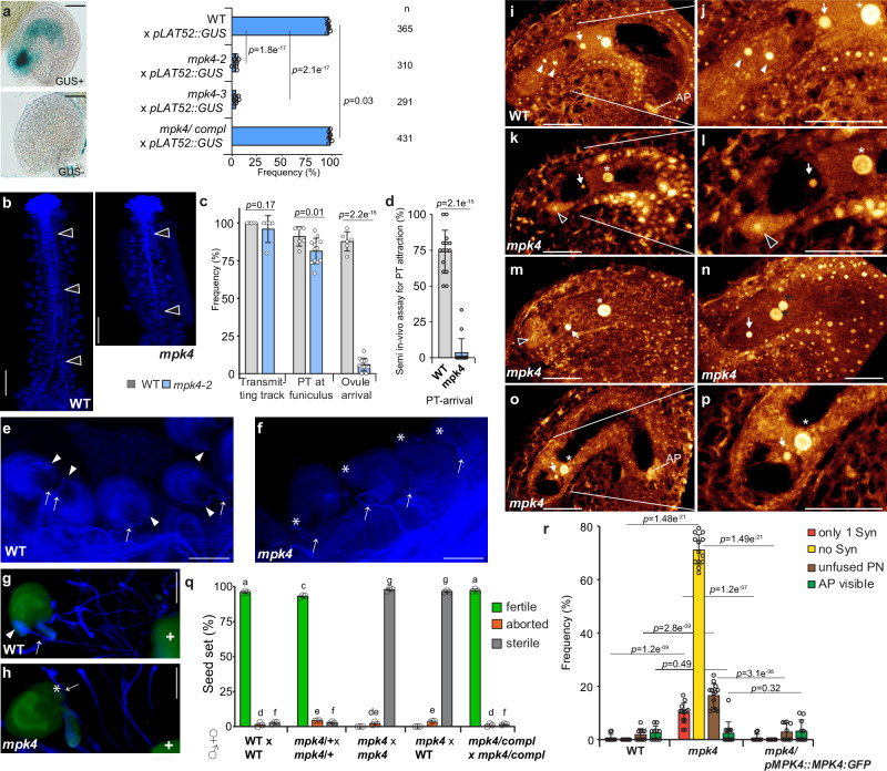
To determine the PT attraction ability of mpk4, we pollinated WT, mpk4, and the complementation line with a PT marker carrying LAT52::GUS originating from a WT source21. After successful PT attraction and reception by the synergids, the blue GUS staining is observable in the respective FGs. We detected GUS-positive ovules in approximately 97-98% of WT and the complementation line samples (Fig. 1a). However, in mpk4-2 only 3.9% and in mpk4-3 4.12% of FGs showed a blue GUS staining. This outcome suggests a defect of mpk4 in PT attraction. Yet, we could not rule out that the PTs are affected on their journey through the transmitting tract and, thus, cannot arrive at the FG. Therefore, we analyzed the ability of WT PTs to grow through the transmitting tract in WT and mpk4 by carrying out aniline blue staining and GUS-stained PT-tracking. We found that WT PTs are fully capable of growing within the transmitting tract in WT and mpk4 and consequently reaching the base of the ovule, the funiculus, in a comparable manner with minor differences (Fig. 1b, c and Supplementary Fig. 1m, n). However, the PTs in mpk4, after having approached the funiculus, did not grow further towards the ovules and seemed to lose guidance (Fig. 1c, e, f). This result suggests a defect in the short-track PT attraction in mpk4. To evaluate this finding, we performed a semi-in-vitro PT attraction assay. We applied WT pollen on a WT stigma and tracked the PT attraction ability of WT and mpk4 ovules which were placed in close vicinity to the stigma. In about 74% of cases the PTs were successfully attracted by the WT ovules, in contrast to mpk4 ovules, which predominantly failed to attract the PTs, with a perception rate of only 3.5% (Fig. 1d, g, h). Together, these findings show that the PT growth towards the ovules is not compromised by the mpk4 mutation, but suggests that the short-track PT-attraction might be affected which is mediated by the synergids of the FG.
Fig. 1. Premature synergid degeneration in mpk4.
a Pollen tube (PT) attraction assay in wild-type (WT), mpk4-2, mpk4-3, and the complementation line expressing pMPK4::MPK4GFP in mpk4-2. Plants were pollinated with pollen of pLAT52::GUS-expressing plants. PT attraction was analyzed 24 hr after pollination. Scale bars, 30 µm. b–c, e–f PT growth assay through the transmitting tract to the ovules in WT and mpk4-2 by aniline blue staining (b, scale bar 0.5 mm; e, f, scale bar 0.1 mm). WT pollen was applied and the growth through stigma, transmitting track towards the ovule was monitored for 24 h. Presented are the results of the 5 biological replicates (n = 26). white-edged black arrowhead, PTs; white arrowhead, PT arrived at the FG; white asterisk, unguided PT; white arrow, PT on the funiculus/ close to the ovule. Error bars show ± SD. (d, g, h) Analysis of the short-track PT attraction ability in WT and mpk4 by semi-in vivo PT assay followed by PT calcofluor white staining. WT pollen were applied on WT-stigmas and ovules of WT (g) and mpk4 (h) were placed below the stigma decapitation site. The PT attraction ability was analyzed after 6 h. White plus refers to the decapitated WT stigma; white arrowhead, PT arrived at the FG; white asterisk, unguided PT; white arrow, PT on the funiculus/ close to the ovule. Presented are the results of 3 biological replicates comprising a total of 69 ovules in WT and 68 ovules in mpk4. Error bars show ± SD. Scale bar 0.1 mm. i–p Representative CLSM images of WT (i–j) and mpk4 (k–p) female gametophytes (FG) 48 h post-emasculation. Depicted are maximum projections of Z-stacks, with a focal plane distance of 1 µm within a total range of about 20 µm, animated 3D-projections are provided in Supplementary movies 1-4. k–m FG of mpk4 that shows prematurely degenerating synergids. n–p FG of mpk4 with completely absent synergids. n FG with polar nuclei and without synergids. White arrowhead, synergid; black arrowhead, degenerating synergid; white arrow, egg cell; white asterisk, central cell nucleus; black asterisks, polar nuclei. Scale bars, 30 µm. q Seed formation in WT (n = 287), mpk4/+ (n = 516), mpk4/- (n = 331), mpk4/- x WT (n = 225) and the MPK4 complementation line (n = 249). Error bars show ± SD. Statistical significance was analyzed by one-way ANOVA. Letters indicate significant differences from the mean (p ≤ 0.05) within the same category of either fertile, aborted or sterile among the different genotypes. r Morphological characterization of the female gametophytic cells in WT (n = 314), mpk4-2 (n = 366) and mpk4 complementation line (n = 306) 48 h post-emasculation. Error bars show ± SD. Syn, Synergids; PN, polar nuclei; AP, antipodal cells. a, c, d, r Statistical significance was analyzed by one-way ANOVA. Source data and further statistical analysis are provided in the source data file.
Thus, we raised the question of whether the formation of the female gametophytic cells is compromised in mpk4. Before PT-reception and fertilization, we found in mpk4 that the egg cell, CC, and antipodal cells are morphologically indistinguishable from WT and the complementation line (Fig. 1i, j, o, p, r, Supplementary Fig. 1h, j and Supplementary movie 1). The CC showed a slight increase of unfused polar nuclei in mpk4 (Fig. 1n, r and Supplementary Fig. 1h, i). However, in the vast majority of mpk4 FGs, we detected a severe defect in the maintenance of the synergid nuclei. In the maximum projection of confocal-laser scanning microscopy (CLSM) Z-stack observations, we detected bright white areas in the synergids which verifies synergid degeneration22,23 (Fig. 1k-m, R and Supplementary movie 2, 3). Moreover, synergids could barely be recognized at all after sample stack observation, whereas the detection of the egg and CC was comparable to WT (Fig. 1n–p, r, Supplementary Fig. 1h-j and Supplementary movie 4). In summary, we predominately observed either only a single synergid nucleus or no synergid nuclei at all, 48 h post-emasculation.
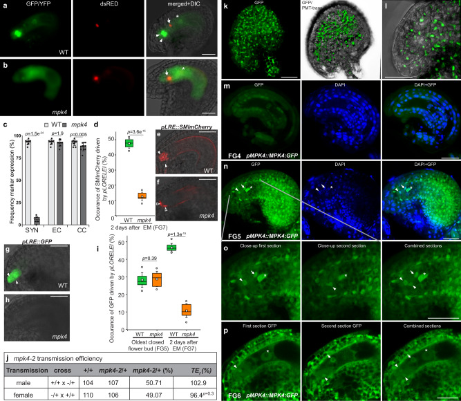
To analyze the synergid degeneration at the molecular level, we introduced a female-gametophyte reporter (FGR7.0) to mpk4. This marker allows simultaneous characterization of synergid cell, egg cell, and CC in a single construct23. Additionally, we analyzed an antipodal marker in mpk4 to characterize these accessory cells. We found the egg cell (pEC1::NLS:dsRED), CC (pDD22::YFP), and antipodal (pAGL62::AGL62:GFP) marker expressions indistinguishable from WT (Fig. 2a–c), Supplementary Fig. 1e, f) which suggests that the development and the molecular profile of these gametophytic cells are not broadly perturbed. However, the synergid marker, driven by the regulatory sequence of AT5G43510/DD224,25, was barely detectable and could be observed in only a small minority of FGs (Fig. 2a–c). Strikingly, the majority of FGs completely lacked the synergid marker expression. Likewise, we analyzed the expression of pHD2B::GFP:HD2B26 which was predominantly found in synergids and CC in WT. However, in mpk4, GFP:HD2B could hardly be captured in the synergids (Supplementary Fig. 1a, b, d). Moreover, we analyzed the expression of the synergid-expressed gene LORELEI with a pLRE::GFP (Fig. 2g–i) and pLRE::SM1-mCherryGolgie reporter (Fig. 2d–f)27. Both reporters were only detected in a minority (10–12%) of mpk4 synergids compared to WT. To analyze whether the synergids initiation in mpk4 resembles WT, we analyzed the pLRE::GFP/- reporter at synergid establishment in the oldest closed flower buds (Fig. 2i) corresponding to the FG5 stage in the FG development22. In WT and mpk4, we captured a GFP signal in about 30% of synergids, indicating a comparable synergid formation in WT and mpk4. Our results demonstrate that before PT reception and fertilization have taken place, synergids in mpk4 are established per se but prematurely degenerate. We conclude that MPK4 guards synergids against premature PCD and thereby enables PT attraction, sperm-cell release, and gamete fertilization.
Fig. 2. Premature synergid degeneration at the molecular level, and MPK4 localization in the embryo sac.
a–c Expression of the female-gametophyte reporter (FGR7.0) in (a) WT and (b) mpk4. c Frequency of FGR7.0/+ expression in WT (n = 321) and mpk4 (n = 285) ovules was determined. Error bars show ± SD. d–i Expression of the LORELEI-reporter (pLRE) coupled to the Golgi-retention peptide sequence SMImCherry (d–f) and a single GFP (g–i). The expression of pLRE::GFP/- was analyzed in the oldest-closed flower bud (FG5 stage) (WT, n = 326; mpk4, n = 240) and 48 h after emasculation (WT, n = 328; mpk4, n = 251; FG7 stage). pLRE::SMImCherry/- was analyzed 48 h after emasculation (WT, n = 293; mpk4, n = 257). Boxes represent the 25th and 75th percentiles, and the inner rectangle highlights the median, whiskers show the SD, and outliers are depicted by dots (Min/max range). j Transmission analysis of mpk4. Transmission efficiency (TEf) indicates the transmission of mpk4 through the male and female gametophyte. k–p Representative images of MPK4:GFP expression and localization in the sporophytic ovule tissue (k–l) and at the female gametophytic developmental stages FG4, FG5, FG6 (m–p). k–l Depicted are maximum projections of Z-stacks, with a focal plane distance of 2 µm within a total range of about 30 µm, animated 3D-projection is provided in Supplementary movie 5. m–p pMPK4::MPK4:GFP-expressing line was analyzed, accompanied by the nuclei-staining dye DAPI. Negative control Supplementary Fig. 1l. Experiments were repeated three times with similar results and presented are representative images. c, d, i, j Statistical significance was analyzed by one-way ANOVA. Source data and further statistical analysis are provided in the source data file. White arrowhead, synergid; black arrowhead, degenerating synergid; white arrow, egg cell; white asterisk, central cell nucleus; black asterisks, polar nuclei. Scale bars, 20 µm (a, b, e, f–h), 30 µm (k–p).
Thus, we raised the question of whether the premature synergid degeneration in mpk4 interferes with seed formation. In mpk4 self-pollinated plants, fertile seed formation could not be observed, solely an amount of 2.11% aborted seeds (n = 331) could be counted (Fig. 1q). This finding is following previous results showing the inability of homozygote mpk4 plants to reproduce. To analyze whether WT pollen might increase the fertilization rate, we hand-pollinated mpk4 with WT pollen. Again, we could not detect fertile seeds and only 3.56% of embryo sacs initiated seed formation before aborting, these were accompanied by an overwhelming number of sterile embryo sacs. As a matter of note, in the few mpk4 gametophytes that truly received a PT, gamete fertilization, embryo, and endosperm initiation were not broadly perturbed suggesting that egg and CC maturation are not adversely affected by the mpk4 knock-out (Supplementary Fig. 3g). The introduction of a complementation construct (pMPK4::MPK4:GFP)20 in mpk4 restored seed formation indicating that the lack of seed production can be traced back to the knock-out of MPK4. Our results indicate a vital function of MPK4 in guarding the synergids against premature degeneration thereby enabling PT attraction and seed formation.
Transmission analysis
To determine whether the premature synergid degeneration is of sporophytic or gametophytic origin, we analyzed the heterozygote mpk4/+ mutant. The occurrence of the synergids, central cell and antipodals was indistinguishable from WT (Supplementary Fig. 3a–c and Supplementary movie 7, 8). Likewise, the expression of the pLRE::SM1-mCherryGolgie reporter in the synergids did not show a significant deviation from WT (Supplementary Fig. 3d, e). The transmission efficiency of self-crossed heterozygote mpk4 mutants resulted in a Mendelian segregation ratio of 28.4% WT, 53.5% heterozygote and 18.1% homozygote mpk4 mutants, suggesting a weak zygotic/embryonic lethal phenotype (Supplementary Fig. 3f). Indeed, when compared to WT, the seed set of heterozygote mpk4 plants showed a slight reduction of fertile seeds in favor of a slight increase of aborted seed formation (Fig. 1q). To further analyze, whether the transmission of the mpk4 mutation through the male and female gametophyte is affected, we performed reciprocal crosses between WT and heterozygote mpk4 mutants and analyzed the mpk4 transmission among the F1 progeny. Both, when mpk4 was introduced from either the male or female site, the transmission of the mpk4 mutation to the next generation was determined very close to 50%, equating a transmission efficiency of almost 100% (Fig. 2j). This result indicates that the gametophytic mpk4 transmission is not affected. Taken together, our results point to a sporophytic origin of the premature synergid degeneration in mpk4/- revealing that the surrounding maternal ovule tissue might specifically determine the formation and function of the PT-attracting synergids.
MPK4:GFP localization in the female gametophytic cells and ovule tissue
To pinpoint the expression and localization of MPK4 in the ovular tissue and during female gametogenesis, we analyzed the line expressing pMPK4::MPK4:GFP20 by CLSM and exploited maximum projections of Z-stack observations. MPK4:GFP showed a strong nuclear signal in the FG-surrounding sporophytic ovule tissue, including the micropylar region of the ovule (Fig. 2k, l and Supplementary movie 5). During the early female gametogenesis at stage FG4, we could not detect MPK4:GFP in the syncytial nuclei (Fig. 2m). However, after cellularization at stage FG5 and FG6, MPK4:GFP was detected in the nucleus of the synergids, egg cell and CC and co-localized with the nucleus-staining agent DAPI (Fig. 2n–p and Supplementary Fig. 1k). In addition, we found MPK4 localization not only in the nucleus but also in the cytoplasm of the female gametophytic cells, particularly in the synergids (Fig. 2p). The negative control by the use of WT plants, did not exhibit a signal under the chosen parameters for CLSM (Supplementary Fig. 1l).
These results revealed the expression of MPK4 and the localization of the encoded protein in the cellularized female gametophyte and the surrounding ovular sporophytic tissue.
Reactive oxygen species accumulation in mpk4 synergids
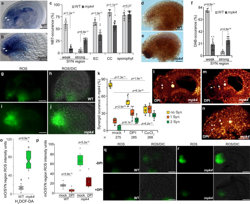
Hydrogen peroxide (H2O2) and superoxide are the main triggers for the immune- and developmental-regulated PCD in plants28–32. Previous studies have revealed that synergid degeneration occurs as a PCD response5,23 and ROS is well known to trigger PCD in plant cells33. However, whether ROS triggers synergid PCD still needs to be deciphered. Furthermore, the MPK4-cascade is essential for ROS-homeostasis and signaling9,34. These previous findings prompted us, to analyze whether the premature synergid cell death in mpk4 might be triggered by a deviated ROS homeostasis. Nitroblue tetrazolium (NBT) is the most commonly employed probe for the detection of in situ superoxide. The NBT staining in WT showed a moderate superoxide accumulation in the inner sporophytic integument cells of the ovule and throughout the female gametophyte within the synergid, egg cell, and CC (Fig. 3a–c). However, in mpk4, we detected very intense NBT staining in the micropylar ovule region while the NBT accumulation in the remaining gametophytic cells was less frequent compared to WT. The sporophytic superoxide concentration in mpk4 was reminiscent of the results in WT (Fig. 3c). To detect in-situ H2O2 levels, we performed diaminobenzidine (DAB) staining. The insoluble brown precipitate that refers to cellular levels of H2O2 could be detected in mpk4 in the egg cell and CC region and the sporophytic ovule tissue in a comparable manner to WT (Fig. 3d–f). Yet, intensely dark DAB staining was mainly captured in mpk4 in the filiform apparatus and the nearby micropylar ovule tissue. Additionally, we applied H2DCF-DA to evaluate the amount of hydroxyl radicals. We detected moderate fluorescence signals in WT synergids, prevalently at the filiform apparatus (Fig. 3g, h). However, the signal intensity was massively enhanced at the micropylar region in mpk4 (Fig. 3i, j, o). These results indicate sustainably higher amounts of the cell-death agents H2O2, superoxide radicals and hydroxyl radicals in the micropylar embryo sac/ synergid cell region in mpk4 that might drive synergid PCD thereby indicating MPK4 as repressor of ROS metabolism in the micropylar ovule region.
Fig. 3. MPK4 inhibits a ROS maximum at the entrance of the female gametophyte.
a–c Superoxide detection in the sporophytic ovule tissue and the female gametophyte following nitroblue tetrazolium (NBT)-staining in WT (n = 281) and mpk4 (n = 272), three biological replicates analyzed. d–f Hydrogen peroxide detection in the sporophytic ovule tissue and the female gametophyte following diaminobenzidine (DAB)-staining in WT (n = 203) and mpk4 (n = 152), three biological replicates analyzed. g–j, o ROS accumulation in WT (g, h) and mpk4 (i, j) in the micropylar ovule (mO) region shown by H2DCF-DA staining. o Quantification of ROS-intensity units (arbitrary) in WT (n = 283) and mpk4 (n = 196) by imageJ. Boxes represent the 25th and 75th percentiles, and the inner rectangle highlights the median, whiskers show the SD, and outliers are depicted by dots (Min/max range). mO/SYN, micropylar ovule/synergid region. k–n Pistil-feeding assay by the use of ROS scavengers: mpk4 pistils of flowers at developmental stage 12b/c (Christensen et al. 22) were treated with mock, DPI (250 µM) or CuCl2 (1 mM) for 24 h and subsequently analyzed for synergid preservation by DIC analysis. Representative images after ROS scavenger-application are shown by maximum projections of Z-stacks l–n. Three biological replicates with a total of 275 (mock), 285 (DPI) and 268 (CuCl2) mpk4 ovules were analyzed. p–r Reduction of micropylar ovular ROS maximum in WT and mpk4 by DPI-application (250 µM). Quantification of ROS-intensity units (arbitrary) in WT and mpk4 by imageJ. Boxes represent the 25th and 75th percentiles, and the inner rectangle highlights the median, whiskers show the SD, and outliers are depicted by dots (Min/max range). mO/SYN, micropylar ovule/synergid area. Three biological replicates were performed with similar results and depicted are the results of the first replicate. Statistical significance was analyzed by one-way ANOVA. Source data and further statistical analysis are provided in the source data file. Error bars show ± SD. White arrowhead, synergid; white arrow, egg cell; white asterisk, the central cell nucleus. Scale bars, 20 µm.
To decipher whether indeed enhanced ROS levels in mpk4 trigger premature synergid PCD, we took advantage of a pistil-feeding approach to manipulate the synergid/ovule environment35. We firstly applied diphenyl iodonium chloride (DPI, 250 µM), an inhibitor of the cell membrane NADPH oxidase-dependent ROS-synthesis that efficiently suppresses ovular ROS production. Secondly, we administered CuCl2 (1 mM), a superoxide scavenger proven of strongly reducing ROS accumulation35. The short-term application of DPI for 15 min reduced the amount of hydroxyl radicals in the sporophytic ovule tissue and synergid region of WT and mpk4 showing a DPI-induced ROS maximum reduction in that part of the ovule (Fig. 3p–r). Feeding mpk4 pistils with DPI or CuCl2 for 24 hrs reduced the premature synergid degeneration and significantly restored the maturation of one or both synergids compared to the mock-treated control (Fig. 3k–n). Synergid-restoration after DPI-treatment shows that NADPH oxidase-dependent ROS synthesis antagonizes synergid preservation. Likewise, CuCl2 application revealed that superoxide crucially determines synergid perpetuation. Taken together, these outcomes show that MPK4-maintained ROS homeostasis in the micropylar ovule/ synergid cell region is critical for regular synergid preservation and that increased ROS levels trigger synergid PCD.
Disruption of the NB-LRR receptor SUMM2 restores PT attraction and synergid maintenance in mpk4
Under regular conditions, MPK4 suppresses the NB-LRR R protein SUMM2 that monitors the integrity of the entire MEKK1-MKK1/2-MPK4 kinase cascade. The NB-LRR receptor-mediated immune response is usually characterized by a rapid and localized cell-death, also known as hypersensitive response29. Thus, the autoimmunity and defense responses in mpk4 are predominantly caused by the activation of SUMM2.
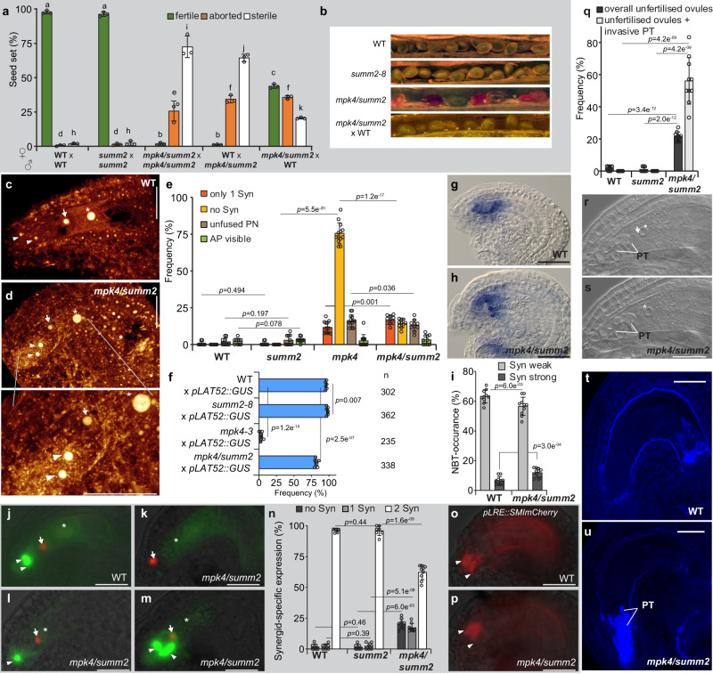
Therefore, we raised the question of whether premature synergid degeneration in mpk4 is subjected to the SUMM2-mediated immune-reaction. Consequently, we analyzed the mpk4/summ2-8 double mutant that restores WT-like defense responses, hydrogen peroxide levels, and regular plant growth16. The hand-self-pollinated double mutant showed a high proportion of sterile seeds and aborted seeds reminiscent of the mpk4 single mutant (Fig. 4a, b and Supplementary Fig 3h). However, we observed a small fraction of fertile seeds giving rise to the next mpk4/summ2 homozygote generation. The fact that the double homozygote mutant generates fertile seeds indicates that the disruption of SUMM2 in mpk4 partly complements the mpk4 mutant defect in seed production. Interestingly, when we pollinated WT plants with mpk4/summ2 originating pollen, then we observed a similar ratio of fertile, aborted, and sterile seeds as compared to the self-pollinated mpk4/summ2 mutant. Yet, the pollination of mpk4/summ2 with WT pollen broadly complemented the defect in the seed formation (Fig. 4a, b). Fertile seed numbers increased by up to 43% and similarly in aborted seeds, giving a total of about 79% fertilized seeds. Notably, the number of sterile seeds dropped to under 21%. These results are intriguing and indicate that the defect in seed formation in self-pollinated mpk4/summ2 mutants is predominately caused by the mpk4/summ2 pollen. It was reported that MPK4 is necessary for male-specific meiotic cytokinesis and MPK4-deficient mutants generate pollen bigger in size and with an aberrant number of nuclei18. We found that pollen of mpk4/summ2 shows a comparable increase in size and a divergent nuclei number as in mpk4 (Supplementary Fig. 3I). These results indicate that the disruption of SUMM2 in mpk4 does not rescue the male-cytokinesis defect, which strongly restricts the transmission through the male gametophyte.
Fig. 4. SUMM2-deficient mpk4 plants restore synergid maintenance and function.
a Seed formation in WT (n = 327), summ2-8 (n = 439), mpk4/summ2 (n = 614), WT x mpk4/summ2 (n = 220), mpk4/summ2 x WT (n = 428). Error bars show ± SD. Error bars show ± SD. Statistical significance was analyzed by one-way ANOVA. Letters indicate significant differences from the mean (p ≤ 0.01) within the same category of either fertile, aborted or sterile among the different genotypes. b Representative images of the seed set in the indicated genomic backgrounds. c, d Representative CLSM images of WT (c) and mpk4/summ2 (d) female gametophytes 48 h post-emasculation. Depicted are maximum projections of Z-stacks, with a focal plane distance of 1 µm within a total range of about 20 µm, animated 3D-projection is provided in Supplementary movie 6. Scale bars, 30 µm. e Morphological characterization of the female gametophytic cells in WT (n = 322), summ2-8 (n = 323), mpk4-2 (n = 340) and mpk4/summ2 (n = 341) 48 h post-emasculation. Syn, Synergids; PN, polar nuclei; AP, antipodal cells; Error bars show ± SD. f Pollen tube (PT) attraction assay in wild-type (WT), summ2-8, mpk4-3 and mpk4/summ2. Plants were pollinated with pollen of pLAT52::GUS-expressing plants. PT attraction was analyzed 24 h after pollination. g–i Superoxide detection in the sporophytic ovule tissue and the female gametophyte following nitroblue tetrazolium (NBT)-staining in WT (n = 269) and mpk4/summ2 (n = 257). j–n Expression of the female gametophyte reporter (FGR7.0/+) in WT (j) and mpk4/summ2 (k–m). n Synergid-marker occurrence was determined in WT (n = 237), summ2 (n = 244) and mpk4/summ2 (n = 294) ovules. Error bars show ± SD. Scale bars, 20 µm. o–p Expression of the LORELEI-reporter (pLRE) coupled to a Golgi-retention peptide sequence SMImCherry. The frequency of the pLRE::SMImCherry/- reporter expression was analyzed in WT (n = 338), summ2 (n = 332) and mpk4/summ2 (n = 307). Statistical analysis of WT, summ2, and mpk4/summ2 is shown in Supplementary Fig. 1g. q–s Frequency of unfertilized ovules (black bars) in WT (n = 269), summ2 (n = 262) and mpk4/summ2 (n = 329), 48 h after pollination with WT pollen. The gray bar depicts the ratio of invasive PT growth within the unfertilized ovules. In WT and summ2, unfertilized ovules that showed invasive PT growth could not be recognized. Error bars show ± SD. Two focal planes of an unfertilized ovule that contains an invasive PT in mpk4/summ2 (r, s), 48 h after pollination with WT pollen. White arrow indicates the egg cell nucleus; white asterisk highlights the central cell nucleus; PT, pollen tube. Shown is a representative image of the results obtained from three biological replicates. t, u Aniline blue staining of an unfertilized ovule in WT and mpk4/summ2 that contains an invasive PT, 48 h after pollination with WT pollen. Shown is a representative image from three biological replicates. Statistical significance was analyzed by one-way ANOVA. Source data and further statistical analysis are provided in the source data file. Error bars show ± SD. White arrowhead, synergid; white arrow, egg cell; white asterisk, the central cell nucleus. Scale bars, 20 µm (j–m, g–h, o, p, r–u).
Subsequently, we analyzed whether mpk4/summ2 restores PT attraction, as a precondition for successful seed formation. We observed that the number of attracted and perceived LAT52::GUS PTs in the single summ2-8 mutant16 was indistinguishable from WT (Fig. 4f). Surprisingly, more than 80% of the mpk4/summ2 ovules received a PT suggesting that the knock-out of SUMM2 rescued the premature synergid-PCD observed in the mpk4 single mutant.
To prove this assumption, we analyzed the synergid integrity in mpk4/summ2 compared to WT, summ2, and mpk4. Compared to mpk4, the synergid morphology and conservation in mpk4/summ2 are mainly restored showing that SUMM2 triggers synergid PCD in the case of MPK4 deficiency (Fig. 4c–e and Supplementary movie 6). However, we also still recognized a small portion of FGs that harbor only one or even no synergids in mpk4/summ2 compared to WT and summ2. This finding indicates that the knock-out of SUMM2 in mpk4 does not fully complement the premature synergid degeneration in mpk4 thereby suggesting that additional factors besides SUMM2 trigger synergid PCD in the case that MPK4 fails.
We captured in mpk4/summ2 a weak signal for ROS accumulation in the synergids of about 57.0% and a strong accumulation of 12% (Fig. 4g–i). These values basically mirror the findings of NBT-staining in WT and are significantly restored when compared to the mpk4 single mutant with 24.2 % strong ROS accumulation (Fig. 3c). Likewise, the NBT-staining and DAB staining at the FG4-FG5 developmental stage at the micropylar region of the female gametophyte and neighboring sporophytic tissue is moderately increased in mpk4/summ2 compared to WT, but also significantly reduced when compared to mpk4 (Supplementary Fig. 2a, b, e, f). After fertilization, NBT-staining of the young seeds showed weakly enhanced ROS levels in mpk4/summ2 compared to WT and summ2 (Supplementary Fig. 2c, d). DAB staining after fertilization resulted in a very diverse outcome from very strongly stained to almost completely unstained seeds in all analyzed genotypes (Supplementary Fig. 2g). Taken together, these results show that the introduction of summ2 in mpk4 partially restores ROS levels in the ovule, during gametogenesis and in the synergids. Moreover, ROS accumulation in mpk4 is caused by SUMM2, which promotes synergid PCD.
Interestingly, we found that the expression of FGR7 in mpk4/summ2 is distinct from the mpk4 single mutant. In mpk4/summ2, the synergid marker was not detectable at all in about 20% of FGs (Fig. 4j, k, n) reminiscent of the mpk4 mutant which exhibited the lack of this marker expression in approximately 95% (Fig. 2c). However, in mpk4/summ2 the synergid marker was visible in a single synergid in 17% of samples and in both synergids in about 62% of FGs (Fig. 4j, l–n). Furthermore, the female gametophyte reporter pLRE::SMImCherry (Fig. 4o, p and Supplementary Fig. 1g) and pHD2B::GFP:HD2B (Supplementary Fig. 1a–d) could be detected in the majority of synergids compared to WT, summ2 and mpk4.
These outcomes suggest that the premature synergid PCD in mpk4 can be considered as an immune-related defense response triggered by the NB-LRR R protein SUMM2. Thus, SUMM2 does not only monitor the integrity of the immune MPK4-cascade concerning basal resistance, it also triggers synergid degeneration in plant reproduction.
Impaired pollen tube rupture in mpk4/summ2
Synergids attract the PT and enable the PT to rupture which results in sperm discharge. We found that synergid maintenance depends on MPK4 function. The disruption of SUMM2 in mpk4 largely restores synergid life-span and function. However, we found that the pollination of mpk4/summ2 with WT pollen led to a significant number of FGs that attracted the PT but remained unfertilized and showed a lack of PT rupture (56.2%) (Fig. 4q–u). In mpk4/summ2, the PT continued growth within the FG, reminiscent of the feronia and lorelei phenotype36,37, without bursting and releasing sperms. This result suggests that the lack of SUMM2 function in mpk4/summ2 partially interferes with male-female communication and prevents synergid degeneration. Furthermore, this result implies that MPK4 does not only determines the synergid lifespan, it might also be critical for the synergid maturation to achieve complete functionality. In mpk4/summ2, the synergid function is restored concerning the short-track PT attraction, but mechanisms that employ PT-perception and burst might be improperly established.
Discussion
We discovered that PCD of the synergids is governed by MPK4 which represses the nucleotide-binding leucine-rich repeat (NLR) receptor-associated cell-death reaction16.
The receptor-like kinase (RLK) FERONIA (FER) is necessary for synergid degeneration after PT reception and exerts vital functions in plant reproduction, hormone signaling, and immunity37–39. The glycosylphosphatidylinositol (GPI)-anchored protein LORELEI (LRE) and LRE-like proteins (LLGs) function as co-receptors for FER. Intriguingly, in the lre mutant, the synergids do not collapse after PT reception reminiscent to fer. The filiform apparatus (FA) of WT synergids and adjacent cells in the micropylar region were found to produce higher levels of ROS in an FER/LRE dependent manner. This ROS maxima is compromised in DPI-treated pistils and in the feronia and lorelei mutant35. It was shown that the FER/LRE-mediated NADPH oxidase-dependent ROS accumulation in this area is crucial to set a ROS environment enabling PT rupture and sperm release after FG arrival35. NADPH oxidases play a pivotal role in ROS production. They transfer electrons from cytosolic NADPH or NADH to apoplastic oxygen, which leads to the production of superoxide (O2-). The produced O2-can be converted to H2O2 by superoxide dismutase40. We detected high levels of various ROS in the synergid area in mpk4 plants showing that MPK4 represses the generation of a ROS maxima in the micropylar region of the ovule. Moreover, the application of the ROS scavenger DPI, an inhibitor of the cell membrane NADPH oxidase-dependent ROS synthesis, partially restore synergid preservation showing that synergid degeneration is intimately coupled to modulations in the ROS homeostasis. Consequently, it is conceivable that an interplay of MPK4 and FER/LRE, with their distinct function in ROS metabolism, give rise to the underlying ROS homeostasis in the micropylar embryo sac and in the FA/SC region. In the mpk4/ summ2 double mutant, an ROS maxima is not being established thereby largely restoring synergid maturation and function.
These findings illustrate that the guardian of MPK4, SUMM2, triggers ROS accumulation and premature synergid degeneration, after being unleashed from MPK4 suppression.
PTs, fungal hyphae secrete and microbial-associated molecular patterns41 stimulate ROS production and the generation of calcium (Ca2+) transients42. ROS signaling is strongly linked to activation of several Ca2+-permeable42 enabling Ca2+ flux and oscillation. Cytosolic calcium ([Ca2+]cyto) oscillations and raise are initiated in synergid cells after the physical contact with the PT apex43. Receptive synergids control the PCD of PTs and themselves by coordinating their distinct calcium signatures in response to the calcium dynamics and growth behavior of the PT44. FER and LRE pathways are required for calcium oscillation in the synergids, which couples the PCD events in the receptive synergid and the PT44. Thus, it is conceivable that the [Ca2+]cyto increase and dynamics in the receptive synergid might interfere with MPK4 functions and might trigger synergid PCD upon SUMM2 activation.
Recently, it was shown that FER and the closely related RLKs, LET1 and LET2, form a specific trimeric Catharanthus roseus RLK1-like (CrRLK1L) module together with the GPI-anchored LLG1 to modulate SUMM2-mediated autoimmunity45. Sensing of cell wall integrity by CrRLK1L-associated pathways has been uncovered as being eminent for cell homeostasis regulating plant immunity, growth, and reproduction46. It is plausible that a similar CrRLK1L complex modulates SUMM2-mediated cell death in the synergids in response to PT-reception. In this respect, PT reception in synergid cells depends on the CrRLK1Ls: HERCULES RECEPTOR KINASE 1 (HERK1) and ANJEA (ANJ)47, that interact with LRE and FER, in a proposed trimeric signaling complex. The proposed FER, LRE, HERK1/ANJ trimeric complex could be the synergid cell counterpart to the immunity FER, LLG1, LET1, LET2 complex thereby modulating SUMM2-mediated ROS accumulation and PT-perception.
In contrast to the summ2 single mutant, we found a PT-overgrowth phenotype in mpk4/summ2, reminiscent of fer, lre, nortia (nta)38 and herk1/anjea. The disruption of MPK4 in summ2 might compromise FER, LRE, NTA or HERK1/ANJEA and eventually the function of the trimeric complex. This effect might not be observable in the mpk4 single mutant owing to the predominant absence of PT-perception. NTA-GFP is homogeneously distributed in synergids in compartments that co-localizes with a cis-Golgi marker48. After PT arrival, NTA-GFP is solely detected in the FA. In fer and lre, NTA-GFP is not translocated to the FA showing that NTA movement and accumulation to the FA depends on FER/LRE function. Yet, forced localization of a chimeric NTA-version (faNTA) in the FA suppresses the fer/lre PT reception phenotype illustrating that NTA accumulation at the FA can bypass the FER/LRE pathway49. We showed that the premature synergid degeneration in mpk4 was partly complemented by introducing summ2. However, to some extend the synergids are still compromised and mechanisms that employ PT-perception might be improperly established, which might be reflected by an unusual distribution of NTA after PT reception. The NTA distribution towards the FA plasma membrane might still be perturbed in mpk4/summ2 resulting in an improper PT-reception reminiscent to the phenotype obtained in mutations of the proposed trimeric complex and nta.
Moreover, introducing mpk4 in summ2 might affect additional guard proteins or regulatory factors that interfere with synergid function and PT-reception. In single summ2, redundant factors, whose function depends on MPK4, might compensate for the lack of SUMM2. Following this notion, disruption of the TNL immune receptor SMN1/RPS650,51 and of the DEAD-Box RNA Helicase SMN2/HEN2 were shown to widely restore mpk450, reminiscent to mpk4/summ2. Mutations in SMN1/RPS6 and SMN2/HEN2 partially suppressed ROS-accumulation and the dwarf, and autoimmune phenotype of mpk4 plants. Thus, the structurally distinct NLR proteins, SMN1/RPS6, SMN2/HEN2 and SUMM2, monitor the integrity of the MPK4 pathway and might exert a distinct impact on PT-perception following the disruption of MPK4. The proper functionality of the MPK4-cascade is guarded by SUMM2 and other NBS-LRRs to hinder infection progression. However, MPK4 also exerts multiple functions in the cell that are distinct from the SUMM2-triggered cell death12. Thus, in mpk4, two types of defects emerge, those caused by the direct lack of MPK4 in the cell, and those of consequence of SUMM2/NBS-LRRs triggered cell death. Therefore, only mpk4 defects that cannot be complemented by mutations of the NBS-LRRs hold potential for unraveling MPK4-specific functions in the plant. In this regard, the pollen tube overgrowth in mpk4/summ2 might refer to a MPK4-specific SUMM2-independent function in plant reproduction. Since MPK4 is expressed both in synergids and integuments, and the SUMM2-induced reproductive impairment is of sporophytic origin, it is possible that MPK4 is critical for FER/LRE/NTA signaling within the synergids, and is guarded by SUMM2 in the integuments as a safekeeping mechanism. Eventually, the precise mechanistic reason for this phenotype remains to be determined, which would be worth tackling in further studies.
Given that both synergids prematurely undergo cell death in the mpk4 mutant, we reasoned that MPK4 maintained the synergid function in both, the receptive and persistent synergid. Thus, the molecular organization triggering PCD in the synergids seems to be conserved, supporting the notion of a uniform synergid fate before PT reception, that is not per se subdivided into either receptive and persistent synergid cell fate.
We found that the synergid pre-maturation defect in mpk4 homozygote mutants is of sporophytic origin and originates from the high ROS accumulation in the predominantly sporophytic micropylar region of the embryo sac right in front of the FG’s synergids. Mutations in the highly glycosylated ARABINOGALACTAN PROTEIN 4 (AGP4)/ JAGGER affects the persistent synergid’s cell death after fertilization and results in polytubey52. JAGGER locates to the cell surface and its sugar moieties have been implicated with the signaling process, as described for the glycosides of the PT activator AMOR19,53. Interestingly, JAGGER is a sporophytic factor and the survival of the persistent synergid after fertilization is also consequently of sporophytic origin in jagger (−/−). The sporophytic origin of their mutant phenotypes, demonstrates direct control of the maternal ovule tissue over the maturation and function of the synergids and consequently on seed production.
JAGGER is deemed to be downstream of the ethylene (ET)-signaling pathway52 which was shown to exclusively govern the disintegration of the persistent synergid. In the ET hyposensitive mutants ein2 and ein3, the persistent synergid is maintained after fertilization, and additional PTs are attracted23. Moreover, EIN3-mediated synergid nuclei disintegration triggers endosperm expansion54. Thus, it is conclusive that two different underlying mechanisms coordinate the degeneration/disintegration of the synergids. On one hand, the guardian of MPK4, SUMM2, induces ROS-triggered cell death in the PT-receipting synergid after the PT-arrival at the ovule. On the other hand, the ethylene-signaling cascade coordinates the disintegration of the persistent synergid in the course of gamete fertilization and seed expansion.
The premature degeneration of the synergids in a SUMM2-dependent manner might refer to an underlying molecular mechanism for the suppression of plant reproduction in the course of systemic NLR-mediated immunity. It is imaginable that acquiring resources beneficial for NLR-triggered defense response, called effector-triggered immunity55, might reasonably be at the expense of seed formation. Following this logic, to negotiate and finally reallocate resources from plant reproduction towards immunity, a tight control over the synergid function might be critical to suspend PT reception, sperm-cell release, and seed formation
Reliable plant reproduction and sustainable defense response to pathogens are the two main cost-intensive processes ensuring plant prosperity and species survival. Considering concurrent seed production and pathogen resistance in agriculture, deciphering underlying signal transduction pathways, that balance both processes, will be beneficial for future food resources.
Methods
Plant material and growth conditions
Arabidopsis plants (Columbia accession) were grown on soil and sugar-free MS-plates under long-day conditions (16 h light: 8 h dark) at 24 °C. Seeds of mpk4-2 (N556245), mpk4-3 (EMS-mutagenized, G186E exchange), summ2-8 (N842411) and mpk4/summ2-8, FGR7.0, pMPK4::MPK4:GFP, pHB2D::GFP:HB2D were received on request by the corresponding authors of the indicated citations. pLAT52::GUS (N16336), pAGL62::AGL62-GFP-GUS (N799498), pLRE::GFP (N69973), pLRE::SMI:mCherry (N69715) containing seeds were obtained from NASC.
Histology and microscopy
For the analysis of the female gametophyte by differential-contrast (DIC) microscopy and confocal-laser scanning microscopy (CLSM), we followed the instructions given by Vӧlz et al.21. The oldest closed flower bud was emasculated and analyzed 48 h later. The flower was cleared in Corney’s solution (9:1 ratio of 100% ethanol and acetic acid) for 24 h. Subsequently, rehydration of the sample was performed by using 80% and 70% ethanol for a half-hour each. The pistil was separated from the flower and mounted on an object slide in 40 µl visikol (optical clearing agent). Afterward, the pistil was opened by the use of fine needles to release the ovules. For CLSM observation, the pistil’s replum was slit opened on both sides using an injection needle and incubated in fixation buffer: 4% glutaraldehyde, 12.5 mM cacodylate buffer, pH 6.9 for 4 h. After dehydration in an ethanol dilution series (10, 20, 40, 60, 80, 95, and 100%), the sample was cleared in a 2:1 ratio of benzyl benzoate: benzyl alcohol for 15 min. The samples were mounted in immersion oil and sealed under a cover slide by the use of colorless nail polish.
Samples expressing GFP, YFP, dsRED, and dtTomato were mounted on 7.5% glycerol and observed on a ZEISS epi-fluorescence microscope. CLSM was performed on the Leica Confocal Laser Scanning Microscope SP8 with an excitation wavelength of 488 and 568 nm laser line.
Histochemical reactive oxygen species detection
In situ detection of H2O2 was performed with the use of 3,3′-diaminobenzidine (DAB). The pistils of flowers 24 h post-emasculation, were laterally opened and submerged in DAB solution (50 mg DAB, 130 mg Na2HPO4, 0.01%v/v Tween 20) and incubated for 4 h. Subsequently, the pistils were mounted in 20% glycerol. The staining of superoxide radicals was carried out by the use of Nitroblue tetrazolium (NBT). The laterally opened pistils were incubated in NBT-solution of 30 min at 37 °C and subsequently mounted in 20% glycerol. For H2DCF-DA-detected ROS accumulation, pistils were carefully laterally opened by the use of a fine needle and immersed in 10 µM H2DCF-DA in ROS-staining buffer (50 mM CaCl2, 5 mM KCl, 10 mM MES, pH 6.15) at room temperature for 5 min. Pistils were washed in ROS-staining buffer for 2 min. Ovules were released and mounted on an object slide in ROS-staining buffer, covered by a cover slide, and observed at a ZEISS epi-fluorescence microscope. ImageJ was used to determine intensity and threshold values.
Pistil-feeding assay for synergid restoration
The oldest closed flower bud was transferred and planted into 1 ml solidified 0.7% agarose in a 1.5 ml Eppendorf vial. Afterward, the vial was closed and stored at long-day conditions at 24 °C. The agarose was either supplemented with DPI (250 µM), to detect NADPH oxidase-dependent ROS production or the superoxide scavenger CuCl2 (1 mM). After 24 h, the flowers were incubated in Corney’s solution for DIC-microscopy or fixation buffer for CLSM observation.
Semi-in vivo pollen tube attraction and calcofluor white staining
1 ml of semi-in vivo pollen tube medium (0.01% H3 BO3, 5 mM CaCl2, 5 mM KCl, 1 mM MgSO4, 15% sucrose, adjusted pH to 7.5 with KOH) was supplemented with 0.012 low-melting agar and carefully heated up in a microwave. 80 µl were spread on an object slide and stored in a moisture box to avoid dehydration of the media. WT stigmas, of flowers 48 h after emasculation, were decapitated and put on the solid growth media. Four to six ovules were placed in front of the excised stigma. Subsequently, the stigma was pollinated with WT pollen. Six hours after pollination the pollen tube attraction rate was determined. Calcofluor white (fluorescence brightener 28, 0.01%w/v) solution plus induction-solution (10% KOH, 10% glycerin, 1:1 v/v) were applied on the semi-in vivo pollen tube approach and analyzed after 10 min by epi-fluorescence microscopy.
GUS staining
Emasculated flowers were pollinated with pLAT52:GUS-carrying pollen. 24 h later, the pistils were slit opened to remove the carpel walls and transferred to GUS staining solution (10 mM EDTA, 0.1% Triton X-100, 2 mM K4Fe(CN)6, 2 mM K3Fe(CN)6, and 1 mg/mL 5-bromo-4-chloro-3-indolyl glucuronide (X-Gluc) in 50 mM sodium phosphate buffer, pH 7.2). A vacuum was applied for 30 min and the pistils were subsequently incubated at 37 °C for 24 h. The pistils were dissected and taken up in 80 µl of 80% glycerol, covered by a coverslip, and inspected under a light microscope.
Aniline blue staining
Pistils were cleared in visikol (optical clearing agent) at 65 °C for 5 min, washed with water, and softened with 5 M NaOH at 65 °C for 5 min. The pistils were washed with water and incubated in aniline blue staining buffer (0.1% aniline blue in 0.1 M K3PO4 buffer, pH 8.3) for 3 h in the dark. Afterward, the pistils were washed with 0.1 M K3PO4 buffer and analyzed in a drop of glycerol by CLSM.
Statistical analysis
Statistical significance was calculated based on one-way ANOVA. Different letters above bars indicate significant differences, p ≤ 0.01. Samples sharing letters are not significantly different. Error bars show ± Standard-deviation of the mean (SD). Three biological replicates (BR) were performed and shown is the average of three BGs, or a representative result of one BG (see also source data file).
Transmission efficiency calculation (TEF)
Reporting summary
Further information on research design is available in the Nature Research Reporting Summary linked to this article.
Supplementary information
Description of Additional Supplementary Files
Acknowledgements
This work was supported by the National Research Foundation of Korea (NRF) grants funded by the Korea government (MSIT) (2020R1A2B5B03096402, 2018R1A5A1023599, and 2021M3H9A1096935). WH is grateful for a graduate fellowship through the Brain Korea 21 Plus Program.
Source data
Author contributions
R.V., H.H., and Y.H.L. designed and analyzed experiments. R.V. and W.H. performed the experiments. R.V. wrote the original draft. H.H., W.H., and Y.H.L. edited the manuscript.
Peer review
Peer review information
Nature Communications thanks Sergio Galindo Trigo and the other, anonymous, reviewer(s) for their contribution to the peer review of this work. Peer reviewer reports are available.
Data availability
All data generated or analyzed during this study are included in this published article (and its supplementary information files). Source data are provided with this paper.
Competing interests
The authors declare no competing interests.
Footnotes
Publisher’s note Springer Nature remains neutral with regard to jurisdictional claims in published maps and institutional affiliations.
Contributor Information
Ronny Völz, Email: Ronnynabu@snu.ac.kr.
Yong-Hwan Lee, Email: yonglee@snu.ac.kr.
Supplementary information
The online version contains supplementary material available at 10.1038/s41467-022-29373-7.
References
- 1.Yadegari R, Drews GN. Female gametophyte development. Plant Cell. 2004;16:S133–S141. doi: 10.1105/tpc.018192. [DOI] [PMC free article] [PubMed] [Google Scholar]
- 2.Okuda S, et al. Defensin-like polypeptide LUREs are pollen tube attractants secreted from synergid cells. Nature. 2009;458:357–361. doi: 10.1038/nature07882. [DOI] [PubMed] [Google Scholar]
- 3.Tekleyohans DG, Nakel T, Gross-Hardt R. Patterning the female gametophyte of flowering plants. Plant Physiol. 2017;173:122–129. doi: 10.1104/pp.16.01472. [DOI] [PMC free article] [PubMed] [Google Scholar]
- 4.Higashiyama T, et al. Pollen tube attraction by the synergid cell. Science. 2001;293:1480–1483. doi: 10.1126/science.1062429. [DOI] [PubMed] [Google Scholar]
- 5.Li DX, Lin MZ, Wang YY, Tian HQ. Synergid: a key link in fertilization of angiosperms. Biol. Plant. 2009;53:401–407. [Google Scholar]
- 6.Maruyama D, et al. Rapid elimination of the persistent synergid through a cell fusion mechanism. Cell. 2015;161:907–918. doi: 10.1016/j.cell.2015.03.018. [DOI] [PubMed] [Google Scholar]
- 7.Pitzschke A, Schikora A, Hirt H. MAPK cascade signalling networks in plant defence. Curr. Opin. Plant Biol. 2009;12:421–426. doi: 10.1016/j.pbi.2009.06.008. [DOI] [PubMed] [Google Scholar]
- 8.Nakagami H, Pitzschke A, Hirt H. Emerging MAP kinase pathways in plant stress signalling. Trends Plant Sci. 2005;10:339–346. doi: 10.1016/j.tplants.2005.05.009. [DOI] [PubMed] [Google Scholar]
- 9.Pitzschke A, Djamei A, Bitton F, Hirt H. A major role of the MEKK1-MKK1/2-MPK4 pathway in ROS signalling. Mol. Plant. 2009;2:120–137. doi: 10.1093/mp/ssn079. [DOI] [PMC free article] [PubMed] [Google Scholar]
- 10.Petersen M, et al. Arabidopsis map kinase 4 negatively regulates systemic acquired resistance. Cell. 2000;103:1111–1120. doi: 10.1016/s0092-8674(00)00213-0. [DOI] [PubMed] [Google Scholar]
- 11.Volz R, et al. The Trihelix transcription factor GT2-like 1 (GTL1) promotes salicylic acid metabolism, and regulates bacterial-triggered immunity. PLoS Genet. 2018;14:e1007708. doi: 10.1371/journal.pgen.1007708. [DOI] [PMC free article] [PubMed] [Google Scholar]
- 12.Bazin J, et al. Role of MPK4 in pathogen-associated molecular pattern-triggered alternative splicing in Arabidopsis. PLoS Pathog. 2020;16:e1008401. doi: 10.1371/journal.ppat.1008401. [DOI] [PMC free article] [PubMed] [Google Scholar]
- 13.Gao M, et al. MEKK1, MKK1/MKK2 and MPK4 function together in a mitogen-activated protein kinase cascade to regulate innate immunity in plants. Cell Res. 2008;18:1190–1198. doi: 10.1038/cr.2008.300. [DOI] [PubMed] [Google Scholar]
- 14.Colcombet J, Berriri S, Hirt H. Constitutively active MPK4 helps to clarify its role in plant immunity. Plant Signal Behav. 2013;8:e22991. doi: 10.4161/psb.22991. [DOI] [PMC free article] [PubMed] [Google Scholar]
- 15.Zhang Z, et al. The NLR protein SUMM2 senses the disruption of an immune signaling MAP kinase cascade via CRCK3. EMBO Rep. 2017;18:292–302. doi: 10.15252/embr.201642704. [DOI] [PMC free article] [PubMed] [Google Scholar]
- 16.Zhang Z, et al. Disruption of PAMP-induced MAP kinase cascade by a Pseudomonas syringae effector activates plant immunity mediated by the NB-LRR protein SUMM2. Cell Host Microbe. 2012;11:253–263. doi: 10.1016/j.chom.2012.01.015. [DOI] [PubMed] [Google Scholar]
- 17.Kosetsu K, et al. The MAP kinase MPK4 is required for cytokinesis in Arabidopsis thaliana. Plant Cell. 2010;22:3778–3790. doi: 10.1105/tpc.110.077164. [DOI] [PMC free article] [PubMed] [Google Scholar]
- 18.Zeng Q, Chen JG, Ellis BE. AtMPK4 is required for male-specific meiotic cytokinesis in Arabidopsis. Plant J. 2011;67:895–906. doi: 10.1111/j.1365-313X.2011.04642.x. [DOI] [PubMed] [Google Scholar]
- 19.Hater F, Nakel T, Gross-Hardt R. Reproductive multitasking: the female gametophyte. Annu Rev. Plant Biol. 2020;71:517–546. doi: 10.1146/annurev-arplant-081519-035943. [DOI] [PubMed] [Google Scholar]
- 20.Berriri S, et al. Constitutively active mitogen-activated protein kinase versions reveal functions of Arabidopsis MPK4 in pathogen defense signaling. Plant Cell. 2012;24:4281–4293. doi: 10.1105/tpc.112.101253. [DOI] [PMC free article] [PubMed] [Google Scholar]
- 21.Völz, R. & Groß-Hardt, R. in Plant Developmental Biology: Methods and Protocols (eds Lars Hennig & Claudia Köhler) 143–153 (Humana Press, 2010).
- 22.Christensen CA, King EJ, Jordan JR, Drews GN. Megagametogenesis in Arabidopsis wild type and the Gf mutant. Sex. Plant Reprod. 1997;10:49–64. [Google Scholar]
- 23.Volz R, Heydlauff J, Ripper D, von Lyncker L, Gross-Hardt R. Ethylene signaling is required for synergid degeneration and the establishment of a pollen tube block. Dev. Cell. 2013;25:310–316. doi: 10.1016/j.devcel.2013.04.001. [DOI] [PubMed] [Google Scholar]
- 24.Steffen JG, Kang IH, Macfarlane J, Drews GN. Identification of genes expressed in the Arabidopsis female gametophyte. Plant J. 2007;51:281–292. doi: 10.1111/j.1365-313X.2007.03137.x. [DOI] [PubMed] [Google Scholar]
- 25.Volz R, et al. LACHESIS-dependent egg-cell signaling regulates the development of female gametophytic cells. Development. 2012;139:498–502. doi: 10.1242/dev.075234. [DOI] [PubMed] [Google Scholar]
- 26.Latrasse D, et al. MAPK-triggered chromatin reprogramming by histone deacetylase in plant innate immunity. Genome Biol. 2017;18:131. doi: 10.1186/s13059-017-1261-8. [DOI] [PMC free article] [PubMed] [Google Scholar]
- 27.Liu X, et al. The role of LORELEI in pollen tube reception at the interface of the synergid cell and pollen tube requires the modified eight-cysteine motif and the receptor-like kinase FERONIA. Plant Cell. 2016;28:1035–1052. doi: 10.1105/tpc.15.00703. [DOI] [PMC free article] [PubMed] [Google Scholar]
- 28.Apel K, Hirt H. Reactive oxygen species: metabolism, oxidative stress, and signal transduction. Annu. Rev. Plant Biol. 2004;55:373–399. doi: 10.1146/annurev.arplant.55.031903.141701. [DOI] [PubMed] [Google Scholar]
- 29.Coll NS, Epple P, Dangl JL. Programmed cell death in the plant immune system. Cell Death Differ. 2011;18:1247–1256. doi: 10.1038/cdd.2011.37. [DOI] [PMC free article] [PubMed] [Google Scholar]
- 30.Volz R, et al. INDETERMINATE-DOMAIN 4 (IDD4) coordinates immune responses with plant-growth in Arabidopsis thaliana. PLoS Pathog. 2019;15:e1007499. doi: 10.1371/journal.ppat.1007499. [DOI] [PMC free article] [PubMed] [Google Scholar]
- 31.Vi Lz R, et al. A chimeric IDD4 repressor constitutively induces immunity in Arabidopsis via the modulation of salicylic acid and jasmonic acid homeostasis. Plant Cell Physiol. 2019;60:1536–1555. doi: 10.1093/pcp/pcz057. [DOI] [PubMed] [Google Scholar]
- 32.Volz R, Park JY, Harris W, Hwang S, Lee YH. Lyso-phosphatidylethanolamine primes the plant immune system and promotes basal resistance against hemibiotrophic pathogens. BMC Biotechnol. 2021;21:12. doi: 10.1186/s12896-020-00661-8. [DOI] [PMC free article] [PubMed] [Google Scholar]
- 33.Singh R, et al. Reactive oxygen species (ROS): beneficial companions of plants’ developmental processes. Front Plant Sci. 2016;7:1299. doi: 10.3389/fpls.2016.01299. [DOI] [PMC free article] [PubMed] [Google Scholar]
- 34.Nakagami H, Soukupova H, Schikora A, Zarsky V, Hirt H. A mitogen-activated protein kinase kinase kinase mediates reactive oxygen species homeostasis in Arabidopsis. J. Biol. Chem. 2006;281:38697–38704. doi: 10.1074/jbc.M605293200. [DOI] [PubMed] [Google Scholar]
- 35.Duan Q, et al. Reactive oxygen species mediate pollen tube rupture to release sperm for fertilization in Arabidopsis. Nat. Commun. 2014;5:3129. doi: 10.1038/ncomms4129. [DOI] [PubMed] [Google Scholar]
- 36.Liu X, et al. The role of LORELEI in pollen tube reception at the interface of the synergid cell and pollen tube requires the modified eight-cysteine motif and the receptor-like kinase FERONIA. Plant Cell. 2016;28:1035–1052. doi: 10.1105/tpc.15.00703. [DOI] [PMC free article] [PubMed] [Google Scholar]
- 37.Escobar-Restrepo JM, et al. The FERONIA receptor-like kinase mediates male-female interactions during pollen tube reception. Science. 2007;317:656–660. doi: 10.1126/science.1143562. [DOI] [PubMed] [Google Scholar]
- 38.Kessler SA, et al. Conserved molecular components for pollen tube reception and fungal invasion. Science. 2010;330:968–971. doi: 10.1126/science.1195211. [DOI] [PubMed] [Google Scholar]
- 39.Huck N, Moore JM, Federer M, Grossniklaus U. The Arabidopsis mutant feronia disrupts the female gametophytic control of pollen tube reception. Development. 2003;130:2149–2159. doi: 10.1242/dev.00458. [DOI] [PubMed] [Google Scholar]
- 40.Kadota Y, Shirasu K, Zipfel C. Regulation of the NADPH Oxidase RBOHD During Plant Immunity. Plant Cell Physiol. 2015;56:1472–1480. doi: 10.1093/pcp/pcv063. [DOI] [PubMed] [Google Scholar]
- 41.Kwaaitaal M, Huisman R, Maintz J, Reinstadler A, Panstruga R. Ionotropic glutamate receptor (iGluR)-like channels mediate MAMP-induced calcium influx in Arabidopsis thaliana. Biochem J. 2011;440:355–365. doi: 10.1042/BJ20111112. [DOI] [PubMed] [Google Scholar]
- 42.Chen J, Gutjahr C, Bleckmann A, Dresselhaus T. Calcium signaling during reproduction and biotrophic fungal interactions in plants. Mol. Plant. 2015;8:595–611. doi: 10.1016/j.molp.2015.01.023. [DOI] [PubMed] [Google Scholar]
- 43.Denninger P, et al. Male-female communication triggers calcium signatures during fertilization in Arabidopsis. Nat. Commun. 2014;5:4645. doi: 10.1038/ncomms5645. [DOI] [PMC free article] [PubMed] [Google Scholar]
- 44.Ngo QA, Vogler H, Lituiev DS, Nestorova A, Grossniklaus U. A calcium dialog mediated by the FERONIA signal transduction pathway controls plant sperm delivery. Dev. Cell. 2014;29:491–500. doi: 10.1016/j.devcel.2014.04.008. [DOI] [PubMed] [Google Scholar]
- 45.Huang Y, et al. A trimeric CrRLK1L-LLG1 complex genetically modulates SUMM2-mediated autoimmunity. Nat. Commun. 2020;11:4859. doi: 10.1038/s41467-020-18600-8. [DOI] [PMC free article] [PubMed] [Google Scholar]
- 46.Franck CM, Westermann J, Boisson-Dernier A. Plant malectin-like receptor kinases: from cell wall integrity to immunity and beyond. Annu. Rev. Plant Biol. 2018;69:301–328. doi: 10.1146/annurev-arplant-042817-040557. [DOI] [PubMed] [Google Scholar]
- 47.Galindo-Trigo S, et al. CrRLK1L receptor-like kinases HERK1 and ANJEA are female determinants of pollen tube reception. EMBO Rep. 2020;21:e48466. doi: 10.15252/embr.201948466. [DOI] [PMC free article] [PubMed] [Google Scholar]
- 48.Jones DS, et al. MILDEW RESISTANCE LOCUS O function in pollen tube reception is linked to its oligomerization and subcellular distribution. Plant Physiol. 2017;175:172–185. doi: 10.1104/pp.17.00523. [DOI] [PMC free article] [PubMed] [Google Scholar]
- 49.Ju Y, et al. Polarized NORTIA accumulation in response to pollen tube arrival at synergids promotes fertilization. Dev. Cell. 2021;56:2938–2951. doi: 10.1016/j.devcel.2021.09.026. [DOI] [PubMed] [Google Scholar]
- 50.Takagi M, et al. Arabidopsis SMN2/HEN2, encoding DEAD-Box RNA helicase, governs proper expression of the resistance gene SMN1/RPS6 and is involved in dwarf, autoimmune phenotypes of mekk1 and mpk4 mutants. Plant Cell Physiol. 2020;61:1507–1516. doi: 10.1093/pcp/pcaa071. [DOI] [PubMed] [Google Scholar]
- 51.Takagi M, et al. Disruption of the MAMP-induced MEKK1-MKK1/MKK2-MPK4 pathway activates the TNL immune receptor SMN1/RPS6. Plant Cell Physiol. 2019;60:778–787. doi: 10.1093/pcp/pcy243. [DOI] [PubMed] [Google Scholar]
- 52.Pereira AM, et al. “Love Is strong, and you’re so sweet”: JAGGER is essential for persistent synergid degeneration and polytubey block in Arabidopsis thaliana. Mol. Plant. 2016;9:601–614. doi: 10.1016/j.molp.2016.01.002. [DOI] [PubMed] [Google Scholar]
- 53.Mizukami AG, et al. The AMOR arabinogalactan sugar chain induces pollen-tube competency to respond to ovular guidance. Curr. Biol. 2016;26:1091–1097. doi: 10.1016/j.cub.2016.02.040. [DOI] [PubMed] [Google Scholar]
- 54.Heydlauff, J. et al. Dual and opposing roles of EIN3 reveal a generation conflict during seed growth. Mol. Plant10.1016/j.molp.2021.11.015 (2021). [DOI] [PMC free article] [PubMed]
- 55.van Wersch S, Tian L, Hoy R, Li X. Plant NLRs: the whistleblowers of plant immunity. Plant Commun. 2020;1:100016. doi: 10.1016/j.xplc.2019.100016. [DOI] [PMC free article] [PubMed] [Google Scholar]
Associated Data
This section collects any data citations, data availability statements, or supplementary materials included in this article.
Supplementary Materials
Description of Additional Supplementary Files
Data Availability Statement
All data generated or analyzed during this study are included in this published article (and its supplementary information files). Source data are provided with this paper.


