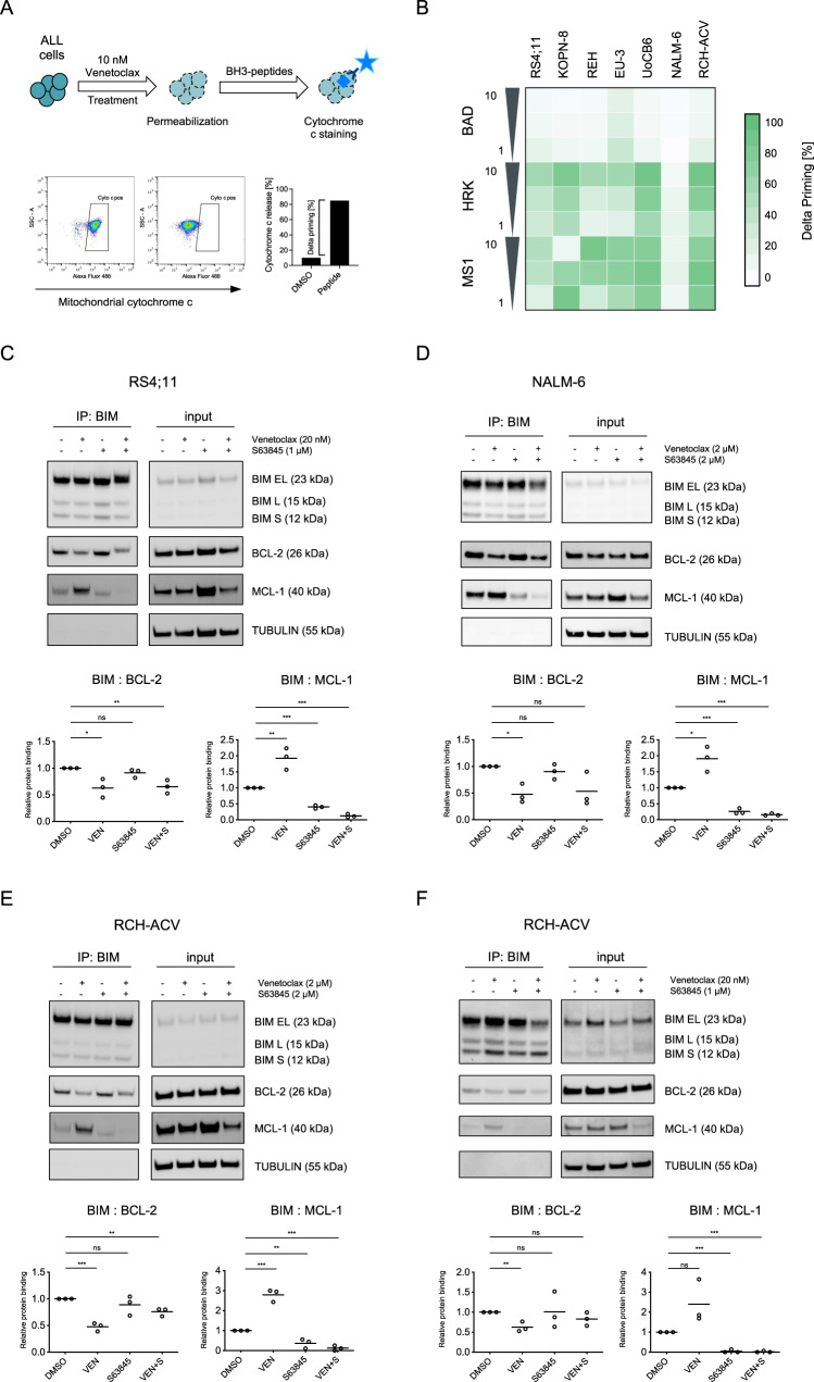
Fig. 3. BCL-XL and MCL-1 mediate resistance of ALL cells to venetoclax.
The interaction of proteins of the BCL-2 family was investigated in BCP-ALL cell lines upon exposure to BH3-mimetics. A Graphical schematic of dynamic BH3 profiling (DBP) analyzing BCP-ALL cell lines. B Cells were exposed to 10 nM venetoclax for 2 h (RS4;11, KOPN-8) or 4 h (all others) followed by permeabilization and incubation with the pro-apoptotic BH3-peptides BAD (indicating BCL-2 dependence), HRK (BCL-XL) and MS1 (MCL-1). Cells were then fixed and stained with an anti-cytochrome c antibody binding exclusively to mitochondrial cytochrome c. Delta priming was calculated as follows: Delta priming (%) = venetoclax-induced cytochrome c release (%) – DMSO control-induced cytochrome c release (%). The heatmap of DBP results shows increased mitochondrial priming in cell lines upon venetoclax-exposure to the MS1 peptide (MCL-1 dependence) and to HRK (BCL-XL). Immunoprecipitation (IP) analysis of BIM and detection of co-precipitated/BIM-bound BCL-2 and MCL-1 upon exposure to venetoclax, S63845 or the combination of both inhibitors for 4 h. C RS4;11, venetoclax sensitive; exposure to 20 nM venetoclax, 1 µM S63845. D NALM-6, venetoclax insensitive; 2 µM venetoclax, 2 µM S63845. E RCH-ACV, venetoclax insensitive; 2 µM venetoclax, 2 µM S63845 and (F) RCH-ACV, low concentrations 20 nM venetoclax, 1 µM S63845. The immunoprecipitation lanes show the interaction of BIM with BCL-2 and MCL-1 and the input lanes show the whole protein lysates. Diagrams show densitometric quantification of co-precipitated BCL-2 (BIM: BCL-2) or MCL-1 (BIM: MCL-1) in the respective condition relative to no inhibitor control summarizing three independent experiments (corresponding replicates see Supplementary Fig. 8). Unpaired two-tailed Student’s t test; significance ***, p < 0.001; **, p < 0.01; *, p < 0.05; ns, not significant.

