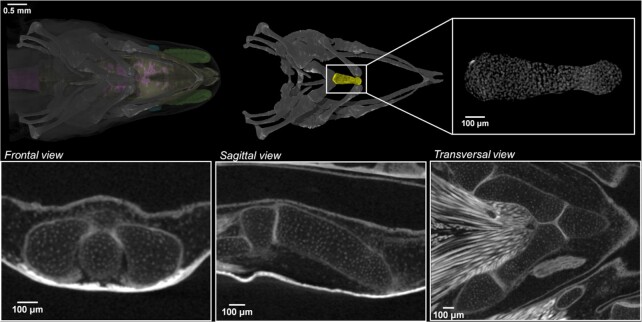
Figure 4:
Images at near cellular-level resolution showing the cartilaginous elements in juvenile P. anguinus obtained by microCT. The white dots represent cell nuclei. 3D detail of the cartilaginous first basibranchial element of the hyobranchial apparatus in ventral view (yellow; top row) with 3 orthogonal CT slices along the frontal, sagittal, and transverse planes (second row).

