Abstract
Background
Axillary lymph node status remains the most powerful prognostic indicator in invasive breast cancer. Ductal carcinoma in situ (DCIS) is a non-invasive disease and does not spread to axillary lymph nodes. The presence of an invasive component to DCIS mandates nodal evaluation through sentinel lymph node biopsy (SLNB). Quantification of the necessity of upfront SLNB for DCIS requires investigation. The aim was to establish the likelihood of having a positive SLNB (SLNB+) for DCIS and to establish parameters predictive of SLNB+.
Methods
A systematic review was performed as per the PRISMA guidelines. Prospective studies only were included. Characteristics predictive of SLNB+ were expressed as dichotomous variables and pooled as odds ratios (o.r.) and associated 95 per cent confidence intervals (c.i.) using the Mantel–Haenszel method.
Results
Overall, 16 studies including 4388 patients were included (mean patient age 54.8 (range 24 to 92) years). Of these, 72.5 per cent of patients underwent SLNB (3156 of 4356 patients) and 4.9 per cent had SLNB+ (153 of 3153 patients). The likelihood of having SLNB+ for DCIS was less than 1 per cent (o.r. <0.01, 95 per cent c.i. 0.00 to 0.01; P < 0.001, I2 = 93 per cent). Palpable DCIS (o.r. 2.01, 95 per cent c.i. 0.64 to 6.24; P = 0.230, I2 = 0 per cent), tumour necrosis (o.r. 3.84, 95 per cent c.i. 0.85 to 17.44; P = 0.080, I2 = 83 per cent), and grade 3 DCIS (o.r. 1.34, 95 per cent c.i. 0.80 to 2.23; P = 0.270, I2 = 0 per cent) all trended towards significance in predicting SLNB+.
Conclusion
While aggressive clinicopathological parameters may guide SLNB for patients with DCIS, the absolute and relative risk of SLNB+ for DCIS is less than 5 per cent and 1 per cent, respectively. Well-designed randomized controlled trials are required to establish fully the necessity of SLNB for patients diagnosed with DCIS.
Registration number
CRD42021284194 (https://www.crd.york.ac.uk/prospero/)
This meta-analysis evaluates the clinical utility of routine sentinel lymph node biopsy for surgery in the cases of ductal carcinoma in situ. The results from this analysis support the de-escalation of sentinel lymph node biopsy in such cases, unless directed otherwise by aggressive clinicopathological data, such as tumour necrosis and palpable disease.
Introduction
Following the widespread establishment and implementation of population-based breast cancer screening programmes and digitalized imaging, detection rates of ductal carcinoma in situ (DCIS) have increased dramatically1,2, with DCIS now constituting 20 to 25 per cent of all breast cancers3. DCIS is a premalignant precursor disease to invasive ductal carcinoma (IDC), which is characterized by abnormal proliferation of epithelial cells confined within the basal membrane of breast glandular tissue4. Theoretically, DCIS is non-invasive, and therefore does not possess any metastatic potential for locoregional spread to axillary lymph nodes. Therefore, routine lymph node sampling to stage the axilla in the setting of DCIS is unnecessary5.
Axillary lymph node status remains the most powerful prognostic indicator in patients diagnosed with breast cancer6. Therefore, sentinel lymph node biopsy (SLNB) is currently mandated in all cases suspected to be invasive breast cancer. In patients with clinically node-negative invasive disease, SLNB is performed and provides non-inferior survival outcomes to axillary lymph node dissection (ALND)7–9. The ACOSOG Z0011 trial demonstrated that patients with invasive breast cancer with limited metastatic disease in the axilla may be spared ALND10. These trials have evolved the paradigm for patients with invasive disease; however, there has been no randomized controlled trial (RCT) published to date investigating the value of performing routine SLNB for patients with DCIS.
At present, a SLNB is only performed in select cases of DCIS, such as cases with large volumes of disease requiring mastectomy, when there is an anticipated risk of upstaging to invasive disease on the specimen following histopathological evaluation. Best practice guidelines, such as those reported by American Society of Clinical Oncology and National Institute for Health and Care Excellence, support SLNB in cases requiring mastectomy, in cases of extensive DCIS (greater than 50 mm), or those with clinical or radiological evidence suggestive of possible invasive disease11,12. However, the evidence supporting such recommendations may be challenged owing the sparsity of data supporting formal staging of the axilla, as well as the absence of concise selection criteria5,13. Thus, it is reasonable to suggest that there is a proportion of patients being treated for DCIS who currently undergo unnecessary upfront SLNB. Moreover, histopathological evaluation of the resected breast specimen is mandatory, which will ultimately dictate the indication for SLNB based on the presence of invasive cancer. Therefore, the rationale for performing upfront SLNB as routine for patients being treated for DCIS should be challenged. Accordingly, the aim of the current systematic review and meta-analysis was to establish the likelihood of having lymph node metastases on SLNB (SLNB+) in patients being treated surgically for DCIS, and to establish clinicopathological parameters that may be useful in predicting those likely to be SLNB+ at the time of breast surgery for DCIS.
Methods
A systematic review was conducted in accordance to the PRISMA checklist14 and Meta-analysis Of Observational Studies in Epidemiology (MOOSE) guidelines15. Given the nature of this review, local institutional ethical approval was not required. The study was registered in PROSPERO (CRD42021284194).
Population, intervention, comparison, outcome (PICO) tool
Using the PICO framework16, the aspects the authors wished to address were:
Population—female patients, aged 18 years or older, with newly diagnosed DCIS breast cancer, with histologically or radiologically confirmed DCIS in the preoperative setting.
Intervention—any patient in the selected group who underwent staging with SLNB and were subsequently found to have positive disease in the axilla at the time of their breast cancer surgery for DCIS.
Comparison—any patient in the selected group who underwent staging with SLNB and were subsequently found not to have positive disease in the axilla at the time of their breast cancer surgery for DCIS.
Outcomes—primary outcomes included SLNB+ (including micro- and/or macrometastatic disease in the axillary lymph nodes) following an initial diagnosis of DCIS. Secondary outcomes included any clinicopathological features predictive of those likely to have SLNB+ following an initial diagnosis of DCIS.
Search strategy
An electronic search was performed of the PubMed, Embase, and Scopus databases on 25 May 2021 for relevant studies that would be suitable for inclusion in this study. The search was performed of all fields under the following headings: (ductal carcinoma in situ), (sentinel lymph node biopsy), which were linked by the Boolean operator ‘AND’. Included studies were limited to those published in the English language and studies were not restricted based on year of publication. For retrieved studies, their titles were initially screened, before the abstracts and full texts which were deemed appropriate were reviewed.
Inclusion and exclusion criteria
Studies meeting the following inclusion criteria were included: studies assessing patients with histologically confirmed DCIS breast cancer in the breast preoperatively with or without positive disease in the axilla (assessed using SLNB); and studies had to include data that were collected prospectively (included studies did not necessarily have to be controlled) and included prospectively collected registry data. Studies meeting any of the following exclusion criteria were excluded: studies with patients with histologically confirmed invasive breast cancer (e.g. IDC histological subtype); studies including data that were not collected prospectively; review articles; studies including fewer than five patients in their series or case reports; or editorial articles.
Data extraction and quality assessment
The literature search was performed by two independent reviewers (M.G.D and C.’OF.) using a predesigned search strategy, which had been developed under the supervision of the senior author (M.J.K.). Duplicate studies were manually removed. Each reviewer then reviewed the titles, abstracts, and/or full texts of the retrieved manuscripts to ensure all inclusion criteria were met, before extracting the following data: first author name; year of publication; study design; level of evidence; study title; number of patients; number of patients who underwent SLNB; number of patients who underwent SLNB and subsequently had axillary lymph nodes positive for cancer cells; clinicopathological and surgical parameters of the entire patient population; and clinicopathological and surgical parameters of the entire patient population with positive axillary lymph nodes. The definition of SLNB+ included ‘micro- and macrometastases only’ in accordance with the AJCC version 8 guidelines for breast cancer17. This definition excluded isolated tumour cells (ITCs). Risk of bias and methodology quality assessment was performed in accordance with the Risk Of Bias In Non-randomised Studies of Interventions (ROBINS-I)18,19. In case of discrepancies in opinion between the reviewers, a third reviewer (E.F.C) was asked to arbitrate.
Statistical analysis
Clinicopathological and treatment characteristics were expressed as descriptive statistics with Fisher’s exact and χ2 tests, as appropriate20, to determine clinicopathological features associated with SLNB+. Determining the likelihood of SLNB+ in cases of DCIS with SLNB and relevance of clinicopathological parameters predictive of SLNB+ were assessed as dichotomous variables, expressed as odds ratios (o.r.) with corresponding 95 per cent confidence intervals (c.i.) using the Mantel–Haenszel method. Either fixed- or random-effects models were applied on the basis of whether significant heterogeneity (I2 > 50 per cent) existed between studies included in the analysis. Symmetry of funnel plots was used to assess publication bias. Statistical heterogeneity was determined using I2 statistics. Statistical significance was determined to be P < 0.050. Statistical analysis was performed using Review Manager (RevMan), Version 5.4 (Nordic Cochrane Centre, Copenhagen, Denmark).
Results
Literature search
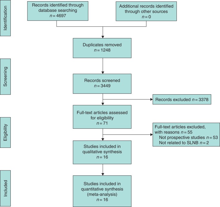
The initial electronic search retrieved 4697 studies. Following removal of the 1248 identified duplicate studies, the remaining 3449 titles were screened for relevance, of which 576 had their abstracts and 71 had their full texts assessed for eligibility. Overall, 16 prospective studies fulfilled the inclusion criteria and were subsequently included in this systematic review21–36, as outlined in Fig. 1. Details of individual included studies are outlined in Table 1.
Fig. 1.
PRISMA flow diagram detailing the systematic search process
SLNB, sentinel lymph node biopsy.
Table 1.
Details of the 16 prospective studies in this systematic review and meta-analysis
| Author | Year | Country | n | Mean patient age (years) | Age range (years) | ROBINS-I |
|---|---|---|---|---|---|---|
| Kelly | 2003 | USA | 420 | 54.3 | — | 2 |
| Mittendorf | 2005 | USA | 85 | 57.0 | 29–85 | 2 |
| Guillot | 2014 | France | 241 | 51.0 | 28–82 | 2 |
| Goyal | 2006 | UK | 367 | 58.0 | 49–81 | 2 |
| Moran | 2007 | ROI | 62 | — | 50–65 | 2 |
| Usmani | 2011 | Kuwait | 23 | 50.0 | 37–78 | 3 |
| Zetterlund | 2014 | Sweden | 1273 | 60.0 | 26–92 | 2 |
| D’Eredita | 2009 | Italy | 90 | 56.0 | 27–86 | 3 |
| Collado | 2010 | Spain | 65 | 51.9 | 38–69 | 3 |
| Klauber-DeMore | 2000 | USA | 76 | — | 3 | |
| Fancellu | 2012 | Italy | 140 | 56.0 | 26–89 | 2 |
| Intra | 2003 | Italy | 854 | — | — | 2 |
| Tunon-de-Lara | 2015 | France | 227 | — | 24–83 | 2 |
| van la Parra | 2008 | Netherlands | 51 | 59.0 | 39–81 | 3 |
| Leidenius | 2006 | Finland | 74 | 56.0 | 38–91 | 3 |
| Park | 2013 | ROK | 340 | 48.5 | 25–78 | 3 |
ROBINS-I, risk of bias in non-randomised studies of interventions; ROI, Republic of Ireland; ROK, Republic of Korea.
Study characteristics
In total, 4388 patients diagnosed with DCIS were included in this study. Mean patient age at diagnosis was 54.8 (range 24 to 92) years. Overall, 67.6 per cent of patients underwent mastectomy (2514 of 3719) and 32.4 per cent underwent breast conservation surgery (1205 of 3719; 13 studies). In total, 72.5 per cent of patients underwent SLNB (3156 of 4356) and 4.9 per cent had SLNB+ (153 of 3153). Of the 4388 patients included in this study, 314 had invasive cancer in the breast present on their final histology (7.2 per cent). Of these, 26.8 per cent had SLNB+ (84 of 314). Pooled clinicopathological and treatment data from the 16 included studies are outlined in Table 2.
Table 2.
Clinicopathological and treatment characteristics of the included patients in this study
| Parameter | Total group | SLNB+ group | P * |
|---|---|---|---|
| Screening detected | 364 | 11 | 0.350 |
| Symptomatic (palpable) | 96 | 5 | |
| Necrosis present | 691 | 30 | <0.001 |
| Necrosis absent | 1127 | 16 | |
| Microcalcification present | 226 | 13 | 0.309 |
| Microcalcification absent | 126 | 15 | |
| Grade 1 | 325 | 7 | 0.969† |
| Grade 2 | 1039 | 24 | |
| Grade 3 | 1614 | 35 | |
| Grade 1/2 | 1727 | 31 | 0.447 |
| Grade 3 | 1614 | 35 | |
| ER+ | 825 | 9 | 0.299 |
| ER− | 381 | 7 | |
| PgR+ | 802 | 9 | 0.386 |
| PgR− | 403 | 7 | |
| HER2+ | 479 | 4 | 0.270 |
| HER2− | 765 | 12 | |
| Ki67 < 20% | 324 | 9 | 0.096 |
| Ki67 > 20% | 446 | 5 | |
| BCS | 1205 | 12 | 0.016 |
| Mastectomy | 2514 | 9 |
SLNB+, metastatic lymph nodes on sentinel lymph node biopsy; ER, oestrogen receptor; PgR, progesterone receptor; HER2, human epidermal growth factor receptor-2; BCS, breast conservation surgery. *P values from Fisher’s exact test unless otherwise stated; †χ2 test.
Axillary lymph node positivity
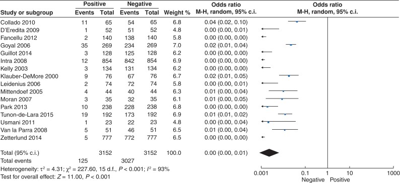
As previously outlined, 4.9 per cent of the 3153 patients who underwent SLNB had positive disease on their SLNB (153 of 3153). Of those reporting type of metastases, 58.4 per cent had micrometastases present on SLNB (66 of 113), while 41.6 per cent had macrometastatic disease present on SLNB (47 of 113). For the 3153 patients undergoing SLNB, the likelihood of having SLNB+ was less than 1 per cent (o.r. < 0.01, 95 per cent c.i. 0.00 to 0.01; P < 0.001, I2 = 93 per cent) (Fig. 2).
Fig. 2.
Forest plot illustrating the likelihood of having metastatic disease in axillary lymph nodes in patients with ductal carcinoma in situ
Of note, ITCs were present in 0.8 per cent of cases (26 of 3153). Overall, 4.7 per cent of patients proceeded to axillary lymph node dissection (148 of 3153). Details in relation to axillary lymph node status are provided in Table 3.
Table 3.
Details in relation to sentinel lymph node biopsies, lymph node status, and axillary lymph node dissection
| Parameter | n (%) |
|---|---|
| Underwent SLNB | 3156 (79.6) |
| Did not undergo SLNB | 1200 (20.4) |
| SLNB− | 3000 (95.1) |
| SLNB+ | 153 (4.9) |
| Not reported | 3 (<0.1) |
| Micrometastases | 66 (43.1) |
| Macrometastases | 47 (30.7) |
| Not reported | 40 (26.1) |
| ITCs | 26 (0.8) |
| ALND | 148 (4.7) |
SLNB, sentinel lymph node biopsy; ITCs, isolated tumour cells; ALND, axillary lymph node dissection.
Clinicopathological predictors of axillary lymph node positivity
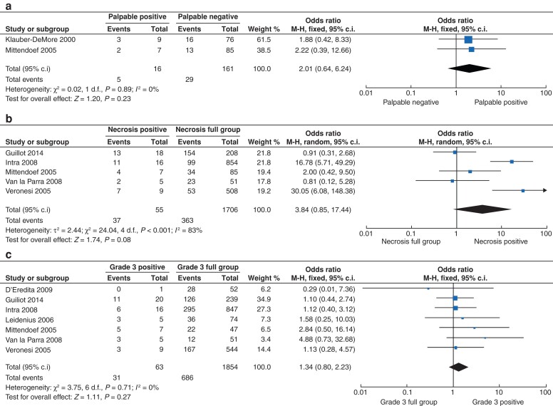
The presence of tumour necrosis (P < 0.001) and undergoing mastectomy (P = 0.016) were both associated with having SLNB+ for DCIS surgery (Table 2). Being symptomatic (or having palpable DCIS (o.r. 2.01, 95 per cent c.i. 0.64 to 6.24; P = 0.230, I2 = 0 per cent)) (Fig. 3a), the presence of tumour necrosis (o.r. 3.84, 95 per cent c.i. 0.85 to 17.44; P = 0.080, I2 = 83 per cent) (Fig. 3b), and the presence of grade 3 disease (o.r. 1.34, 95 per cent c.i. 0.80 to 2.23; P = 0.270, I2 = 0 per cent) (Fig. 3c) all trended towards significance in predicting patients likely to have SLNB+. Forest plots for other clinicopathological parameters and predicted value for SLNB+ are outlined in Fig. S1.
Fig. 3.
Forest plot illustrating the ability of
a palpable disease, b tumour necrosis, and c grade 3 ductal carcinoma in situ in predicting metastatic disease in axillary lymph nodes.
Discussion
This systematic review and meta-analysis assessed the value of performing routine SLNB in patients being treated surgically for DCIS. For decades, the surgical conundrum surrounding the appropriateness of SLNB for cases of DCIS has been debated by surgical oncologists, owing to a lack of clear consensus. The results of the current meta-analysis were derived from the highest level of evidence available (prospectively collected data only). Similarly to the work of El Hage Chehade et al.37, the overall absolute likelihood of capturing metastatic disease in axillary lymph nodes following SLNB was approximately 5 per cent, with an estimated relative detection rate of less than 1 per cent. Although this illustrates there is the potential to detect metastatic disease in the axilla at SLNB, the data do not support the performance of a priori lymph node sampling as routine in all cases of DCIS. Therefore, the clear message from this meta-analysis is that the surgical oncologist, at their own discretion, should avoid performing SLNB for DCIS surgery, unless there is high suspicion for invasive disease.
In this analysis, 72.5 per cent of patients underwent SLNB for DCIS, yet less than 5 per cent of these had SLNB+. This suggests that there is a tendency for breast surgeons to stage the axilla in cases of DCIS, despite acknowledgement that this is a non-invasive disease38. Debate fuelling the controversy of sentinel node mapping as routine management of DCIS is based on the following fundamental concepts. Primarily, the resecting surgeon is aware that there is a proportion of patients with DCIS who will ultimately progress to develop IDC39,40. Additionally, sentinel lymph node status remains the most crucial predictor of prognosis in invasive carcinoma6, and if invasive disease is detected, axillary staging is fundamental to therapeutic decision-making in the adjuvant setting41,42. These principles remain at the crux of the argument supporting routine SLNB for DCIS. Nevertheless, the real-world data presented in the current analysis highlight that there is a less than a 5 per cent absolute risk of invasive cancer being detected on SLNB on final histology. Therefore, judicious use of SLNB is required within the setting of DCIS, with limited exceptions.
This study may be challenged by being perceived as oversimplifying the requirement for routine axillary staging in cases of DCIS. However, these data illustrate that there are certain clinicopathological parameters associated with SLNB+, which may be useful in guiding preoperative decision-making in relation to SLNB. These data suggest that having palpable disease (o.r. 2.01), the presence of tumour necrosis (o.r. 3.84), and having grade 3 DCIS (o.r. 1.34) are useful tumour characteristics for predicting SLNB+. This is somewhat unsurprising. Palpable DCIS has been associated with aggressive clinicopathological features, such as high-grade and comedo necrosis43, as well as invasive cancer in approximately 25 per cent of cases44. Therefore, it is fair to expect that such cases may require mastectomy, particularly when palpable DCIS (or large-volume DCIS, which will require mastectomy) is a reasonable parameter for which SLNB may be considered. Furthermore, Kerlikowske et al. reported that palpable DCIS, combined with high-grade histology, independently predicts DCIS recurrence as invasive disease45. High-grade DCIS (or grade 3 DCIS) shows large-sized, pleomorphic neoplastic cells, with large and irregularly shaped nuclei, with multiple, prominent nucleoli and high mitotic indices, indicating high proliferative potential46. Moreover, these cancers often show a necrotic core46, and recent prospective data from the Sloane Project illustrated that high-grade DCIS correlated with poorer oncological outcome than those with low–intermediate grade DCIS after more than 9 years of follow-up47. Additionally, comedo necrosis (or central necrosis) occurs in highly proliferative cancers that outgrow their supply of nutrients and oxygen, causing deprivation and tumour apoptosis48. Unsurprisingly, comedo necrosis has been correlated with aggressive tumour features such as increased tumour burden, higher proliferative potential, and poorer anticipated prognosis49, with strong associations with ipsilateral invasive cancer recurrence50. This suggests that caution is required when deciding on the appropriate staging of such cases. It is acknowledged that grade and necrosis are contemporary characteristics in the College of American Pathologists reporting protocol for the histopathological specimens of DCIS51, which further emphasizes their importance in cases of DCIS. Therefore, when the breast multidisciplinary team meeting is faced with a case of palpable, grade 3, necrosing DCIS, consideration for SLNB is justified, to ensure adequate staging of the axilla in the incidence that the resected tumour is upstaged to invasive disease on final histology.
While the era of molecular profiling and minimally invasive surgery have revolutionized the approach to the management of invasive breast cancer7,8,10,41,52–54, the translational research efforts to progress the management of DCIS have lagged behind considerably. For example, multigene assays, such as the 21-gene and 70-gene signatures, have become embedded into the paradigm for certain early-stage invasive cancers41,53,55–57. In contrast, the uptake of the clinically validated 12-gene DCIS recurrence assay has been less successful58,59. With respect to surgical management of the axilla in cases of DCIS, there is currently just one ongoing clinical trial focused on enhancing surgical practice for patients with DCIS: the SentiNot 2.0 trial (NCT04722692) is currently randomizing patients to either radioisotope (control) or superparamagnetic iron oxide (SPIO) tracing of the axillary nodes at delayed SLNB in patients with a preoperative diagnosis of DCIS, who are subsequently found to have invasive disease on final histology60. Similar to the message of the current meta-analysis, SentiNot 2.0 proposes a delay in performing SLNB in patients undergoing mastectomy for DCIS. Therefore, the next generation of prospective trials should look to evaluate the necessity of upfront SLNB for DCIS, in order to provide clear consensus to the debate regarding the most appropriate management of the axilla in such circumstances.
This meta-analysis is subject to several limitations. In the absence of well-designed RCTs evaluating the necessity of SLNB in DCIS surgery, cautious interpretation of these results is required. Observational studies of a non-randomized design, in particular those where retrospective analysis of prospectively collected data is performed, are subject to the inherent risk of selection and confounding biases. In this study, surgical procedures performed in those with SLNB+ were outlined in just 13.7 per cent of cases (21 of 153), meaning the full necessity of SLNB in cases requiring mastectomy for DCIS has not been fully evaluated. In such circumstances, axillary staging may be appropriate at the time of resection12, as small invasive cancers are occasionally present on final histology. Detecting clinicopathological characteristics predictive of SLNB+ was the secondary outcome in this study; however, the paucity of such data may bring the validity of these results into question (as outlined in Table 2). Despite these limitations, this meta-analysis provides the highest quality of available prospective data reflecting current management strategies of the axilla in cases of DCIS.
This systematic review and meta-analysis illustrates an absolute likelihood of less than 5 per cent of having metastatic disease following SLNB for DCIS, with an estimated relative risk of less than 1 per cent. It therefore suggests that there is limited premise for upfront axillary lymph node sampling in the setting of DCIS. However, aggressive clinicopathological characteristics, such as having a clinically palpable tumour, or possessing comedo necrosis and/or high-grade DCIS on diagnostic core biopsy, may be useful to guide preoperative decision-making as to when SLNB may be required. The provision of well-designed prospective studies are essential to evaluate properly the de-escalation of upfront SLNB in patients being treated surgically for DCIS.
Supplementary Material
Funding
M.G.D., C.O’F., A.N., J.P., and E.R. received funding from the National Breast Cancer Research Institute, Ireland.
Disclosure. The authors declare no conflict of interest
Supplementary material
Supplementary material is available at BJS Open online.
Data availability
Data are available upon request at the discretion of the corresponding author.
References
- 1. Virnig BA, Wang S-Y, Shamilyan T, Kane RL, Tuttle TM. Ductal carcinoma in situ: risk factors and impact of screening. J Natl Cancer Inst Monogr 2010;2010:113–116 [DOI] [PMC free article] [PubMed] [Google Scholar]
- 2. Neal CH, Joe AI, Patterson SK, Pujara AC, Helvie MA. Digital mammography has persistently increased high-grade and overall DCIS detection without altering upgrade rate. AJR Am J Roentgenol 2021;216:912–918 [DOI] [PubMed] [Google Scholar]
- 3. Siegel RL, Miller KD, Jemal A. Cancer statistics, 2018. CA Cancer J Clin 2018;68:7–30 [DOI] [PubMed] [Google Scholar]
- 4. Vaidya Y, Vaidya P, Vaidya T. Ductal carcinoma in situ of the breast. Indian J Surg 2015;77:141–146 [DOI] [PMC free article] [PubMed] [Google Scholar]
- 5. Shin YD, Lee H-M, Choi YJ. Necessity of sentinel lymph node biopsy in ductal carcinoma in situ patients: a retrospective analysis. BMC Surg 2021;21:159. [DOI] [PMC free article] [PubMed] [Google Scholar]
- 6. Andersson Y, Bergkvist L, Frisell J, de Boniface J. Long-term breast cancer survival in relation to the metastatic tumor burden in axillary lymph nodes. Breast Cancer Res Treat 2018;171:359–369 [DOI] [PubMed] [Google Scholar]
- 7. Giuliano AE, Haigh PI, Brennan MB, Hansen NM, Kelley MC, Ye Wet al. Prospective observational study of sentinel lymphadenectomy without further axillary dissection in patients with sentinel node–negative breast cancer. J Clin Oncol 2000;18:2553–2559 [DOI] [PubMed] [Google Scholar]
- 8. Veronesi U, Paganelli G, Viale G, Luini A, Zurrida S, Galimberti Vet al. A randomized comparison of sentinel-node biopsy with routine axillary dissection in breast cancer. N Engl J Med 2003;349:546–553 [DOI] [PubMed] [Google Scholar]
- 9. Davey MG, Ryan ÉJ, Burke D, McKevitt K, McAnena PF, Kerin MJet al. Evaluating the clinical utility of routine sentinel lymph node biopsy and the value of adjuvant chemotherapy in elderly patients diagnosed with oestrogen receptor positive, clinically node negative breast cancer. Breast Cancer (Auckl) 2021;15:11782234211022203. [DOI] [PMC free article] [PubMed] [Google Scholar]
- 10. Giuliano AE, Hunt KK, Ballman KV, Beitsch PD, Whitworth PW, Blumencranz PWet al. Axillary dissection vs no axillary dissection in women with invasive breast cancer and sentinel node metastasis: a randomized clinical trial. JAMA 2011;305:569–575 [DOI] [PMC free article] [PubMed] [Google Scholar]
- 11. Lyman GH, Temin S, Edge SB, Newman LA, Turner RR, Weaver DLet al. Sentinel lymph node biopsy for patients with early-stage breast cancer: American Society of Clinical Oncology clinical practice guideline update. J Clin Oncol 2014;32:1365–1383 [DOI] [PubMed] [Google Scholar]
- 12. NICE . Early and locally advanced breast cancer: diagnosis and management. https://www.nice.org.uk/guidance/ng101/resources/early-and-locally-advanced-breast-cancer-diagnosis-and-management-pdf-66141532913605 (accessed 1 October 2021)
- 13. Gojon H, Fawunmi D, Valachis A. Sentinel lymph node biopsy in patients with microinvasive breast cancer: a systematic review and meta-analysis. Eur J Surg Oncol 2014;40:5–11 [DOI] [PubMed] [Google Scholar]
- 14. Moher D, Liberati A, Tetzlaff J, Altman DG. Preferred reporting items for systematic reviews and meta-analyses: the PRISMA statement. BMJ 2009;339:b2535. [DOI] [PMC free article] [PubMed] [Google Scholar]
- 15. Stroup DF, Berlin JA, Morton SC, Olkin I, Williamson GD, Rennie Det al. Meta-analysis of observational studies in epidemiology: a proposal for reporting. Meta-analysis Of Observational Studies in Epidemiology (MOOSE) group. JAMA 2000;283:2008–2012 [DOI] [PubMed] [Google Scholar]
- 16. Armstrong EC. The well-built clinical question: the key to finding the best evidence efficiently. WMJ 1999;98:25–28 [PubMed] [Google Scholar]
- 17. Giuliano AE, Connolly JL, Edge SB, Mittendorf EA, Rugo HS, Solin LJet al. Breast cancer—major changes in the American Joint Committee on Cancer eighth edition cancer staging manual. CA Cancer J Clin 2017;67:290–303 [DOI] [PubMed] [Google Scholar]
- 18. Higgins JPT, Altman DG, Gøtzsche PC, Jüni P, Moher D, Oxman ADet al. The Cochrane Collaboration’s tool for assessing risk of bias in randomised trials. BMJ 2011;343:d5928. [DOI] [PMC free article] [PubMed] [Google Scholar]
- 19. Sterne JAC, Hernán MA, Reeves BC, Savović J, Berkman ND, Viswanathan Met al. ROBINS-I: a tool for assessing risk of bias in non-randomised studies of interventions. BMJ 2016;355:i4919. [DOI] [PMC free article] [PubMed] [Google Scholar]
- 20. Kim H-Y. Statistical notes for clinical researchers: chi-squared test and Fisher’s exact test. Restor Dent Endod 2017;42:152–155 [DOI] [PMC free article] [PubMed] [Google Scholar]
- 21. Kelly TA, Kim JA, Patrick R, Grundfest S, Crowe JP. Axillary lymph node metastases in patients with a final diagnosis of ductal carcinoma in situ. Am J Surg 2003;186:368–370 [DOI] [PubMed] [Google Scholar]
- 22. Mittendorf EA, Arciero CA, Gutchell V, Hooke J, Shriver CD. Core biopsy diagnosis of ductal carcinoma in situ: an indication for sentinel lymph node biopsy. Curr Surg 2005;62:253–257 [DOI] [PubMed] [Google Scholar]
- 23. Guillot E, Vaysse C, Goetgeluck J, Falcou MC, Couturaud B, Fitoussi Aet al. Extensive pure ductal carcinoma in situ of the breast: identification of predictors of associated infiltrating carcinoma and lymph node metastasis before immediate reconstructive surgery. Breast 2014;23:97–103 [DOI] [PubMed] [Google Scholar]
- 24. Goyal A, Douglas-Jones A, Monypenny I, Sweetland H, Stevens G, Mansel RE. Is there a role of sentinel lymph node biopsy in ductal carcinoma in situ?: analysis of 587 cases. Breast Cancer Res Treat 2006;98:311–314 [DOI] [PubMed] [Google Scholar]
- 25. Moran CJ, Kell MR, Flanagan FL, Kennedy M, Gorey TF, Kerin MJ. Role of sentinel lymph node biopsy in high-risk ductal carcinoma in situ patients. Am J Surg 2007;194:172–175 [DOI] [PubMed] [Google Scholar]
- 26. Usmani S, Khan HA, Al Saleh N, abu Huda F, Marafi F, Amanguno HGet al. Selective approach to radionuclide-guided sentinel lymph node biopsy in high-risk ductal carcinoma in situ of the breast. Nucl Med Commun 2011;32:1084–1087 [DOI] [PubMed] [Google Scholar]
- 27. D’Eredità G, Giardina C, Napoli A, Ingravallo G, Troilo VL, Fischetti Fet al. Sentinel lymph node biopsy in patients with pure and high-risk ductal carcinoma in situ of the breast. Tumori J 2009;95:706–711 [DOI] [PubMed] [Google Scholar]
- 28. Collado MV, Ruiz-Tovar J, García-Villanueva A, Rojo R, Latorre L, Rioja MEet al. Sentinel lymph node biopsy in selected cases of ductal carcinoma in situ. Clin Transl Oncol 2010;12:499–502 [DOI] [PubMed] [Google Scholar]
- 29. Klauber-DeMore N, Tan LK, Liberman L, Kaptain S, Fey J, Borgen Pet al. Sentinel lymph node biopsy: is it indicated in patients with high-risk ductal carcinoma-in-situ and ductal carcinoma-in-situ with microinvasion? Ann Surg Oncol 2000;7:636–642 [DOI] [PubMed] [Google Scholar]
- 30. Fancellu A, Cottu P, Feo CF, Bertulu D, Giuliani G, Mulas Set al. Sentinel node biopsy in early breast cancer: lessons learned from more than 1000 cases at a single institution. Tumori J 2012;98:413–420 [DOI] [PubMed] [Google Scholar]
- 31. Intra M, Veronesi P, Mazzarol G, Galimberti V, Luini A, Sacchini Vet al. Axillary sentinel lymph node biopsy in patients with pure ductal carcinoma in situ of the breast. Arch Surg 2003;138:309–313 [DOI] [PubMed] [Google Scholar]
- 32. Tunon-de-Lara C, Chauvet MP, Baranzelli MC, Baron M, Piquenot J, Le-Bouédec Get al. The role of sentinel lymph node biopsy and factors associated with invasion in extensive DCIS of the breast treated by mastectomy: the Cinnamome prospective multicenter study. Ann Surg Oncol 2015;22:3853–3860 [DOI] [PMC free article] [PubMed] [Google Scholar]
- 33. van la Parra RFD, Ernst MF, Barneveld PC, Broekman JM, Rutten MJCM, Bosscha K. The value of sentinel lymph node biopsy in ductal carcinoma in situ (DCIS) and DCIS with microinvasion of the breast. Eur J Surg Oncol 2008;34:631–635 [DOI] [PubMed] [Google Scholar]
- 34. Leidenius M, Salmenkivi K, von Smitten K, Heikkilä P. Tumour-positive sentinel node findings in patients with ductal carcinoma in situ. J Surg Oncol 2006;94:380–384 [DOI] [PubMed] [Google Scholar]
- 35. Park HS, Park S, Cho J, Park JM, Kim SI, Park BW. Risk predictors of underestimation and the need for sentinel node biopsy in patients diagnosed with ductal carcinoma in situ by preoperative needle biopsy. J Surg Oncol 2013;107:388–392 [DOI] [PubMed] [Google Scholar]
- 36. Zetterlund L, Stemme S, Arnrup H, de Boniface J. Incidence of and risk factors for sentinel lymph node metastasis in patients with a postoperative diagnosis of ductal carcinoma in situ. Br J Surg 2014;101:488–494 [DOI] [PubMed] [Google Scholar]
- 37. El Hage Chehade H, Headon H, Wazir U, Abtar H, Kasem A, Mokbel K. Is sentinel lymph node biopsy indicated in patients with a diagnosis of ductal carcinoma in situ? A systematic literature review and meta-analysis. Am J Surg 2017;213:171–180 [DOI] [PubMed] [Google Scholar]
- 38. Hong YK, McMasters KM, Egger ME, Ajkay N. Ductal carcinoma in situ current trends, controversies, and review of literature. Am J Surg 2018;216:998–1003 [DOI] [PubMed] [Google Scholar]
- 39. Lamb LR, Kim G, Oseni TO, Bahl M. Noncalcified ductal carcinoma in situ (DCIS): rate and predictors of upgrade to invasive carcinoma. Acad Radiol 2021;28:e71–e76 [DOI] [PubMed] [Google Scholar]
- 40. van Seijen M, Lips EH, Thompson AM, Nik-Zainal S, Futreal A, Hwang ESet al. Ductal carcinoma in situ: to treat or not to treat, that is the question. Br J Cancer 2019;121:285–292 [DOI] [PMC free article] [PubMed] [Google Scholar]
- 41. Kalinsky K, Barlow WE, Meric-Bernstam F, Gralow JR, Albain KS, Hayes Det al. Abstract GS3-00: first results from a phase III randomized clinical trial of standard adjuvant endocrine therapy (ET) +/- chemotherapy (CT) in patients (pts) with 1–3 positive nodes, hormone receptor-positive (HR+) and HER2-negative (HER2-) breast cancer (BC) with recurrence score (RS) <25: SWOG S1007 (RxPonder). Cancer Res 2021;81:GS3 [Google Scholar]
- 42. Thomssen C, Balic M, Harbeck N, Gnant M. St. Gallen/Vienna 2021: a brief summary of the consensus discussion on customizing therapies for women with early breast cancer. Breast Care 2021;16:135–143 [DOI] [PMC free article] [PubMed] [Google Scholar]
- 43. Sundara Rajan S, Verma R, Shaaban AM, Sharma N, Dall B, Lansdown M. Palpable ductal carcinoma in situ: analysis of radiological and histological features of a large series with 5-year follow-up. Clin Breast Cancer 2013;13:486–491 [DOI] [PubMed] [Google Scholar]
- 44. Lee RJ, Vallow LA, McLaughlin SA, Tzou KS, Hines SL, Peterson JL. Ductal carcinoma in situ of the breast. Int J Surg Oncol 2012;2012:123549. [DOI] [PMC free article] [PubMed] [Google Scholar]
- 45. Kerlikowske K, Molinaro A, Cha I, Ljung B-M, Ernster VL, Stewart Ket al. Characteristics associated with recurrence among women with ductal carcinoma in situ treated by lumpectomy. J Natl Cancer Inst 2003;95:1692–1702 [DOI] [PubMed] [Google Scholar]
- 46. Salvatorelli L, Puzzo L, Vecchio GM, Caltabiano R, Virzì V, Magro G. Ductal carcinoma in situ of the breast: an update with emphasis on radiological and morphological features as predictive prognostic factors. Cancers 2020;12:609. [DOI] [PMC free article] [PubMed] [Google Scholar]
- 47. Shaaban AM, Hilton B, Clements K, Provenzano E, Cheung S, Wallis MGet al. Pathological features of 11,337 patients with primary ductal carcinoma in situ (DCIS) and subsequent events: results from the UK Sloane Project. Br J Cancer 2021;124:1009–1017 [DOI] [PMC free article] [PubMed] [Google Scholar]
- 48. Karsch-Bluman A, Feiglin A, Arbib E, Stern T, Shoval H, Schwob Oet al. Tissue necrosis and its role in cancer progression. Oncogene 2019;38:1920–1935 [DOI] [PubMed] [Google Scholar]
- 49. Lee SY, Ju MK, Jeon HM, Jeong EK, Lee YJ, Kim CHet al. Regulation of tumor progression by programmed necrosis. Oxid Med Cell Longev 2018;2018:3537471. [DOI] [PMC free article] [PubMed] [Google Scholar]
- 50. Hanna WM, Parra-Herran C, Lu FI, Slodkowska E, Rakovitch E, Nofech-Mozes S. Ductal carcinoma in situ of the breast: an update for the pathologist in the era of individualized risk assessment and tailored therapies. Mod Pathol 2019;32:896–915 [DOI] [PubMed] [Google Scholar]
- 51. Cho SY, Park SY, Bae YK, Kim JY, Kim EK, Kim WGet al. Standardized pathology report for breast cancer. J Pathol Transl Med 2021;55:1–15 [DOI] [PMC free article] [PubMed] [Google Scholar]
- 52. Davey MG, Davey CM, Ryan ÉJ, Lowery AJ, Kerin MJ. Combined breast conservation therapy versus mastectomy for BRCA mutation carriers—a systematic review and meta-analysis. Breast 2021;56:26–34 [DOI] [PMC free article] [PubMed] [Google Scholar]
- 53. Sparano JA, Gray RJ, Makower DF, Pritchard KI, Albain KS, Hayes DFet al. Adjuvant chemotherapy guided by a 21-gene expression assay in breast cancer. N Engl J Med 2018;379:111–121 [DOI] [PMC free article] [PubMed] [Google Scholar]
- 54. Davey MG, Ryan ÉJ, Abd Elwahab S, Elliott JA, McAnena PF, Sweeney KJet al. Clinicopathological correlates, oncological impact, and validation of Oncotype DX™ in a European Tertiary Referral Centre. Breast J 2021;27:521–528 [DOI] [PubMed] [Google Scholar]
- 55. Cardoso F, van’t Veer LJ, Bogaerts J, Slaets L, Viale G, Delaloge Set al. 70-gene signature as an aid to treatment decisions in early-stage breast cancer. N Engl J Med 2016;375:717–729 [DOI] [PubMed] [Google Scholar]
- 56. Goldhirsch A, Winer EP, Coates AS, Gelber RD, Piccart-Gebhart M, Thürlimann Bet al. Personalizing the treatment of women with early breast cancer: highlights of the St Gallen International Expert Consensus on the Primary Therapy of Early Breast Cancer 2013. Ann Oncol 2013;24:2206–2223 [DOI] [PMC free article] [PubMed] [Google Scholar]
- 57. Davey MG, Lowery AJ, Miller N, Kerin MJ. MicroRNA expression profiles and breast cancer chemotherapy. Int J Mol Sci 2021;22:10812. [DOI] [PMC free article] [PubMed] [Google Scholar]
- 58. Rakovitch E, Nofech-Mozes S, Hanna W, Baehner FL, Saskin R, Butler SMet al. A population-based validation study of the DCIS Score predicting recurrence risk in individuals treated by breast-conserving surgery alone. Breast Cancer Res Treat 2015;152:389–398 [DOI] [PMC free article] [PubMed] [Google Scholar]
- 59. Solin LJ, Gray R, Baehner FL, Butler SM, Hughes LL, Yoshizawa Cet al. A multigene expression assay to predict local recurrence risk for ductal carcinoma in situ of the breast. J Natl Cancer Inst 2013;105:701–710 [DOI] [PMC free article] [PubMed] [Google Scholar]
- 60. Karakatsanis A, Warnberg F, Thompson A, Kwong A, Christenson G, Mohamed Iet al. Sentinel lymph node biopsy in ductal cancer in situ or unclear lesions of the breast and how to not do it. an open-label, phase 3, randomised controlled trial. (SentiNot 2.0).https://clinicaltrials.gov/ct2/show/NCT04722692 (accessed 1 October 2021)
Associated Data
This section collects any data citations, data availability statements, or supplementary materials included in this article.
Supplementary Materials
Data Availability Statement
Data are available upon request at the discretion of the corresponding author.


