Abstract
Single-molecule fluorescence techniques have revolutionized our ability to study proteins. However, the presence of a fluorescent label can alter the protein structure and/or modify its reaction with other species. To avoid the need for a fluorescent label, the intrinsic autofluorescence of proteins in the ultraviolet offers the benefits of fluorescence techniques without introducing the labelling drawbacks. Unfortunately, the low autofluorescence brightness of proteins has greatly challenged single molecule detection so far. Here we introduce optical horn antennas, a dedicated nanophotonic platform enabling the label-free detection of single proteins in the UV. This design combines fluorescence plasmonic enhancement, efficient collection up to 85° angle and background screening. We detect the UV autofluorescence from immobilized and diffusing single proteins, and monitor protein unfolding and dissociation upon denaturation. Optical horn antennas open up a unique and promising form of fluorescence spectroscopy to investigate single proteins in their native states in real time.
Subject terms: Single-molecule biophysics, Fluorescence spectroscopy, Nanophotonics and plasmonics
The authors introduce optical horn antennas, a nanophotonic platform combining plasmonic enhancement, efficient collection and background screening, for detection of UV autofluorescence from single proteins. They demonstrate label-free monitoring of protein unfolding and dissociation upon denaturation.
Introduction
One of the ultimate goals of molecular biology is to watch how single proteins work in their native state. While single-molecule fluorescence techniques have achieved impressive results towards this goal1,2, the requirement for fluorescent markers can potentially lead to severe issues altering the protein structure or modifying its reaction with other species3–8. Therefore, label-free alternatives to detect single molecules are actively investigated9–13. The protein autofluorescence in the ultraviolet (UV) is an appealing route to rule out all the issues related to external fluorescence labeling14,15. More than 90% of all proteins contain some tryptophan or tyrosine aminoacid residues which are naturally fluorescent in the UV15. Being able to detect the UV autofluorescence from a single (label-free) protein would be a disruptive method offering many benefits of fluorescence techniques (signal-to-noise ratio, temporal dynamics, sensitivity…) without introducing the labeling drawbacks.
However, proteins are orders of magnitude dimmer as compared to conventional fluorescent dyes, so that single protein UV detection has remained a major challenge so far16–18. Hence, new nanotechnology tools need to be introduced to intensify the emission from single proteins. One of the main limiting issues is that close to a planar dielectric interface, a significant fraction of the light from a single dipole is emitted at large angles above 65°. This fundamental phenomenon is known as supercritical or forbidden light19,20. Microscopes operating in the visible spectral range use objectives of high numerical apertures of 1.4 or above to maximize the fluorescence collected from a single molecule. In the UV, however, the choice of microscope objectives is strongly restricted14. UV objectives have a numerical aperture typically below 0.8, which corresponds to a maximum collection angle of 33° into the quartz substrate of 1.48 refractive index. Collecting the forbidden UV light emitted at high angles is crucial to maximize the autofluorescence signal and unlock single label-free protein detection.
In analogy to radiofrequency antennas, optical antennas offer a way to control and intensify the emission of single quantum emitters21. Intense fluorescence enhancement factors have been achieved with strongly absorbing dyes in the visible22–27, but most optical antennas designs remain unsuitable for UV protein detection due to their narrowband spectral response28,29, challenging nanofabrication24,27, or requirement for solid-state integration30,31. Alternative designs must be developed to offer a highly efficient platform, reaching the needs of high photon count rates, microsecond time resolution, background-free operation, and full compatibility with the UV detection of proteins.
Here, we introduce an optical horn antenna platform for label-free detection of single proteins in the UV with unprecedented resolutions and sensitivity. Our approach combines (i) a conical horn reflector for fluorescence collection at ultrahigh angles with (ii) a metal nanoaperture for fluorescence enhancement and background screening. To experimentally demonstrate the usefulness of our approach and its direct application to biochemical challenges, we detect the UV autofluorescence signal from immobilized and diffusing single proteins, and we monitor the unfolding and dissociation upon denaturation of a widely used protein. Optical horn antennas open up a promising form of fluorescence spectroscopy to investigate single proteins in their native states in real time. As additional advantage of our dedicated design, the improved brightness achieved with our optical horn antenna enables a 100-fold reduction of the experiment integration time as compared to the confocal reference. While the horn antennas are primarily developed here for UV protein detection, the concept is intrinsically broadband, and is straightforward to extend into the visible range to improve molecular sensing, single-photon sources, and non-linear light-emitting devices.
Results
Optical performance and fluorescence enhancement assessment
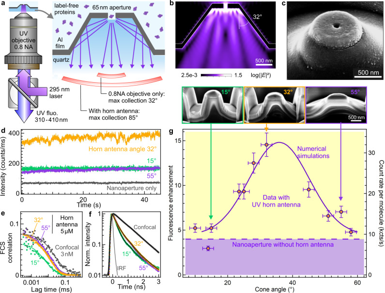
Our optical horn antenna platform addresses specifically the challenges of label-free single protein UV detection. It combines a reflective unit with a nanoaperture (Fig. 1a–c) and is the UV analog of a microwave horn antenna (Supplementary Fig. S1). The central nanoaperture of 65 nm diameter concentrates the light in an attoliter detection volume to isolate a single protein and enhance its autofluorescence32,33, while the reflective conical unit covered with a 100 nm thick aluminum layer steers the autofluorescence light toward the microscope objective. Contrarily to the Yagi Uda or Bull’s eyes resonant designs28,29, the conical horn is intrinsically broadband, covering the full 300–400 nm bandwidth, independently of resonance or interference effects. The detection volume provided by the 65 nm central aperture is three orders of magnitude below that of a diffraction-limited confocal microscope24,33, enabling single-molecule detection at micromolar physiological conditions, and circumventing the need for sub-nanomolar dilutions in conventional confocal microscopy34.
Fig. 1. Ultraviolet horn antenna to enhance the autofluorescence detection of single label-free proteins.
a Scheme of the experiment. b Numerical simulation of the emission pattern of a dipole located in the center of the nanoaperture, averaging the contributions from horizontal and vertical dipole orientations. c Scanning electron microscope image of a horn antenna. Similar images could be reproduced more than 10 times using the same milling parameters. d Fluorescence intensity time traces recorded on a 5 µM solution of p-terphenyl using horn antennas of different cone angles. e FCS correlation functions corresponding to the traces shown in (d), the case for the isolated nanoaperture is equivalent to the horn antennas with cone angles 32 and 55°. f Normalized fluorescence lifetime decay traces acquired simultaneously to the data in (d, e). IRF indicates the instrument response function. Black lines are numerical fits. g Fluorescence enhancement of the brightness per molecule as a function of the horn antenna cone angle. The right axis shows the corresponding count rate per p-terphenyl molecule at 80 µW of the 266 nm laser. The level achieved with a nanoaperture without any horn antenna is indicated by the dashed horizontal line. The solid line shows the numerical simulations results accounting for the collection efficiency gain into the 0.8 NA microscope objective. The SEM images in the inset show the antenna geometry after a cross-section has been cut by focused ion beam. Data are presented as mean values +/− one standard deviation determined from a pool of at least three different samples.
The horn antenna performance is assessed using p-terphenyl, a 93% quantum yield UV fluorescent dye (Fig. 1d–g). The conical reflector angle mainly determines the antenna collection efficiency into the 0.8 NA objective (Supplementary Figs. S2, S3). We have fabricated horn antennas with various cone angles from 10 to 55° (Supplementary Fig. S4–S6). The raw fluorescence time trace already shows a 4× larger signal with 32° horn as compared to the bare nanoaperture, indicating an improved collection efficiency by the same ratio (Fig. 1d). Fluorescence correlation spectroscopy (FCS) analysis measures the fluorescence brightness per molecule (Fig. 1e and Table S1) from which we compute the fluorescence enhancement as compared to the confocal reference24,29. The enhancement factor clearly depends on the cone angle with an optimum around 35° (Fig. 1g). While a single nanoaperture improves the p-terphenyl brightness by 4×33, the horn reflector brings it to 15×. These values are lower than previous reports using gold antennas in the red part of the spectrum22–25, yet this is explained by the ultraviolet range and the simple non-resonant design of the horn antenna. The major goal here is not to compete with plasmonics in the visible, but rather to enable UV autofluorescence detection of single proteins above the background noise. The collection efficiency gain has been numerically simulated using finite-difference time-domain (FDTD, solid curve in Fig. 1g). The simulation results match well with the experimental data trend, confirming the dependence with the cone angle.
The fluorescence lifetime measurements on Fig. 1f and Table S2 indicate that the p-terphenyl fluorescence lifetime is reduced by 3× in the antennas as compared to the confocal reference. This lifetime reduction is independent of the cone angle and is similar to the lifetime reduction found for the single aperture without the conical reflector. This shows that the emitter’s fluorescence lifetime (and thus the local density of optical states) is mainly set by the aperture diameter. Having a similar local density of optical states between the nanostructured samples, we can conclude that the supplementary gain brought by the optimized horn antenna is directly related to the increase in directivity as compared to the bare nanoaperture. This confirms the idea of the conical reflector as a collection unit to steer the emitted light towards the microscope objective. Based on the gain respective to the bare nanoaperture, the maximum collection angle is estimated to be around 85° for our best system with 32° cone angle. Our nanophotonic platform collects the fluorescence light emitted at high angles, even beyond the supercritical angle. For a description of the aluminum nanoaperture influence on the fluorescence process, our group has recently published a detailed characterization using label-free proteins in the UV35. As shown by the data in Fig. 1, the presence of the horn reflector improves the collection efficiency, but the fluorescence excitation and emission enhancements occurring in the nanoaperture are not affected. We independently confirm the fluorescence enhancement by quantifying the noise reduction in the correlation data (Supplementary Fig. S7). The high brightness observed with the optimized horn antenna directly improves the FCS signal-to-noise ratio allowing to reduce the experiment integration time as compared to the confocal reference while keeping the same accuracy.
Single immobilized protein detection
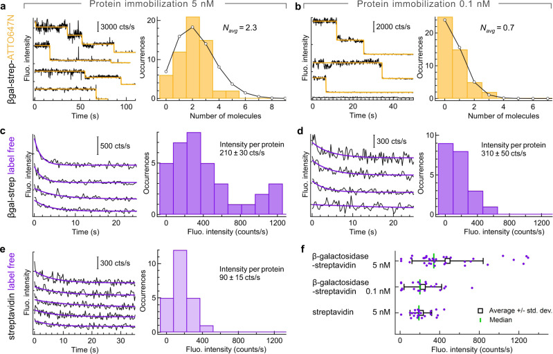
We next focus on the label-free detection of single immobilized proteins using our optimized UV horn antennas with 32° cone angle (Fig. 2). The surface of the central nanoaperture is functionalized with silane-polyethylene glycol-biotin to graft individual β-galactosidase-streptavidin proteins. β-galactosidase from Escherichia coli (156 tryptophans) has been modified to bear a streptavidin anchor (24 tryptophans). We first assess the distribution of the number of proteins inside the central nanoaperture using control experiments where the proteins are labeled with an Atto647N-biotin red fluorescent dye (Fig. 2a, b). Specific care was taken to ensure a 1:1 labeling ratio before the surface immobilization so that on average every protein carries a single fluorescent label (see “Methods” for details). The experiments are repeated for two protein concentrations of 5 and 0.1 nM, allowing to report the evolution with the protein concentration. With the Atto647N label, the fluorescence intensity time traces show fast step decays typical of single-molecule fluorescence photobleaching (Fig. 2a, b). These traces are analyzed using the automated software AutoStepfinder36 to count the number of bleaching steps from which we deduce the number of proteins. The histogram of the number of molecules per horn antenna follow the expected Poisson distribution, with an average number of molecules of 2.3 for the 5 nM concentration and 0.7 for the 0.1 nM case. The evolution of the average number of molecules does not scale exactly with the concentration (although there is an obvious dependence) as the 30 min incubation time and possible steric hindrance between neighboring proteins may limit the number of proteins able to bind on the horn antenna surface. Importantly, for the 0.1 nM protein concentration, there is less than one protein per horn antenna on average, so this case is representative of single-molecule experiments.
Fig. 2. Horn antenna-enhanced label-free detection of immobilized single proteins.
The surface of the horn antenna has been functionalized with biotin-PEG-silane to bind β-galactosidase-streptavidin and pure streptavidin proteins. In (a, c, e), the total protein concentration is 5 nM while it is reduced to 0.1 nM in (b, d). a, b Additional Atto647N-biotin red fluorescent label is added as a control to quantify the number of proteins inside individual horn antennas. Typical Atto647N fluorescence time traces are shown on the left panels in (a, b), together with their step-function fit (yellow line) allowing to count single molecules. The traces are vertically shifted for clarity. The right panels in (a, b) display the histogram of the number of detected molecules (bars) together with a Poisson distribution fit (line and dots). For (c–f), the experiments are performed with label-free proteins in the UV using 5 nM β-galactosidase-streptavidin (c), 0.1 nM β-galactosidase-streptavidin (d) and 5 nM streptavidin (e). In (c–e), the left panels show autofluorescence time traces and their exponential decay fit recorded on different individual horn antennas (the traces are vertically shifted for clarity). The right panels represent the histogram of the exponential fit amplitudes. For (c) 35 horn antennas were probed, for (d) 21 and (e) 23. (f) Scatter plot of the exponential fit amplitudes corresponding to the histograms in (c–e). The points are vertically shifted using a uniform statistical distribution to better view the results. The white square marker denotes the average value with the bars extending to one standard deviation. The green vertical line indicates the median. Respectively, 35, 21, and 23 antennas were probed for the different cases from top to bottom. Throughout these experiments, the cone angle is 32° and the aperture diameter is 200 nm (see Supplementary Fig. S13 for a discussion on the aperture diameter).
UV autofluorescence time traces are shown in Fig. 2c–e and Figs. S8–S10. These label-free experiments have been performed in exactly the same surface immobilization conditions as for Fig. 2a, b, so it is fair to consider that the distributions of the number of proteins are unchanged. Due to the high number of tryptophan residues in each protein, the autofluorescence time traces no longer bear step-like decays but follow an exponential decay due to photobleaching. Control experiments performed in the absence of the protein using only the photostabilizing buffer show that the background noise does not show the exponential signal decay upon UV illumination (Supplementary Fig. S11). In presence of the proteins, each trace stemming from a different horn antenna is fitted with an exponential function to extract the decay amplitude. The histograms of the autofluorescence signal amplitudes are displayed in Fig. 2c, d for 5 and 0.1 nM protein concentration, while the raw data, average, standard deviation, and median values are summarized in Fig. 2f. Dividing the average fluorescence intensity (Fig. 2f) by the average number of proteins (determined from Fig. 2a, b), we estimate the average brightness per β-galactosidase-streptavidin protein in the horn antenna. We find 210 ± 30 counts/s for the 5 nM concentration and 310 ± 50 counts/s for the 0.1 nM concentration, which yield statistically quite comparable values (the difference is twice the standard deviation).
The experiments are reproduced with pure streptavidin, which has 24 tryptophan residues as compared to the 156 residues of β-galactosidase. A separate calibration using diffusing molecules shows that the average brightness per protein is 3× lower for streptavidin as compared to β-galactosidase35. Although streptavidin has a 6.5× lower absolute number of tryptophan residues, their average quantum yield is estimated to be around 3.5% while it is only of 1.6% in β-galactosidase due to a higher quenching by nearby aminoacids18,35. The experiments on immobilized streptavidin proteins are performed using a 5 nM concentration to work in the same conditions as for Fig. 2a, c. The average amplitude found for streptavidin is 2.5× lower than for β-galactosidase (Fig. 2e, f), confirming the expected evolution of the signal with the number of tryptophan residues and their average quantum yield. Finally, the experiments with immobilized proteins are reproduced for single nanoapertures without the horn microreflector (Supplementary Fig. S12). The signal is about 3× brighter with the horn antenna as compared to the nanoaperture on a flat substrate, which goes along with the results found for p-terphenyl in Fig. 1g and the improved collection performance of the horn antenna.
Altogether, the data presented in Fig. 2 and S8–S12 demonstrate that the autofluorescence from a single protein can be recorded on a horn antenna. The signal scales with the protein concentration, the number of tryptophan residues, and the collection efficiency. Experiments using a fluorescent marker allow an independent measurement of the number of proteins. This realizes label-free single protein detection in the UV. In addition, fluorescence lifetime histograms can be extracted for traces corresponding to a single protein (Supplementary Fig. S14), a highly challenging task owing to the limited total photon budget that has to be distributed among the histogram time bins.
Detecting single diffusing proteins
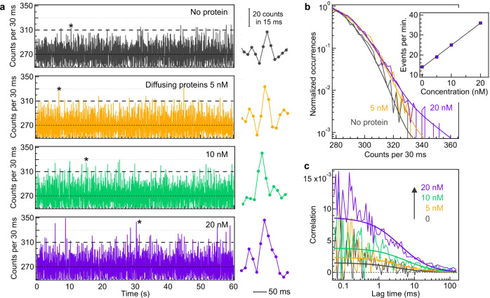
To confirm the single-molecule sensitivity claim, we perform experiments with diffusing β-galactosidase-streptavidin proteins at very low concentrations so that the average number of proteins present in the detection volume is significantly below 1 (Fig. 3). For these experiments, we use a 200 nm diameter nanoaperture to increase the protein residency time inside the horn antenna. For the highest concentration of 20 nM, the calculated average number of proteins inside the 200 nm nanoaperture is 0.1233, which corresponds well to the regime required to observe fluorescence bursts from single molecules23,37. Moreover, we add 55% sucrose to the buffer solution to increase the viscosity and ensure the proteins stay a sufficiently long time of several milliseconds inside the nanoaperture volume. Without sucrose or any other viscous medium, the diffusion time of proteins across the nanoaperture would be below 1 ms which is not sufficient to record enough photons and clearly resolve the UV autofluorescence bursts stemming from a single protein. The presence of impurities in sucrose leads to a higher background noise level and limits the maximum amount of sucrose that we can use (glycerol mixtures lead to similar observations). We find that a 55% w/w sucrose mixture is a good compromise between increased viscosity and tolerable noise level.
Fig. 3. Label-free detection of single diffusing proteins across the UV horn antenna.
55% sucrose was added to the buffer to slow down the protein diffusion by 30× and ease observing the autofluorescence bursts. a Autofluorescence time traces with increasing β-galactosidase-streptavidin concentrations. The binning time is 30 ms. The stars indicate selected autofluorescence bursts which are displayed on the right panel with 15 ms bin time to better view individual bursts. b Normalized photon counts histograms computed over the full trace duration of 200 s. The thick lines are numerical fits using the sum of a Gaussian and an exponential distribution to account for the noise and the autofluorescence bursts respectively. The inset shows the number of detected events per minute above the threshold corresponding to 2.5× the standard deviation of the noise when no protein is present (dashed lines at 310 counts in (a). c FCS correlation functions of the traces displayed in (a). The thick lines are numerical fits.
Figure 3a shows typical autofluorescence time traces recorded with increasing protein concentrations from 0 (background only) to 20 nM. Without the β-galactosidase-streptavidin proteins, we detect 14 events per minute above the threshold of 310 counts per 30 ms bin time (corresponding to 2.5× the standard deviation of the background noise, dashed horizontal line in Fig. 3a). In the presence of the protein sample, the number of events exceeding this threshold increases with the protein concentration (Fig. 3a, b inset). Zooming in on some selected bursts (star markers in Fig. 3a), the signal is above the average for several binning times, which indicates that these bursts are not spurious single-time bin events. The photon count histograms confirm an increasing difference with the background level as the protein concentration is increased. Comparing the respective maxima, we estimate that the brightness for diffusing molecules is about 600 counts per second, which stands in agreement with an independent FCS calibration at micromolar concentration (Supplementary Fig. S15). To confirm that the bursts seen on the autofluorescence traces stem from the proteins and are not just random noise, we compute the temporal correlations of the traces in Fig. 3a and obtain the FCS correlograms on Fig. 3c. In the presence of the proteins, the FCS correlation is significantly higher than the residual background noise correlation. The FCS amplitude increases with the concentration because of the dominating presence of the background noise at such low concentrations38. The positive correlation amplitude and the 5 ms diffusion time (due to the presence of sucrose) indicate that the bursts seen on Fig. 3a stem from single β-galactosidase-streptavidin proteins. Altogether, the data in Fig. 3 demonstrate the ability of the UV horn antenna to resolve the autofluorescence bursts from diffusing single label-free proteins.
Label-free protein denaturation and unfolding
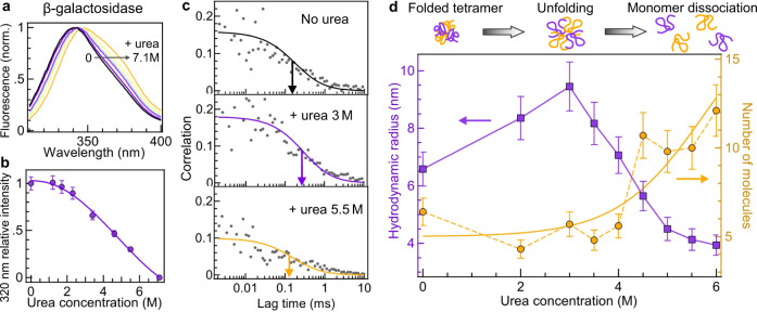
β-galactosidase has gained importance as a model system to study protein folding39. Ensemble-level spectroscopy measurements such as the data in Fig. 4a, b and Supplementary Fig. S16 are commonly used to follow β-galactosidase denaturation in presence of urea40. It is believed that the tetrameric β-galactosidase first unfolds into a globular structure which then dissociates into unfolded monomers as the urea concentration is increased39,40. However, this pathway has never been studied at the single-molecule level and only indirect ensemble-averaged measurements are available. Single-molecule resolution is important here as it allows to clearly distinguish the dissociation into monomers by counting the number of molecules. An approach using fluorescence labeling would be very complicated as it would require all the β-galactosidase monomers to be labeled with a fluorescent dye, which is challenging to achieve and would lead to inter-chromophoric quenching due to the close proximity of dyes41. Another motivation for single-molecule resolution is to be able to simultaneously measure the protein hydrodynamic radius to clearly evidence unfolding.
Fig. 4. Application of UV horn antenna to study denaturation of label-free proteins.
a Autofluorescence spectra of β-galactosidase in presence of increasing concentrations of urea. b Following the approach in40, the autofluorescence intensity at 320 nm is used to monitor the denaturation of β-galactosidase as a function of the urea concentration. Data are presented as mean values, with the error bars corresponding to a 7% deviation accounting for the measurement uncertainty. The measurements have been repeated twice. c FCS correlation functions of β-galactosidase with increasing urea concentrations. The color lines are numerical fits. The arrows indicate the half-width of the correlation. d Evolution of the protein average hydrodynamic radius (left axis) and the mean number of detected molecules (right axis) as a function of the urea concentration. Error bars are standard deviations of the FCS measurements. Each measurement has been reproduced independently at least two times. The yellow line is a guide to the eyes based on a sigmoid function fit where c is the urea concentration in molar.
Here we use label-free FCS enhanced by the optical horn antenna to study β-galactosidase denaturation (Fig. 4c, d). Without the horn antenna, the FCS data is too noisy to reliably measure unfolding and monomer dissociation (Supplementary Fig. S15). Thanks to the horn antenna, the UV autofluorescence brightness of β-galactosidase tetramers is increased by 10× which directly reduces the FCS noise by the same factor (Supplementary Fig. S15). In these conditions, the number of detected proteins and their hydrodynamic radius can be assessed as the urea concentration is increased (Fig. 4c, d and Supplementary Fig. S17). The number of molecules informs about the dissociation of the β-galactosidase tetramers into monomers while the hydrodynamic radius indicates expansion or compaction of the protein structure. As additional advantage, the attoliter detection volume of the horn antenna seems beneficial to avoid observing large aggregates which are a perturbation source for ensemble measurements40. While the autofluorescence spectrum of β-galactosidase is red-shifted up to 10 nm by the presence of urea (Fig. 4a and Fig. S16a, b), the total integrated intensity in the 310–410 nm detection range remains almost unaffected (Fig. S16c) as well as the autofluorescence lifetime (Fig. S16d).
Our experimental data show that below 3.5 M urea, β-galactosidase remains as a tetramer (the number of molecules is constant) while its hydrodynamic radius increases from 6.5 to 9.5 nm indicating protein unfolding (Fig. 4d). Our analysis takes into account the viscosity change as the urea concentration is increased (Supplementary Fig. S17) as well as the influence of the nanoaperture calibrated in ref. 33. In the absence of urea, our 6.5 ± 0.6 nm value for the hydrodynamic radius corresponds well to the 6.85 nm (experimental) and 6.7 nm (calculated) values determined previously in ref. 42. Above 3.5 M urea, the hydrodynamic radius decreases down to 4 nm and the number of molecules increases by more than 2×. These are clear evidences of the tetramer partial dissociation into unfolded monomers, providing a confirmation of the β-galactosidase denaturation pathway established using ensemble methods39,40.
Discussion
While label-free alternatives to fluorescence labeling are actively searched9–11, fluorescence spectroscopy remains by far the most widely used approach for single-molecule detection. The optical horn antennas developed here make a significant step forward by enabling the direct detection of single label-free proteins via their natural ultraviolet fluorescence. This dedicated design combines simultaneously plasmonic fluorescence enhancement, efficient fluorescence collection, attoliter detection volume, and strong background rejection, allowing to achieve unprecedented protein autofluorescence brightness. Improving the net detected UV photon count rate is key to enable the biophysical applications investigating single proteins in their native state in real time. Photodamage of aminoacids due to UV illumination is a potential issue that may affect the protein structure. For diffusing proteins, the short illumination time balances the negative impact of the UV photodegradation. However, for immobilized proteins, the risk of photodamage limits the maximum UV power exciting the protein. We have used so far the lowest possible power of 0.3 µW, yet this phenomenon is currently setting the limit for the achievable signal-to-noise ratio. UV detection of single molecules is still at its infancy, and we hope that this work will stimulate more studies in this direction. Moreover, optical horn antennas are also beneficial to improve the collection efficiency in the visible regime28,29, and analyze single fluorescent molecules in a crowded and confined environment reproducing the physiological conditions34.
Methods
Optical horn antenna fabrication
The fabrication process involves several steps (Supplementary Fig. S4). Briefly, we first mill the horn antenna by focused ion beam (FIB). Then a 100 nm aluminum layer is deposited to ensure a good UV reflectivity of the horn antenna walls. Last, we carve a 65 nm or a 200 nm diameter nanoaperture by FIB in the center of the top plateau of the horn antenna. The substrates are cleaned NEGS1 quartz microscope coverslips of 150 µm thickness (Neyco). Aluminum layers are deposited by electron-beam evaporation (Bühler Syrus Pro 710) with 10 nm/s rate at a chamber pressure of 10−6 mbar. FIB milling is performed using a gallium-based system (FEI dual beam DB235 Strata) with 30 kV acceleration voltage and 300 pA current for milling the horn antenna and 10 pA current for milling the central nanoaperture. All nanoapertures have a 50 nm deep undercut into the quartz substrate to maximize the signal enhancement. Last, a 12-nm-thick SiO2 layer is deposited by plasma-enhanced chemical vapor protection (PECVD, PlasmaPro NGP80 from Oxford Instruments) to protect the aluminum surface against corrosion43,44.
Protein samples and photostabilizing buffer
β-galactosidase from Escherichia coli (156 tryptophan residues, PDB 1DP0), β-galactosidase-streptavidin conjugate (180 tryptophan residues), and streptavidin from Streptomyces avidinii (24 tryptophan residues) are purchased from Sigma-Aldrich. The proteins are dissolved in a Hepes buffer (25 mM Hepes, 300 mM NaCl, 0.1 v/v% Tween20, 1 mM DTT, and 1 mM EDTA 1 mM at pH 6.1) which was reported to stabilize β-galactosidase conformation and avoid aggregate formation40. All the protein stock solutions have been centrifuged for 12 min at 142,000 × g (Airfuge). Just before the optical measurements, GODCAT oxygen scavenger (100 nM glucose oxidase, 830 nM catalase, 10 w/v% D-glucose) with 10 mM DABCO (1,4-Diazabicyclo[2.2.2]octane) is added to the solution to improve the UV photostability18. p-Terphenyl is also used as received from Sigma-Aldrich and diluted in HPLC-grade cyclohexane.
Surface immobilization of proteins
The horn antennas are thoroughly cleaned with UV-ozone (Novascan PSD-UV 100 W), ethanol rinsing, and oxygen-plasma (Diener Zepto 50 W, 0.6 mbar, 10 min). The silica surface is then coated with silane-modified polyethylene glycol (PEG-silane) by immersion into an ethanol solution containing 0.75 mg PEG-silane 1000 Da and 1.2 mg biotin-PEG-silane 2000 Da. PEG-silane and biotin-PEG-silane are purchased from Nanocs. After overnight incubation, the sample is rinsed with ethanol and dried. The protein solution with either 5 or 0.1 nM concentration is placed on the surface for 30 min allowing the streptavidin anchor to bind the surface-grafted biotin. Before the optical measurements, the surface is rinsed three times with water and covered with the GODCAT photostabilizing buffer.
Control experiments with fluorescent labels
A solution of β-galactosidase-streptavidin is mixed with a solution of Atto647N-biotin in a 1:1 ratio and kept at 4 °C overnight. The concentrations were carefully checked using a spectrofluorometer (Tecan Spark 10 M) to ensure proper labeling of the proteins with a single Atto647N dye. In this configuration, on average each protein is expected to bind only one fluorescent tag via the streptavidin-biotin bridge. Assuming that each β-galactosidase-streptavidin has 3 binding sites for the biotinylated fluorescent tag (one binding site is taken by the anchor between β-galactosidase and streptavidin), the probability that a protein bears more than 1 fluorescent label is estimated to be below 4%. The surface grafting of the labeled β-galactosidase-streptavidin is performed using exactly the same protocol used for the label-free counterpart. The fluorescence readout is performed on a confocal microscope described in ref. 26 using 1 µW laser power at 635 nm. To promote Atto647N photostability and minimize blinking, we use the GODCAT oxygen scavenger system together with 1 mM Trolox ((±)−6-Hydroxy-2,5,7,8-tetramethylchromane-2-carboxylic acid)45. Prior to the experiments, the Trolox stock solution in DMSO is illuminated with a UV lamp for 25 min to ensure a proper ratio of Trolox and its Trolox-quinone derivative45.
Urea denaturation
A 1.7 µM β-galactosidase protein solution is incubated with urea at various concentrations from 0 to 6 M (pH 7) for 90 min at room temperature. 0.4 v/v% Tween20 is added to the buffer to minimize aggregation of denaturated proteins. To avoid non-specific adsorption of proteins, the horn antenna surface is passivated with PEG-silane 1000 Da (Nanocs) by immersion into a 1 mg/ml PEG-silane solution ethanol with 1% acetic acid for 3–4 h followed by ethanol rinsing. GODCAT photostabilizing system is added to the protein buffer just before the optical measurements and the urea concentration is adjusted to keep a constant value to avoid protein refolding.
UV microscope
We operate a custom-built confocal microscope with a LOMO 58x, 0.8 NA, water immersion objective. Experiments on p-terphenyl use a 266 nm picosecond laser (Picoquant LDH-P-FA-266, 70 ps pulse duration, 80 MHz repetition rate) with 80 µW average power, while experiments on proteins use a 295 nm picosecond laser (Picoquant VisUV-295-590, 70 ps pulse duration, 80 MHz repetition rate). The 295 nm wavelength selectively excites tryptophan residues, as tyrosine and phenylalanine have negligible absorption above 290 nm. The laser power for immobilized protein detection is 0.3 µW, while for diffusing molecules we use 8 µW. The urea denaturation experiments on diffusing proteins are performed at 10 µW. Both laser beams are spatially filtered to ensure a Gaussian profile filling the objective back aperture, they pass through a short-pass filter (Semrock FF01-311/SP-25) and are reflected by a dichroic mirror (Semrock FF310-Di01-25-D). The optical horn antenna is positioned at the laser focus with a 3-axis piezoelectric stage (Physik Instrumente P-517.3CD). For the immobilized protein experiment, we use the microscope LED illumination to localize the horn antenna. The laser illumination is turned on immediately at the start of the acquisition to avoid bleaching the proteins while scanning the sample.
The fluorescence light is collected back by the microscope objective and separated from the laser light by the dichroic mirror and two emission filters (Semrock FF01-300/LP-25 and Semrock FF01-375/110-25). The spectral range for fluorescence detection goes from 310 to 410 nm. Confocal detection is performed using a 200 mm focal length doublet lens (Thorlabs ACA254-200-UV) and a 80 μm pinhole. Single-photon counting uses a photomultiplier tube (Picoquant PMA 175) connected to a photon counting module (Picoquant Picoharp 300 with time-tagged time-resolved mode). The integration time is 2 to 3 min.
Fluorescence time trace analysis
The fluorescence data is analyzed with Symphotime 64 (Picoquant) and Igor Pro 7 (Wavemetrics). For the fluorescence time traces from immobilized label-free single proteins, the temporal bin width is set to 300 ms to optimize the signal-to-noise ratio while still providing enough time resolution. Each trace stemming from a different horn antenna is fitted using and exponential decay model where A is the decay amplitude (used for the histrograms), τB is the bleaching time and y0 is the background level set by the dark counts of the photodetector and the residual photoluminescence from the nanostructure. For the control experiments using Atto647N labels, the traces are analyzed using AutoStepfinder, a recently developed software for automatic step detection36.
For the experiments on diffusing proteins at very low concentrations (average numbers of proteins in the nanoaperture <0.12, Fig. 3), we apply a 1 Hz high-pass filter to remove all long-term drifts and fluctuations. These drifts have a small amplitude (typically 5 to 10 counts per 30 ms) and long periods (several seconds), yet due to the low signal-to-noise ratio at low protein concentrations, the long-term fluctuations have to be removed before computing the photon count histogram. The average value of the initial trace is then added to retrieve the filtered count information.
FCS correlations are fitted using a three-dimensional Brownian diffusion model with a blinking term18,33:
| 1 |
where Nmol is the total number of detected molecules, B the background noise intensity, F the total fluorescence intensity, nT and τT are the blinking amplitude and characteristic time, τd is the mean diffusion time and κ the aspect ratio of the axial to transversal dimensions of the detection volume (κ = 8 for the confocal illumination and κ = 1 for the horn antenna). Note that the fast kinetics components determined by nT and τT may not be only related to triplet blinking, they could also account for residual afterpulsing from the photon counting detector and/or metal quenching when the protein diffuses in nanometer proximity to the aluminum surface. The FCS fit results are summarized in the Supplementary information.
The fluorescence lifetime decays are fitted by an iterative reconvolution taking into account the measured instrument response function (IRF). As noted in our previous work on single apertures33, a three exponential model is needed to correctly interpolate the experimental data. A fixed 10 ps component accounts for the metal photoluminescence and Raman scattering background, while a long component with a lifetime similar to the confocal reference corresponds to a residual fluorescence stemming from molecules away from the optical antenna. The intermediate lifetime component (which has a dominating intensity) corresponds to the lifetime of molecules inside the horn antenna. All the fit results are detailed in the supplementary information.
Numerical simulations
The electric field intensity radiated by a point dipole is calculated with finite-difference time-domain (FDTD) method using RSoft Fullwave software. We set a fixed emission wavelength at 350 nm. Horizontal and vertical orientations of the source dipole are computed separately and averaged for the final output to represent the emission of a molecule with nanosecond rotational time. Each simulation is run with 2 nm mesh size and is checked for convergence after several optical periods. To compute the enhancement displayed in Fig. 1g, 10 horn antennas with different cone angle are simulated. For each of them, we compute the gain into the 0.8 collection NA of the microscope objective as compared to the emission from a single aperture without horn antenna (Fig. S2). The data points are then fitted with a Gaussian function.
Reporting summary
Further information on research design is available in the Nature Research Reporting Summary linked to this article.
Supplementary information
Acknowledgements
The authors thank Antonin Moreau, Julien Lumeau, Satyajit Patra, Manos Mavrakis, Redha Abdeddaim, and Marco Abbarchi for stimulating discussions and technical help. This project has received funding from the European Research Council (ERC) under the European Union’s Horizon 2020 research and innovation program (grant agreement No. 723241, A.B., P.R., J.-B.C., J.W.).
Author contributions
A.B. performed and analyzed measurements, P.R. performed and analyzed measurements and simulations, J.B.C. performed nanofabrication, J.W. designed and supervised research and prepared manuscript with the help of all authors.
Peer review
Peer review information
Nature Communications thanks Marek Piliarik and the other anonymous reviewers for their contribution to the peer review of this work.
Data availability
All relevant data are available from the corresponding author upon request.
Competing interests
The authors declare no competing interests.
Footnotes
Publisher’s note Springer Nature remains neutral with regard to jurisdictional claims in published maps and institutional affiliations.
Supplementary information
The online version contains supplementary material available at 10.1038/s41467-022-29546-4.
References
- 1.Joo C, Balci H, Ishitsuka Y, Buranachai C, Ha T. Advances in single-molecule fluorescence methods for molecular biology. Annu. Rev. Biochem. 2008;77:51–76. doi: 10.1146/annurev.biochem.77.070606.101543. [DOI] [PubMed] [Google Scholar]
- 2.Lerner E, et al. Toward dynamic structural biology: two decades of single-molecule Förster resonance energy transfer. Science. 2018;359:eaan1133. doi: 10.1126/science.aan1133. [DOI] [PMC free article] [PubMed] [Google Scholar]
- 3.Cabantous S, Terwilliger TC, Waldo GS. Protein tagging and detection with engineered self-assembling fragments of green fluorescent protein. Nat. Biotechnol. 2005;23:102–107. doi: 10.1038/nbt1044. [DOI] [PubMed] [Google Scholar]
- 4.Wigley WC, Stidham RD, Smith NM, Hunt JF, Thomas PJ. Protein solubility and folding monitored in vivo by structural complementation of a genetic marker protein. Nat. Biotechnol. 2001;19:131–136. doi: 10.1038/84389. [DOI] [PubMed] [Google Scholar]
- 5.Dietz MS, Wehrheim SS, Harwardt M-LIE, Niemann HH, Heilemann M. Competitive binding study revealing the influence of fluorophore labels on biomolecular interactions. Nano Lett. 2019;19:8245–8249. doi: 10.1021/acs.nanolett.9b03736. [DOI] [PubMed] [Google Scholar]
- 6.Riback JA, et al. Commonly used FRET fluorophores promote collapse of an otherwise disordered protein. Proc. Natl Acad. Sci. USA. 2019;116:8889–8894. doi: 10.1073/pnas.1813038116. [DOI] [PMC free article] [PubMed] [Google Scholar]
- 7.Zanetti-Domingues LC, Tynan CJ, Rolfe DJ, Clarke DT, Martin-Fernandez M. Hydrophobic fluorescent probes introduce artifacts into single molecule tracking experiments due to non-specific binding. PLoS ONE. 2013;8:e74200. doi: 10.1371/journal.pone.0074200. [DOI] [PMC free article] [PubMed] [Google Scholar]
- 8.Hughes LD, Rawle RJ, Boxer SG. Choose your label wisely: water-soluble fluorophores often interact with lipid bilayers. PLoS ONE. 2014;9:e87649. doi: 10.1371/journal.pone.0087649. [DOI] [PMC free article] [PubMed] [Google Scholar]
- 9.Arroyo JO, Kukura P. Non-fluorescent schemes for single-molecule detection, imaging and spectroscopy. Nat. Photonics. 2016;10:11–17. [Google Scholar]
- 10.Zijlstra P, Paulo PMR, Orrit M. Optical detection of single non-absorbing molecules using the surface plasmon resonance of a gold nanorod. Nat. Nanotechnol. 2012;7:379–382. doi: 10.1038/nnano.2012.51. [DOI] [PubMed] [Google Scholar]
- 11.Hanay MS, et al. Single-protein nanomechanical mass spectrometry in real time. Nat. Nanotechnol. 2012;7:602–608. doi: 10.1038/nnano.2012.119. [DOI] [PMC free article] [PubMed] [Google Scholar]
- 12.Baaske MD, Neu PS, Orrit M. Label-free plasmonic detection of untethered nanometer-sized Brownian particles. ACS Nano. 2020;14:14212–14218. doi: 10.1021/acsnano.0c07335. [DOI] [PMC free article] [PubMed] [Google Scholar]
- 13.Garoli D, Yamazaki H, Maccaferri N, Wanunu M. Plasmonic nanopores for single-molecule detection and manipulation: toward sequencing applications. Nano Lett. 2019;19:7553–7562. doi: 10.1021/acs.nanolett.9b02759. [DOI] [PubMed] [Google Scholar]
- 14.Kumamoto Y, Taguchi A, Kawata S. Deep-ultraviolet biomolecular imaging and analysis. Adv. Opt. Mater. 2019;7:1801099. [Google Scholar]
- 15.Lakowicz, J. R. Principles of Fluorescence Spectroscopy. (Springer US, 2006).
- 16.Li Q, Seeger S. Label-free detection of single protein molecules using deep UV fluorescence lifetime microscopy. Anal. Chem. 2006;78:2732–2737. doi: 10.1021/ac052166u. [DOI] [PubMed] [Google Scholar]
- 17.Lippitz M, Erker W, Decker H, van Holde KE, Basché T. Two-photon excitation microscopy of tryptophan-containing proteins. Proc. Natl Acad. Sci. USA. 2002;99:2772–2777. doi: 10.1073/pnas.052662999. [DOI] [PMC free article] [PubMed] [Google Scholar]
- 18.Barulin A, Wenger J. Ultraviolet photostability improvement for autofluorescence correlation spectroscopy on label-free proteins. J. Phys. Chem. Lett. 2020;11:2027–2035. doi: 10.1021/acs.jpclett.0c00209. [DOI] [PubMed] [Google Scholar]
- 19.Novotny, L. & Hecht, B. Principles of Nano-Optics. (Cambridge University Press, 2012).
- 20.Ruckstuhl T, Enderlein J, Jung S, Seeger S. Forbidden light detection from single molecules. Anal. Chem. 2000;72:2117–2123. doi: 10.1021/ac991358k. [DOI] [PubMed] [Google Scholar]
- 21.Novotny L, van Hulst N. Antennas for light. Nat. Photonics. 2011;5:83–90. [Google Scholar]
- 22.Kinkhabwala A, et al. Large single-molecule fluorescence enhancements produced by a bowtie nanoantenna. Nat. Photonics. 2009;3:654–657. [Google Scholar]
- 23.Acuna GP, et al. Fluorescence enhancement at docking sites of DNA-directed self-assembled nanoantennas. Science. 2012;338:506–510. doi: 10.1126/science.1228638. [DOI] [PubMed] [Google Scholar]
- 24.Punj D, et al. A plasmonic ‘antenna-in-box’ platform for enhanced single-molecule analysis at micromolar concentrations. Nat. Nanotechnol. 2013;8:512–516. doi: 10.1038/nnano.2013.98. [DOI] [PubMed] [Google Scholar]
- 25.Puchkova A, et al. DNA origami nanoantennas with over 5000-fold fluorescence enhancement and single-molecule detection at 25 μM. Nano Lett. 2015;15:8354–8359. doi: 10.1021/acs.nanolett.5b04045. [DOI] [PubMed] [Google Scholar]
- 26.Flauraud V, et al. In-plane plasmonic antenna arrays with surface nanogaps for giant fluorescence enhancement. Nano Lett. 2017;17:1703–1710. doi: 10.1021/acs.nanolett.6b04978. [DOI] [PubMed] [Google Scholar]
- 27.Khatua S, et al. Resonant plasmonic enhancement of single-molecule fluorescence by individual gold nanorods. ACS Nano. 2014;8:4440–4449. doi: 10.1021/nn406434y. [DOI] [PubMed] [Google Scholar]
- 28.Curto AG, et al. Unidirectional emission of a quantum dot coupled to a nanoantenna. Science. 2010;329:930–933. doi: 10.1126/science.1191922. [DOI] [PubMed] [Google Scholar]
- 29.Aouani H, et al. Bright unidirectional fluorescence emission of molecules in a nanoaperture with plasmonic corrugations. Nano Lett. 2011;11:637–644. doi: 10.1021/nl103738d. [DOI] [PubMed] [Google Scholar]
- 30.Lee KG, et al. A planar dielectric antenna for directional single-photon emission and near-unity collection efficiency. Nat. Photonics. 2011;5:166–169. [Google Scholar]
- 31.Morozov S, Gaio M, Maier SA, Sapienza R. Metal–dielectric parabolic antenna for directing single photons. Nano Lett. 2018;18:3060–3065. doi: 10.1021/acs.nanolett.8b00557. [DOI] [PubMed] [Google Scholar]
- 32.Levene MJ, et al. Zero-mode waveguides for single-molecule analysis at high concentrations. Science. 2003;299:682–686. doi: 10.1126/science.1079700. [DOI] [PubMed] [Google Scholar]
- 33.Barulin A, Claude J-B, Patra S, Bonod N, Wenger J. Deep ultraviolet plasmonic enhancement of single protein autofluorescence in zero-mode waveguides. Nano Lett. 2019;19:7434–7442. doi: 10.1021/acs.nanolett.9b03137. [DOI] [PubMed] [Google Scholar]
- 34.Grommet AB, Feller M, Klajn R. Chemical reactivity under nanoconfinement. Nat. Nanotechnol. 2020;15:256–271. doi: 10.1038/s41565-020-0652-2. [DOI] [PubMed] [Google Scholar]
- 35.Barulin A, Roy P, Claude J-B, Wenger J. Purcell radiative rate enhancement of label-free proteins with ultraviolet aluminum plasmonics. J. Phys. Appl. Phys. 2021;54:425101. [Google Scholar]
- 36.Loeff L, Kerssemakers JWJ, Joo C, Dekker C. AutoStepfinder: a fast and automated step detection method for single-molecule analysis. Patterns. 2021;2:100256. doi: 10.1016/j.patter.2021.100256. [DOI] [PMC free article] [PubMed] [Google Scholar]
- 37.Baibakov M, et al. Extending single-molecule förster resonance energy transfer (FRET) range beyond 10 nanometers in zero-mode waveguides. ACS Nano. 2019;13:8469–8480. doi: 10.1021/acsnano.9b04378. [DOI] [PubMed] [Google Scholar]
- 38.Rüttinger, S. Confocal Microscopy and Quantitative Single Molecule Techniques for Metrology in Molecular Medicine. (Technischen Universität Berlin, 2006).
- 39.Juers DH, Matthews BW, Huber RE. LacZ β-galactosidase: structure and function of an enzyme of historical and molecular biological importance. Protein Sci. 2012;21:1792–1807. doi: 10.1002/pro.2165. [DOI] [PMC free article] [PubMed] [Google Scholar]
- 40.Nichtl A, Buchner J, Jaenicke R, Rudolph R, Scheibel T. Folding and association of β-galactosidase. J. Mol. Biol. 1998;282:1083–1091. doi: 10.1006/jmbi.1998.2075. [DOI] [PubMed] [Google Scholar]
- 41.Schröder T, Scheible MB, Steiner F, Vogelsang J, Tinnefeld P. Interchromophoric interactions determine the maximum brightness density in DNA origami structures. Nano Lett. 2019;19:1275–1281. doi: 10.1021/acs.nanolett.8b04845. [DOI] [PubMed] [Google Scholar]
- 42.Fleming PJ, Fleming KG. HullRad: fast calculations of folded and disordered protein and nucleic acid hydrodynamic properties. Biophys. J. 2018;114:856–869. doi: 10.1016/j.bpj.2018.01.002. [DOI] [PMC free article] [PubMed] [Google Scholar]
- 43.Barulin A, et al. Preventing aluminum photocorrosion for ultraviolet plasmonics. J. Phys. Chem. Lett. 2019;10:5700–5707. doi: 10.1021/acs.jpclett.9b02137. [DOI] [PubMed] [Google Scholar]
- 44.Roy P, et al. Preventing corrosion of aluminum metal with nanometer-thick films of Al2O3 capped with TiO2 for ultraviolet plasmonics. ACS Appl. Nano Mater. 2021;4:7199–7205. [Google Scholar]
- 45.Cordes T, Vogelsang J, Tinnefeld P. On the mechanism of trolox as antiblinking and antibleaching reagent. J. Am. Chem. Soc. 2009;131:5018–5019. doi: 10.1021/ja809117z. [DOI] [PubMed] [Google Scholar]
Associated Data
This section collects any data citations, data availability statements, or supplementary materials included in this article.
Supplementary Materials
Data Availability Statement
All relevant data are available from the corresponding author upon request.


