Abstract
We evaluated the activity of quinupristin-dalfopristin (Q-D) against three clinical strains of Staphylococcus aureus susceptible to Q (MIC, 8 μg/ml) and Q-D (MICs, 0.5 to 1 μg/ml) but displaying various levels of susceptibility to D. D was active against S. aureus HM 1054 (MIC, 4 μg/ml) and had reduced activity against S. aureus RP 13 and S. aureus N 95 (MICs, 32 and 64 μg/ml, respectively). In vitro, Q-D at a concentration two times the MIC (2×MIC) produced reductions of 4.3, 3.9, and 5.8 log10 CFU/ml after 24 h of incubation for HM 1054, RP 13, and N 95, respectively. Comparable killing was obtained at 8×MIC. Q-D-resistant mutants were selected in vitro at a frequency of 2 × 10⁻⁸ to 2 × 10⁻⁷ for the three strains on agar containing 2×MIC of Q-D; no resistant bacteria were detected at 4×MIC. Rabbits with aortic endocarditis were treated for 4 days with Q-D at 30 mg/kg of body weight intramuscularly (i.m.) three times a day (t.i.d.) or vancomycin at 50 mg/kg i.m. t.i.d. In vivo, Q-D and vancomycin were similarly active and bactericidal against the three tested strains compared to the results for control animals (P < 0.01). Among animals infected with RP 13 and treated with Q-D, one rabbit retained Q-D-resistant mutants that were resistant to Q and to high levels of D (MICs, 64, >256, and 8 μg/ml for Q, D, and Q-D, respectively). We conclude that the bactericidal activity of Q-D against strains with reduced susceptibility to D and susceptible to Q-D is retained and is comparable to that of vancomycin. Acquisition of resistance to both Q and D is necessary to select resistance to Q-D.
Staphylococcus aureus resistant to methicillin is a major cause of nosocomial infections. Most strains are now also resistant to fluoroquinolones, rifampin, tetracyclines, and aminoglycosides (9, 20, 24). Vancomycin remains the standard agent for the therapy of systemic infections due to such strains of S. aureus. However, because of the recent report of emergence of resistance to vancomycin in S. aureus (23), the need for new antibiotics is increasing.
Quinupristin-dalfopristin (Q-D; Synercid), a semisynthetic injectable streptogramin, is a combination of two synergistic components (6), a streptogramin A type antibiotic (dalfopristin) and a streptogramin B type antibiotic (quinupristin), in a 70:30 ratio. Q-D is active in vitro against methicillin-resistant S. aureus; the MIC for 99% of tested strains is ≤1 μg/ml (1, 8, 18).
The interesting aspect of an antibiotic association such as Q-D is that the synergy displayed by the combination allows resistance to either compound to be overcome. However, it is not known if the in vitro observation that Q-D remains active despite resistance to quinupristin or dalfopristin holds true in vivo. Cross-resistance to macrolides, lincosamides, and streptogramin B type antibiotics by target modification is the most common mechanism of resistance to these antibiotics in S. aureus (16). The expression of this resistance may be inducible or constitutive. We previously demonstrated that if this resistance is inducible, the bacteriostatic and bactericidal effects of the streptogramin complex are retained both in vitro and in vivo (14). In contrast, when this resistance is constitutive, the bactericidal activity of Q-D may be altered in vitro and in vivo (10, 14).
Resistance to streptogramin A type antibiotics is uncommon (5, 17, 18, 19). Three genes coding for resistance to the A type compounds have been detected in S. aureus: vga, coding for an ATP-binding protein involved in the active efflux of A type compounds (2), and vat and vatB, coding for acetyltransferases which inactivate streptogramin A (3, 4, 19). However, not all determinants conferring resistance to the A type compounds are known (5). In staphylococci, resistance to the streptogramin complex is always associated with resistance to the A type compound (5, 19). In contrast, Allignet et al. did not find any relationship between the presence of a given streptogramin A resistance gene or a given combination of genes and the level of resistance to dalfopristin or Q-D (5). Therefore, resistance to streptogramin A-type antibiotics seems necessary but not sufficient for resistance to the Q-D combination (19).
In order to investigate the relationship between resistance to streptogramin A type antibiotics and the activity of Q-D, we studied the activity of Q-D in vitro and in experimental endocarditis against strains of S. aureus susceptible or resistant to dalfopristin in terms of bactericidal effect and emergence of resistant mutants.
MATERIALS AND METHODS
In vitro studies. (i) Organisms.
Three clinical strains of S. aureus with various dalfopristin MICs but susceptible to erythromycin, to quinupristin, and to Q-D were used for in vitro and in vivo studies (Table 1). None of the patients from whom the strains were isolated had previously received Q-D. S. aureus HM 1054 was susceptible to dalfopristin, whereas dalfopristin was less active against S. aureus RP 13 and S. aureus N 95. The mechanism of resistance of S. aureus RP 13 and S. aureus N 95 was not revealed; vat (3), vatB (4), and vga (2) sequences were not detected by the PCR method (7). The resistance phenotypes displayed by the bacteria were characterized by the agar diffusion technique with disks of erythromycin, lincomycin, quinupristin, dalfopristin, and Q-D as previously described (16, 18).
TABLE 1.
Characteristics of the three strains of S. aureus used for in vitro and in vivo studiesa
| Strain | Origin | Resistance phenotype | MIC (μg/ml) of:
|
Frequency of mutation to Q-D at two times the MIC | |||
|---|---|---|---|---|---|---|---|
| Q | D | Q-D | VAN | ||||
| HM 1054 | Blood culture | Oxa | 8 | 4 | 0.5 | 1 | 2 × 10⁻⁸ |
| RP 13 | Blood culture | SgA | 8 | 32 | 1 | 1 | 7 × 10⁻⁸ |
| N 95 | Wound infection | Oxa SgA | 8 | 64 | 0.5 | 1 | 2 × 10⁻⁷ |
Q, quinupristin; D, dalfopristin; VAN, vancomycin; Oxa, oxacillin; SgA, streptogramin A.
(ii) Media and antibiotics.
Trypticase soy agar was used for subcultures, Mueller-Hinton (MH) agar was used for MIC determinations, brain heart infusion (BHI) agar was used for selection of resistant mutants, and BHI was used for overnight cultures. All media were from Difco (Detroit, Mich.). Antibiotics were provided by their respective manufacturers: quinupristin, dalfopristin, and Q-D by Aventis, Vitry sur Seine, France (lot no. GRV1205 for quinupristin, GRV1204 for dalfopristin, and CB06069 and CB06489 for Q-D), and vancomycin by Lilly, Saint-Cloud, France (lot no. 2NE30MA).
(iii) In vitro susceptibilities to antibiotics.
MICs of quinupristin, dalfopristin, Q-D, and vancomycin were determined by the agar dilution method (21). For each strain, five or six independent determinations were performed.
(iv) In vitro selection of mutants.
BHI was inoculated with one colony of S. aureus and incubated overnight at 37°C. The culture was centrifuged (3,000 × g, 10 min, 4°C) and resuspended in fresh BHI to yield an inoculum of 1010 CFU/ml. Concentrated bacteria were plated on agar containing Q-D at two times the MIC and four times the MIC. After 48 h of incubation, the CFU were enumerated and the MIC of Q-D was determined.
(v) Study of Q-D bactericidal activity.
Time-kill curves were used to test the bactericidal activity of Q-D against the three strains. Overnight cultures were diluted in fresh MH broth to yield an inoculum of 5 × 10⁶ CFU/ml. Q-D was used at two and eight times the MIC. After 0, 3, 6, and 24 h of incubation at 37°C, serial dilutions of 50-μl samples were subcultured on Trypticase soy agar plates by use of a spiral plater (Spiral Systems Inc., Cincinnati, Ohio) and incubated for 24 h at 37°C before CFU were counted. CFU were counted for two different dilutions in which colonies were enumerable. The lower limit of detection of the assay with the spiral plater was 20 CFU/ml (1.3 log10 CFU/ml). In preliminary experiments, antibiotic carryover was ruled out by plating samples of bacterial suspension containing 101 to 103 CFU/ml in the presence or absence of antibiotics (13). Bactericidal activity was defined as a decrease of at least 3 log10 CFU/ml in the original inoculum.
Experimental staphylococcal endocarditis.
All investigations were performed according to the European Community guidelines for animal experimentation (12). Investigations were performed with New Zealand White rabbits (2.2 to 2.5 kg). Aortic endocarditis was induced in groups of 8 to 12 rabbits by insertion of a polyethylene catheter through the right carotid artery into the left ventricle to induce the formation of vegetations (22). Twenty-four hours after catheter insertion, each rabbit was inoculated via the ear vein with 1 × 10⁶ to 5 × 10⁶ CFU of S. aureus in 1 ml of sterile saline. This inoculum produced endocarditis in all rabbits with proper placement of the catheter (14). The catheter was left in place throughout the experiment.
Untreated controls were killed at the start of therapy. For all strains but N 95, the sacrifice of control animals and the start of therapy were performed 48 h after bacterial inoculation. For strain N 95, because of the lower growth rate of this strain, leading to a bacterial concentration at 48 h of less than 6 log10 CFU/g of vegetation, the delay between inoculation and the start of therapy was prolonged to 72 h. The weights of the vegetations at the start of therapy were then the same for the three strains, even if the bacterial concentrations in the vegetations still remained lower for strain N 95. Animals were treated for 4 days with one of the following regimens: Q-D at 30 mg/kg of body weight intramuscularly (i.m.) every 8 hours (t.i.d.) or vancomycin at 50 mg/kg i.m. t.i.d. The vancomycin regimen produced peak and trough levels in plasma that were comparable to those recommended for humans. The Q-D regimen produced levels in plasma and areas under the concentration-time curves that were close to those obtained for humans after a 7.5-mg/kg intravenous (i.v.) injection (11).
Animals were killed by i.v. injection of phenobarbital. The heart was removed; all vegetations from each rabbit were excised, rinsed in saline, pooled, and weighed. They were homogenized in 1 ml of sterile saline. Vegetation homogenates were diluted 10-fold (up to 10−6) and were plated on Trypticase soy agar to count surviving bacteria after 24 h of incubation. Colony counts were expressed as mean ± standard deviations (SD) log10 CFU per gram of vegetation. An in vivo bactericidal effect was defined as a decrease of at least 3 log10 CFU/g of vegetation in treated animals compared with controls. Vegetations were considered sterile when no colony grew after plating of 100 μl of undiluted vegetation homogenate. For calculation of the mean CFU per gram of vegetation, sterile vegetations were considered to contain 1 CFU, corresponding to 1.6 to 2.7 log10 CFU/g of vegetation, according to the weight of the vegetations.
In vivo selection of mutants.
Portions (0.1 ml) of each undiluted vegetation homogenate were also plated on BHI agar containing quinupristin, dalfopristin, or Q-D at two and four times the MIC. After 48 h of incubation, CFU were counted and MICs of quinupristin, dalfopristin, and Q-D were determined.
Determination of antibiotic concentrations in plasma. (i) Samples.
Antibiotic concentrations in plasma from healthy rabbits were determined in order to decrease the variability between the animals and increase the reproducibility of the data. Vancomycin levels in three rabbits were determined 1 h (peak) and 8 h (trough) after an injection. Plasma quinupristin and dalfopristin levels in three rabbits were determined 0.25, 0.5, 0.75, 1, 1.5, 2, 3, 4, 6, and 8 h after an i.m. injection of 30 mg of Q-D per kg. For each sample, 2 ml of blood was drawn from the ear vein, placed in a tube containing 0.5 ml of 0.25 N hydrochloric acid, gently stirred, and then centrifuged (10 min at 3,000 × g and 4°C) as previously described (13). The upper phase was immediately stored at −80°C.
(ii) Assays.
Vancomycin was measured by a fluorescence polarization immunoassay. Plasma quinupristin and dalfopristin concentrations were measured by use of two bacterial strains with different susceptibilities to these compounds as previously described (10). The assay for quinupristin was performed with S. aureus HBD 511, which was susceptible to quinupristin and resistant to dalfopristin. This strain harbors a gene encoding an acetylase. The assay was performed with MH agar no. 2 in the presence of 20 μg of dalfopristin per ml. The assay for dalfopristin was performed with S. epidermidis HBD 523, harboring an erm gene; this strain was thus constitutively resistant to quinupristin and susceptible to dalfopristin. The assay for dalfopristin was performed with medium no. 5 from Difco in the presence of 20 μg of quinupristin per ml. The limits of detection of the assays were 1 μg/ml for vancomycin, 0.07 μg/ml for quinupristin, and 0.3 μg/ml for dalfopristin.
Pharmacokinetics.
Pharmacokinetic parameters were generated with the SIPHAR program (SIMED, Créteil, France) (15). The area under the concentration-time curve was determined by the trapezoidal rule from zero time to the time of the last measurable concentration and then extrapolated to infinity by dividing the concentration by the elimination rate constant k. The elimination half-life was calculated by nonlinear regression from the terminal portion of the concentration-time curve.
Statistics.
All results were expressed as mean ± SD. Variance analysis followed by the Scheffe test for multiple comparisons was used to compare bacterial counts in vegetations from groups of animals infected with the same strain and treated with different regimen. Proportions of sterile rabbits in Q-D- and vancomycin-treated groups were compared by the Fisher exact test. A P value of <0.05 was considered significant.
RESULTS
Susceptibility to antibiotics.
The MICs of Q-D and vancomycin against the three study strains are shown in Table 1. The three strains remained susceptible to Q-D (MICs, 0.5 to 1 μg/ml), whatever the MIC of dalfopristin. Synergy between quinupristin and dalfopristin was retained against the three strains, since the MIC of Q-D alone was at least eightfold lower than the MIC of the most effective component of the combination used alone (Table 1).
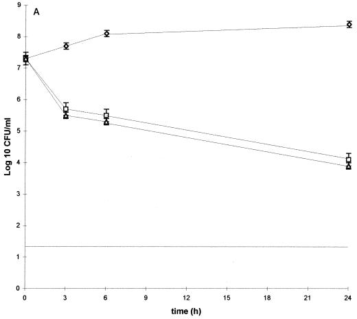
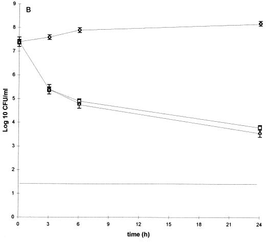
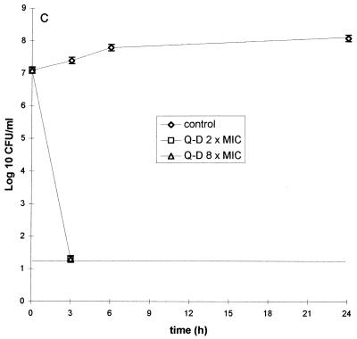
Time-kill curves showed that Q-D was as bactericidal against the strains with reduced susceptibility to dalfopristin as against the susceptible strain (Fig. 1). At concentrations of two times the MIC (i.e., 1 μg/ml for strains HM 1054 and N 95 and 2 μg/ml for strain RP 13), Q-D was bactericidal after 24 h of incubation. Q-D produced reductions of 4.3, 3.9, and 5.8 log10 CFU/ml for strains HM 1054, RP 13, and N 95, respectively. The bactericidal effects against strains HM 1054 and RP 13 were similar (Fig. 1A and B). Against strain N 95, the bactericidal effect was achieved after 3 h of exposure (Fig. 1C).
FIG. 1.
Time-kill curves for S. aureus HM 1054 (A), RP 13 (B), and N 95 (C) grown in MH broth in the absence (control) or presence of Q-D at two or eight times the MIC. Each point is the mean of at least two determinations. Vertical bars represent SD of the means.
In vitro selection of streptogramin-resistant mutants.
Spontaneous mutants of the three strains were obtained on media containing Q-D at two times the MIC for the parental strain with frequencies ranging from 7 × 10⁻⁸ to 2 × 10⁻⁷ (Table 1). The MIC of Q-D against the colonies growing on the selective agar ranged between 1 and 4 μg/ml. However, whatever the parental strain, these mutants were not stable, since the MIC of Q-D decreased after one or two subcultures. Neither the level of susceptibility of these mutants to Q-D nor their frequency of mutation was influenced by the MIC of dalfopristin for the parental strain. No mutant was detected on selective media containing four times the MIC of Q-D.
Antibiotic concentrations.
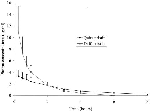
The concentrations of quinupristin and dalfopristin in plasma after an i.m. injection of 30 mg of Q-D (30:70) per kg are shown in Fig. 2. Peak levels were achieved within 0.25 and 0.5 h after antibiotic administration. Mean maximum concentrations of quinupristin and dalfopristin were 3.32 ± 0.97 and 10.9 ± 4.5 μg/ml, respectively. These values were in the same ratio as the administered drugs. Due to a more rapid apparent elimination half-life of dalfopristin than of quinupristin (0.97 ± 0.28 versus 2.79 ± 1.00 h, respectively), the ratios of AUCs for quinupristin and dalfopristin were higher than those of the administered drug. Indeed, the AUCs of quinupristin and dalfopristin were 9.9 ± 2.0 and 11.5 ± 3.4 h · μg/ml, respectively. Quinupristin was still present in plasma 8 h after administration at a concentration near 0.20 μg/ml, while dalfopristin was no longer detected 6 h after administration. Vancomycin concentrations were 40 ± 8 μg/ml (peak) and 12 ± 5 μg/ml (trough).
FIG. 2.
Mean plasma concentration-time curves of quinupristin and dalfopristin for uninfected rabbits after an i.m. injection of 30 mg of Q-D per kg. Each point is the mean for three different animals. Vertical bars represent SD of the means.
Activities of antimicrobial agents in staphylococcal endocarditis.
The activities of the different antibiotic regimens are shown in Table 2. Despite the fact that the delay between bacterial inoculation and the sacrifice of control animals at the start of therapy was up to 72 h for S. aureus N 95, bacterial titers in control animals were the lowest for this strain, indicating that it was less virulent than the other two strains. Q-D and vancomycin significantly reduced bacterial counts in vegetations compared to those in controls (P < 0.01). Against the three tested strains, Q-D was as active as vancomycin (P > 0.2) and was bactericidal. Overall, the proportions of animals with sterile vegetations at the end of treatment were not different in the two treatment groups with the three tested strains (P > 0.05).
TABLE 2.
Activities of Q-D and vancomycin against three strains of S. aureus with various levels of susceptibility to dalfopristin after 4 days of treatment of rabbit endocarditis
| Regimen | Log10 CFU/g of vegetation (no. of sterile animals/no. of treated animals) with strain:
|
||
|---|---|---|---|
| HM 1054 | RP 13 | N 95 | |
| Control | 10.2 ± 0.6 (—/14)a | 9.1 ± 0.5 (—/8) | 6.4 ± 1.8 (—/8) |
| Q-D (30 mg/kg t.i.d.) | 4.3 ± 1.2 (1/9)b | 5.6 ± 1.5 (1/12)b | 2.3 ± 0.6 (6/8)b |
| Vancomycin (50 mg/kg t.i.d.) | 3.8 ± 2.6 (2/8)b | 4.4 ± 2.2 (4/9)b | 2.9 ± 1.1 (2/7)b |
—, controls were sacrificed at the start of therapy.
The P value for comparison against the control was <0.05.
In vivo selection of streptogramin-resistant mutants.
No vegetations isolated from animals infected with S. aureus HM 1054 or N 95 and treated with Q-D contained resistant mutants.
However, in nine rabbits infected with S. aureus RP 13, mutants resistant to dalfopristin, quinupristin, and Q-D were selected on agar containing two times the MIC of the considered antibiotic (Table 3). All the rabbits tested retained 1% of bacteria that were resistant to high concentrations of dalfopristin (>128 μg/ml). In five of nine rabbits, mutants selected on two times the MIC of quinupristin also had increased quinupristin MICs. However, synergy between quinupristin and dalfopristin was retained against the mutants selected on dalfopristin- or quinupristin-containing agar, with an MIC of Q-D of 1-2 μg/ml (Table 3). None of the mutants selected on dalfopristin or quinupristin at two times the MIC was stable (Table 3). However, one of nine animals retained a mutant resistant to Q-D; this mutant was stable after five subcultures (Table 3). This mutant was selected on agar containing two times the MIC of Q-D. The MICs of quinupristin, dalfopristin, and Q-D were 64, >256, and 8 μg/ml, respectively. In the agar diffusion method, this mutant exhibited cross-resistance to all the tested macrolide-lincosamide-streptogramin antibiotics (erythromycin, clarythromycin, spiramycin, and lincomycin). The phenotype of this mutant for aminoglycosides, rifampin, and tetracyclines was the same as that of its parental strain, S. aureus RP 13. The pulsed-field gel electrophoresis patterns of S. aureus RP 13 and its mutant were similar (data not shown).
TABLE 3.
Mutants of S. aureus RP 13 selected in cardiac vegetations after a 4-day treatment with Q-D in nine rabbits with aortic endocarditisa
| Selective mediumb | No. of animals with mutants | No. of mutants in the vegetations | Stability of the mutants | MIC (μg/ml) of:
|
||
|---|---|---|---|---|---|---|
| Q | D | Q-D | ||||
| D | 9 | 1 × 10⁻² | Unstable | 8 | >128 | 1 |
| Q | 5 | 2.8 × 10⁻³ | Unstable | 64 | >128 | 2 |
| Q-D | 1 | 1 × 10⁻² | Stable | 64 | >256 | 8 |
Q, quinupristin; D, dalfopristin.
Drugs were used at two times the MIC.
DISCUSSION
We previously showed that quinupristin and dalfopristin have synergistic and bactericidal effects in vitro and in experimental endocarditis due to S. aureus susceptible to quinupristin and dalfopristin (13). However, the bactericidal activity of the streptogramin complex may be altered in vitro and in vivo against strains of S. aureus resistant to quinupristin, harboring a constitutive macrolide-lincosamide-streptogramin B (MLSB) resistance phenotype, a very common phenotype among clinical strains of methicillin-resistant S. aureus (14, 19).
In this study, we investigated the influence of resistance to dalfopristin alone, a feature which is much more uncommon in clinical strains (19). Synergy between quinupristin and dalfopristin was retained in vitro against two strains of S. aureus with reduced susceptibility to dalfopristin, which thus remained susceptible to Q-D. In vitro, the synergy between quinupristin and dalfopristin has already been described for such strains. Allignet et al. (5) reported several strains of S. aureus for which the MICs of dalfopristin ranged from 16 to 256 μg/ml and which were susceptible to the streptogramin combination (MICs of Q-D ranging from 0.5 to 1 μg/ml). None of these strains harbored a known gene for resistance to dalfopristin. The mechanism of resistance of our two study strains, S. aureus RP 13 and S. aureus N 95, also was not revealed. vat, vatB, and vga sequences were not detected by a PCR method. Preliminary experiments showed that ribosomal binding of dalfopristin was not altered and that this drug was not inactivated (data not shown), suggesting that an efflux mechanism rather than ribosomal modification or enzymatic inactivation could be responsible for dalfopristin resistance.
Q-D remained bactericidal in vitro and in vivo against strains of S. aureus with reduced susceptibility to dalfopristin. In vitro, the magnitude of the bactericidal effect against the two dalfopristin-resistant strains was similar to that against the susceptible strain. In vivo, the bactericidal activity of Q-D against the dalfopristin-resistant strains was retained, despite the fact that the diffusion of dalfopristin into cardiac vegetations is less important than that of quinupristin (13). This result might be explained by the fact that synergy between quinupristin and dalfopristin has been observed in vitro and in vivo for a wide range of ratios of the two components (6).
However, among the nine rabbits infected with S. aureus RP 13 and treated with Q-D, one rabbit retained 200 CFU of a mutant resistant to Q-D, which represented 1% of the surviving bacteria from the cardiac vegetations. Since each rabbit was inoculated with a maximum of 5 × 10⁶ CFU and the spontaneous frequency of mutation of RP 13 in vitro was 7 × 10⁻⁸ (Table 3), it can be concluded that mutants developed in the cardiac vegetations under the selective pressure of Q-D. This mutant of RP 13 remained stable after five subcultures and had the same pulsed-field gel electrophoresis pattern as the parental strain. It was resistant not only to dalfopristin but also to the macrolide, lincosamide, and streptogramin B type (quinupristin) antibiotics. Thus, acquisition of resistance to both quinupristin and dalfopristin is necessary to acquire resistance to Q-D, since synergy between quinupristin and dalfopristin is retained when either drug alone is inactive. This finding was confirmed by the fact that the acquisition of high-level resistance to dalfopristin was not sufficient to obtain such mutants. Indeed, we obtained in vivo mutants which were resistant to a high level of dalfopristin (MIC > 128 μg/ml) but which remained susceptible to Q-D, the MIC being the same as that for the parental strain (Table 3). This in vivo result is in agreement with a recent report showing that clinical strains of S. aureus resistant to Q-D accumulated vatB and ermA genes (19). In contrast, most strains that were resistant to streptogramin A only remained susceptible to Q-D (5, 19). The absence of in vitro selection of mutants to Q-D in a single step suggested that in vivo acquisition of resistance to Q-D in experimental endocarditis is a multiple-step process.
We have previously explained the decreased bactericidal activity of Q-D against constitutive MLSB-resistant strains of S. aureus in endocarditis by the different diffusion behaviors of quinupristin and dalfopristin in vegetations, which are representative of fibrin-rich foci of infections (13, 14). We believe that the same difference in tissue distribution may explain the emergence of resistance to Q-D in dalfopristin-resistant strains. Since the diffusion of dalfopristin is limited in the core of the vegetation, in contrast to that of quinupristin, treatment with Q-D leads to exposure of the bacteria in the center of the vegetation mainly to quinupristin. If the strain is susceptible to both quinupristin and dalfopristin, the limited amount of dalfopristin present in the center of the vegetation may be sufficient to display a synergistic effect with quinupristin. If the strain is resistant to quinupristin (constitutive MLSB resistance phenotype), bacteria present in the center of the vegetation are exposed to quinupristin, to which they are resistant, leading to a decrease in bactericidal activity, as previously shown (14). If the strain is resistant to dalfopristin, bacteria in the core of the vegetation are exposed to low concentrations of dalfopristin, which might lead to the rapid selection of highly dalfopristin-resistant mutants. In a further step, double mutants resistant to both quinupristin and dalfopristin could be selected, leading at least in some cases to the loss of synergy between quinupristin and dalfopristin and resistance to the combination.
Finally, it is important to point out that our in vivo results were obtained with areas under the concentration-time curves of quinupristin and dalfopristin in plasma that were at least comparable to those achieved in humans (Fig. 2) (11). Therefore, selection of strains of S. aureus resistant to the Q-D combination may occur in vivo despite the use of appropriate dosage of Q-D, provided that the initial strain harbors resistance to streptogramin A and that the size of the initial inoculum is sufficient.
We conclude that the synergy and the bactericidal effect of Q-D are retained in vitro and in vivo against strains resistant to dalfopristin and susceptible to quinupristin. Our study demonstrated that the acquisition of resistance to both quinupristin and dalfopristin is necessary to select mutants resistant to Q-D. Therefore, in staphylococcal infections with a large inoculum and reduced susceptibility to dalfopristin, therapy with Q-D may select mutants resistant to Q-D. Combination of Q-D with another antibiotic might be of interest in preventing the emergence of resistance.
ACKNOWLEDGMENTS
We thank Marie-Laure Ozoux and Jean-Paul Plard from Aventis for their technical assistance and Jérôme Etienne for kindly providing S. aureus strain N 95.
Virginie Zarrouk was supported by a grant from la Fondation pour la Recherche Médicale. This work was supported by a grant from Aventis.
REFERENCES
- 1.Aldridge K, Schiro D D, Varner L M. In vitro antistaphylococcal activity and testing of RP 59500, a new streptogramin, by two methods. Antimicrob Agents Chemother. 1992;36:854–855. doi: 10.1128/aac.36.4.854. [DOI] [PMC free article] [PubMed] [Google Scholar]
- 2.Allignet J, Loncle V, El Sohl N. Sequence of a staphylococcal plasmid gene, vga, encoding a putative ATP-binding protein involved in resistance to virginiamycin-A like antibiotics. Gene. 1992;117:45–51. doi: 10.1016/0378-1119(92)90488-b. [DOI] [PubMed] [Google Scholar]
- 3.Allignet J, Loncle V, Simenel C, Delepierre M, El Sohl N. Sequence of a staphylococcal gene, vat, encoding an acetyltransferase inactivating the A-type compounds of virginiamycin-like antibiotics. Gene. 1993;130:91–98. doi: 10.1016/0378-1119(93)90350-c. [DOI] [PubMed] [Google Scholar]
- 4.Allignet J, El Sohl N. Diversity among the gram-positive acetyltransferases inactivating streptogramin A and structurally related compounds and characterization of a new staphylococcal determinant, vatB. Antimicrob Agents Chemother. 1995;39:2027–2036. doi: 10.1128/aac.39.9.2027. [DOI] [PMC free article] [PubMed] [Google Scholar]
- 5.Allignet J, Aubert S, Morvan A, El Sohl N. Distribution of genes encoding resistance to streptogramin A and related compounds among staphylococci resistant to these antibiotics. Antimicrob Agents Chemother. 1996;40:2523–2528. doi: 10.1128/aac.40.11.2523. [DOI] [PMC free article] [PubMed] [Google Scholar]
- 6.Bouanchaud D H. In-vitro and in-vivo synergic activity and fractional inhibitory concentration (FIC) of the components of a semisynthetic streptogramin, RP 59500. J Antimicrob Chemother. 1992;30(Suppl. A):95–99. doi: 10.1093/jac/30.suppl_a.95. [DOI] [PubMed] [Google Scholar]
- 7.Bozdogan B, Leclercq R. Effects of genes encoding resistance to streptogramins A and B on the activity of quinupristin-dalfopristin against Enterococcus faecium. Antimicrob Agents Chemother. 1999;43:2720–2725. doi: 10.1128/aac.43.11.2720. [DOI] [PMC free article] [PubMed] [Google Scholar]
- 8.Brumfitt W, Hamilton Miller J M T, Shah S. In vitro activity of RP 59500, a new semisynthetic streptogramin against Gram-positive bacteria. J Antimicrob Chemother. 1992;30(Suppl. A):29–37. doi: 10.1093/jac/30.suppl_a.29. [DOI] [PubMed] [Google Scholar]
- 9.Brumfitt W, Hamilton-Miller J. Methicillin resistant Staphylococcus aureus. N Engl J Med. 1992;320:1188–1196. doi: 10.1056/NEJM198905043201806. [DOI] [PubMed] [Google Scholar]
- 10.Entenza J M, Drugeon H, Glauser M P, Moreillon P. Treatment of experimental endocarditis due to erythromycin-susceptible or -resistant methicillin-resistant Staphylococcus aureus with RP 59500. Antimicrob Agents Chemother. 1995;39:1419–1424. doi: 10.1128/aac.39.7.1419. [DOI] [PMC free article] [PubMed] [Google Scholar]
- 11.Etienne S D, Montay G, Le Liboux A, Frydman A, Garaud J J. A phase I, double-blind, placebo-controlled study of the tolerance and pharmacokinetic behaviour of RP 59500. J Antimicrob Chemother. 1992;30(Suppl. A):123–131. doi: 10.1093/jac/30.suppl_a.123. [DOI] [PubMed] [Google Scholar]
- 12.European Commission. Directive for the protection of vertebrate animals used for experimental and other scientific purposes (86/609/EEC) Off J C Eur Comm L. 1986;358:1–29. [Google Scholar]
- 13.Fantin B, Leclercq R, Ottaviani M, Vallois J M, Mazière B, Duval J, Pocidalo J J, Carbon C. In vivo activities and penetration of the two components of the streptogramin RP 59500 in cardiac vegetations of experimental endocarditis. Antimicrob Agents Chemother. 1994;38:432–437. doi: 10.1128/aac.38.3.432. [DOI] [PMC free article] [PubMed] [Google Scholar]
- 14.Fantin B, Leclercq R, Merlé Y, Saint-Julien L, Veyrat C, Duval J, Carbon C. Critical influence of resistance to streptogramin B-type antibiotics on activity of RP 59500 (quinupristin-dalfopristin) in experimental endocarditis due to Staphylococcus aureus. Antimicrob Agents Chemother. 1995;39:400–405. doi: 10.1128/aac.39.2.400. [DOI] [PMC free article] [PubMed] [Google Scholar]
- 15.Gomeni R. An interactive programme for individual and population parameter estimation. In: Von Bommek M J, Ball N, Wigertz N, editors. Medinfo 83. Amsterdam, The Netherlands: North-Holland Publishing Co.; 1983. pp. 1022–1025. [Google Scholar]
- 16.Leclercq R, Courvalin P. Bacterial resistance to macrolide, lincosamide, and streptogramin antibiotics by target modification. Antimicrob Agents Chemother. 1991;35:1267–1272. doi: 10.1128/aac.35.7.1267. [DOI] [PMC free article] [PubMed] [Google Scholar]
- 17.Leclercq R, Courvalin P. Intrinsic and unusual resistance to macrolide, lincosamide, and streptogramin antibiotics in bacteria. Antimicrob Agents Chemother. 1991;35:1273–1276. doi: 10.1128/aac.35.7.1273. [DOI] [PMC free article] [PubMed] [Google Scholar]
- 18.Leclercq R, Nantas L, Soussy C J, Duval J. Activity of RP 59500, a new parenteral semisynthetic streptogramin, against staphylococci with various mechanisms of resistance to macrolide-lincosamide-streptogramin antibiotics. J Antimicrob Chemother. 1992;30(Suppl. A):67–75. doi: 10.1093/jac/30.suppl_a.67. [DOI] [PubMed] [Google Scholar]
- 19.Lina G, Quaglia A, Reverdy M E, Leclercq R, Vandenesch F, Etienne J. Distribution of genes encoding resistance to macrolides, lincosamides, and streptogramins among staphylococci. Antimicrob Agents Chemother. 1999;43:1062–1066. doi: 10.1128/aac.43.5.1062. [DOI] [PMC free article] [PubMed] [Google Scholar]
- 20.Mapple P A C, Hamilton-Miller J M T, Brumfitt W. World-wide antibiotic resistance in methicillin resistant Staphylococcus aureus. Lancet. 1989;i:537–539. doi: 10.1016/s0140-6736(89)90076-7. [DOI] [PubMed] [Google Scholar]
- 21.Pearson R D, Steibigel R T, Davis H T, Chapman S W. Method for reliable determination of minimal lethal antibiotic concentrations. Antimicrob Agents Chemother. 1980;18:699–708. doi: 10.1128/aac.18.5.699. [DOI] [PMC free article] [PubMed] [Google Scholar]
- 22.Perlman B B, Freedman L R. Experimental endocarditis. II. Staphylococcal infection of the aortic valve following placement of a polyethylene catheter in the left side of the heart. Yale J Biol Med. 1971;44:206–213. [PMC free article] [PubMed] [Google Scholar]
- 23.Smith T L, Pearson M L, Wilcox K R, Cruz C, Lancaster M V, Robinson-Dunn B, Tenover F C, Zervos M T, Band J D, White E, Jarvis W R. Emergence of vancomycin resistance in Staphylococcus aureus. Glycopeptide-Intermediate Staphylococcus aureus Working Group. N Engl J Med. 1999;340:493–501. doi: 10.1056/NEJM199902183400701. [DOI] [PubMed] [Google Scholar]
- 24.Voss A, Milatovic D, Wallrauch-Schwarz C, Rosdahl V T, Braveny I. Methicillin-resistant Staphylococcus aureus in Europe. Eur J Clin Microbiol Infect Dis. 1994;13:50–55. doi: 10.1007/BF02026127. [DOI] [PubMed] [Google Scholar]


