Abstract
Background
According to the latest the World Health Organization (WHO) classification in 2015, invasive mucinous adenocarcinoma (IMA) is defined as a new pathological subtype of lung adenocarcinoma (LUAD). However, whether this rare subtype of lung pathology has any difference in prognosis than conventional LUAD is debatable. Our study attempted to compare clinical characteristics and prognosis of IMA vs. noninvasive mucinous adenocarcinomas (NMA).
Methods
A total of 1,857 patients with LUAD who underwent radical resection were screened from 2010 to 2015 at Zhejiang Cancer Hospital. Patients with pulmonary IMA were matched 1:1 by using propensity scores with LUAD adjusted for clinicopathological characteristics. After follow-up, overall survival (OS) and disease-free survival (DFS) were explored by Kaplan-Meier and Cox regression analyses. Forest plots were used for subgroup analyses.
Results
Following screening, 499 patients with LUAD were enrolled, with 97 IMA and 402 NMA. Compared to NMA of the lung, IMA was proportionately lower in women (50.5% vs. 63.4%; P=0.026) and nonsmokers (P<0.001). IMA was also associated with earlier tumor stage I (68.0% vs. 55.5%; P=0.033) and lower frequency of upper lobe tumors compared to NMA (P=0.007). Following propensity score matching, 97 pairs were selected, among which we found that patients with pulmonary IMA had a longer OS than those with NMA (P=0.014). According to the subgroup analysis, improved OS in the IMA cohort versus the NMA cohort was observed across various factors, including the absence of lymphovascular invasion or perineural invasion.
Conclusions
In this study, we found that resectable IMA patients had a better OS than NMA patients. This study contributes to the understanding of IMA in depth, but it needs to be validated through additional multicenter studies.
Keywords: Non-small cell lung cancer (NSCLC), pulmonary invasive mucinous adenocarcinoma, propensity-matched analysis, prognosis
Introduction
Lung cancer is the leading cause of cancer-related deaths worldwide. The majority of cases present with advanced disease at the time of diagnosis. Consequently, cancer statistics from the United States for 2019 show that the overall 5-year relative survival rate for lung cancer is low at 16% and 22% for men and women, respectively (1). Non-small cell lung cancer (NSCLC) accounts for more than 80% of all lung cancers with adenocarcinoma being the most common pathological subtype. Primary invasive mucinous adenocarcinoma (IMA), formerly known as bronchioloalveolar carcinoma (BAC), is a subtype of adenocarcinoma according to the current World Health Organization (WHO) classification of lung cancer (2). The prevalence of IMA is low compared to noninvasive mucinous adenocarcinoma (NMA), accounting for only 1.5–10% of all lung adenocarcinomas (LUADs) (3-5). In the 2011 edition of the pathological classification jointly developed by the International Association for the Study of Lung Cancer (IASLC), the American Thoracic Society (ATS), and the European Respiratory Society (ERS), the name was changed from mucinous fine bronchoalveolar carcinoma to IMA (6).
The clinical presentation, imaging, pathology, and genetic features of IMA are different from those of non-mucinous adenocarcinoma. Patients with IMA lack specific clinical manifestations in the early stage, and most of them are found during a routine physical examination. Clinically, IMA often presents with cough, sputum, bloody sputum, and chest pain, which is easily misdiagnosed as pneumonia or tuberculosis (7). As a subtype of LUAD, the most important treatment for IMA in the early stage is surgical resection (8), but many IMA patients have advanced beyond surgical resectability at the time of first diagnosis.
Due to the low incidence of pulmonary IMA, there are only a few, albeit conflicting, statistical reports regarding its prognosis. A retrospective study conducted by Casali et al. (9) showed that early non-mucinous BAC had a better prognosis, while mucinous BAC had a worse prognosis. Similarly, a study by Russell et al. (10) reported similar findings, with a 5-year survival rate of 51% for IMA and close to 100% for other new subtypes of in situ adenocarcinomas, such as microinvasive adenocarcinoma and predominant lymphoid adenocarcinoma. A retrospective study by Yoshizawa et al. (11), which analyzed the survival of 440 Japanese patients with LUAD, showed that the 5-year disease-free survival (DFS) rate of IMA was 88.8%, which was between that of the low- and high-grade adenocarcinoma groups. However, a retrospective study of 1,699 LUAD patients by Cai et al. (12) showed that there was no significant difference in recurrence-free survival (RFS) or overall survival (OS) between IMA patients and NMA patients, but the subtype of IMA had an impact on patient prognosis. Furthermore, in another retrospective study conducted by Shim et al. in the United States and Korea (13), there was no statistically significant difference in 5-year survival and OS between IMA and NMA patients, and the recurrence sites of all IMA patients were intrapulmonary. Finally, the study by Warth et al. (14) yielded completely different results, with patients with IMA having a better prognosis than most patients with noninvasive mucinous adenocarcinoma. Therefore, research based on more precise statistical methods is urgently needed to explore the prognosis of IMA patients. In this study, we retrospectively analyzed previous cases of IMA and excluded confounding factors by propensity score matching (PSM) to better reveal the prognostic features of IMA. We present the following article in accordance with the STROBE reporting checklist (available at https://tlcr.amegroups.com/article/view/10.21037/tlcr-22-190/rc).
Methods
Study design and patients
Between January 2010 and December 2015, 1,857 patients with LUAD underwent surgery at Zhejiang Cancer Hospital. The inclusion criteria of the IMA patients were as follows: (I) a pathological diagnosis of pulmonary invasive adenocarcinoma and surgical samples with complete patient medical records; (II) no antitumor therapy, such as chemoradiotherapy, biotherapy, or immunotherapy, had been administered before surgery; (III) follow-up time ≥3 years; (IV) age ≥18 years. The exclusion criteria were as follows: other concurrent types of malignant tumors within 5 years; metastases from other tumors; and neoadjuvant chemotherapy. Finally, a total of 97 IMA patients were enrolled in the current study. A control group was established by enrolling NSCLC patients with NMA. The study was conducted in accordance with the Declaration of Helsinki (as revised in 2013). The study was approved by the Ethics Committee of Zhejiang Cancer Hospital (No. IRB-2021-14). In accordance with national legislation and institutional requirements, written informed consent was not required for this study.
All LUAD patients received surgical therapy. Clinicopathological data were collected for analyses of the association with LUAD. Gender, age, smoking history, T stage, N stage, primary stage, lymphovascular invasion, perineural invasion, pleural involvement, laterality, operation, and location were included. Based on the new IASLC/ATS/ERS classification, patients were classified into IMA and NMA group, respectively.
PSM
In order to minimize the effect of potential confounders, we performed a matched analysis of patients with pulmonary IMA via PSM. We included as many variables as possible in our propensity score model to maximize the balance of propensity among the variables. The variables included gender, age, smoking history, T stage, N stage, primary stage, Lymphovascular invasion, perineural invasion, pleural involvement, laterality, operation, and location. We calculated the propensity scores for each case regardless of the statistical significance of the independent variables in the model. Cases were finally matched 1:1 without the use of a neighboring method with caliper restrictions.
Follow-up
The outcomes included disease-free status (DFS) and overall survival (OS). If DFS was confirmed at the time of the last visit, the patient was regarded to be alive and free of recurrence. All patients were followed for at least 36 months, or until recurrence, death or lost to follow-up. OS was defined as the period of time from surgery to death of any cause. DFS is defined as the time interval from surgery and the first locoregional recurrence, distant progression, or death from any cause. The routine telephone follow-up visits was performed every 3 months. The follow-up end point was the death date or September 30, 2021.
Statistical methods
Statistical analysis was performed using SPSS 26.0 (IBM Corp., Armonk, NY, USA) and R 3.6.2 software (The R foundation for Statistical Computing, Vienna, Austria; https://www.r-project.org/), and the statistical data were compared using Pearson’s chi-square test for continuous data and the Mann–Whitney rank-sum test for count data. The study endpoint event was OS, defined as the time from surgery to death or last follow-up. We defined DFS as the time from radical resection to recurrence of the lesion or death. Both OS and DFS were analyzed using the Kaplan-Meier log-rank test, and prognostic correlates were explored using a Cox proportional risk regression model. The factors identified in univariate analyses with a P value of less than 0.05 were then further analyzed by multivariate analysis. This study used the R package “MatchIt” for PSM analysis.
Results
Patient characteristics
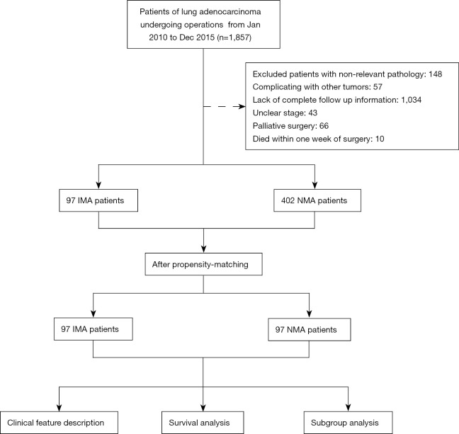
In total, 1,857 patients with LUAD underwent operations from January 2010 to December 2015. Among these, 1,358 patients were excluded due to nonrelevant pathology (n=148), complications with other tumors (n=57), a lack of complete follow-up information (n=1,034), unclear stage (n=43), palliative surgery (n=66), or death within 1 week of surgery (n=10) (Figure 1). Then, a total of 499 patients with NSCLC were enrolled, among whom 97 had invasive mucinous adenocarcinoma and the remaining 402 had noninvasive mucinous adenocarcinoma. The clinicopathological characteristics are shown in Table 1. After PSM, 97 patients with NMA were screened and balanced with 97 patients with pulmonary IMA. The PSM analysis yielded well-balanced IMA and NMA cohorts (Table 1).
Figure 1.
Study flow diagram of the selection process of eligible cases. NMA, non-mucinous adenocarcinoma; IMA, invasive mucinous adenocarcinoma.
Table 1. Baseline characteristics of the matched IMA and NMA patient cohorts.
| Variables | All (N=499) | Before PSM | After PSM | |||||
|---|---|---|---|---|---|---|---|---|
| IMA (N=97) | NMA (N=402) | P value | IMA (N=97) | NMA (N=97) | P value | |||
| Gender, n (%) | 0.026* | 0.886 | ||||||
| Female | 304 (60.9) | 49 (50.5) | 255 (63.4) | 49 (50.5) | 51 (52.6) | |||
| Male | 195 (39.1) | 48 (49.5) | 147 (36.6) | 48 (49.5) | 46 (47.4) | |||
| Age, n (%) | 0.563 | 1.000 | ||||||
| ≤60 years | 278 (55.7) | 51 (52.6) | 227 (56.5) | 51 (52.6) | 51 (52.6) | |||
| >60 years | 221 (44.3) | 46 (47.4) | 175 (43.5) | 46 (47.4) | 46 (47.4) | |||
| Smoke, n (%) | <0.001* | 0.072 | ||||||
| No | 363 (72.7) | 56 (57.7) | 307 (76.4) | 56 (57.7) | 69 (71.1) | |||
| Yes | 136 (27.3) | 41 (42.3) | 95 (23.6) | 41 (42.3) | 28 (28.9) | |||
| T, n (%) | 0.462 | 1.000 | ||||||
| T1/T2 | 473 (94.8) | 90 (92.8) | 383 (95.3) | 90 (92.8) | 91 (93.8) | |||
| T3/T4 | 26 (5.2) | 7 (7.2) | 19 (4.7) | 7 (7.2) | 6 (6.2) | |||
| N, n (%) | 0.070 | 0.506 | ||||||
| N0 | 305 (61.1) | 69 (71.1) | 236 (58.7) | 69 (71.1) | 66 (68.0) | |||
| N1 | 64 (12.8) | 8 (8.2) | 56 (13.9) | 8 (8.2) | 13 (13.4) | |||
| N2/N3 | 130 (26.1) | 20 (20.6) | 110 (27.4) | 20 (20.6) | 18 (18.6) | |||
| Stage, n (%) | 0.033 | 0.461 | ||||||
| I | 289 (57.9) | 66 (68.0) | 223 (55.5) | 66 (68.0) | 64 (66.0) | |||
| II | 72 (14.4) | 7 (7.2) | 65 (16.2) | 7 (7.2) | 12 (12.4) | |||
| III | 138 (27.7) | 24 (24.7) | 114 (28.4) | 24 (24.7) | 21 (21.6) | |||
| Lymphovascular invasion, n (%) | 0.131 | 0.668 | ||||||
| No | 414 (83.0) | 86 (88.7) | 328 (81.6) | 86 (88.7) | 83 (85.6) | |||
| Yes | 85 (17.0) | 11 (11.3) | 74 (18.4) | 11 (11.3) | 14 (14.4) | |||
| Perineural invasion, n (%) | 1.000 | 0.745 | ||||||
| No | 467 (93.6) | 91 (93.8) | 376 (93.5) | 91 (93.8) | 93 (95.9) | |||
| Yes | 32 (6.4) | 6 (6.2) | 26 (6.5) | 6 (6.2) | 4 (4.1) | |||
| Pleural involvement, n (%) | 1.000 | 1.000 | ||||||
| No | 300 (60.1) | 58 (59.8) | 242 (60.2) | 58 (59.8) | 58 (59.8) | |||
| Yes | 199 (39.9) | 39 (40.2) | 160 (39.8) | 39 (40.2) | 39 (40.2) | |||
| Laterality, n (%) | 0.180 | 0.286 | ||||||
| Left | 203 (40.7) | 44 (45.4) | 159 (39.6) | 44 (45.4) | 52 (53.6) | |||
| Right | 285 (57.1) | 53 (54.6) | 232 (57.7) | 53 (54.6) | 44 (45.4) | |||
| Bilateral | 11 (2.2) | 0 (0.0) | 11 (2.7) | 0 (0.0) | 1 (1.0) | |||
| Location, n (%) | 0.007* | 0.607 | ||||||
| Upper | 270 (54.2) | 40 (41.2) | 230 (57.4) | 40 (41.2) | 43 (44.3) | |||
| Middle | 40 (8.0) | 13 (13.4) | 27 (6.7) | 13 (13.4) | 9 (9.3) | |||
| Lower | 182 (36.5) | 44 (45.4) | 138 (34.4) | 44 (45.4) | 44 (45.4) | |||
| Other | 6 (1.2) | 0 (0.0) | 6 (1.5) | 0 (0.0) | 1 (1.0) | |||
| Operation, n (%) | 0.134 | 0.592 | ||||||
| Sublobar resection | 36 (7.2) | 4 (4.1) | 32 (8.0) | 4 (4.1) | 2 (2.1) | |||
| Lobectomy | 459 (92.0) | 91 (93.8) | 368 (91.5) | 91 (93.8) | 94 (96.9) | |||
| Pneumonectomy | 4 (0.8) | 2 (2.1) | 2 (0.5) | 2 (2.1) | 1 (1.0) | |||
*, P value is less than 0.05. PSM, propensity score matching; IMA, pulmonary invasive mucinous adenocarcinoma group; NMA, pulmonary non-invasive mucinous adenocarcinoma group.
Compared to NMA of the lung, IMA had a lower proportion of women (P=0.026) and nonsmokers (P<0.001) and a higher proportion with early-stage tumor pathology (P=0.033), with fewer tumor lesions located in the upper lobes (P=0.007) (Table 1).
Survival analysis
The median follow-up times were 41.3 (0.7–60.6) and 46.85 (11.2–64.2) months in the IMA and NMA groups, respectively.
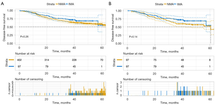
Before PSM, the 3- and 5-year DFS rates for the IMA patients were 73.1% and 54.8%, respectively, compared to 65.5% and 58.9% for NMA patients [hazard ratio (HR) =0.80; 95% confidence interval (CI): 0.55–1.18; P=0.260] (Figure 2A). After PSM, IMA cases had a slightly longer DFS than NMA cases. The 3- and 5-year DFS rates for IMA patients were 73.1% and 54.8%, respectively, compared to 59.5% and 53.1% for NMA patients (HR =0.70; 95% CI: 0.44–1.13; P=0.140) (Figure 2B).
Figure 2.
Disease-free survival (DFS) in non-mucinous adenocarcinoma (IMA) and non-mucinous adenocarcinoma (NMA) group. Shown are Kaplan-Meier curves estimates of DFS between patients with IMA and NMA before (A) and after (B) propensity score matching (PSM).
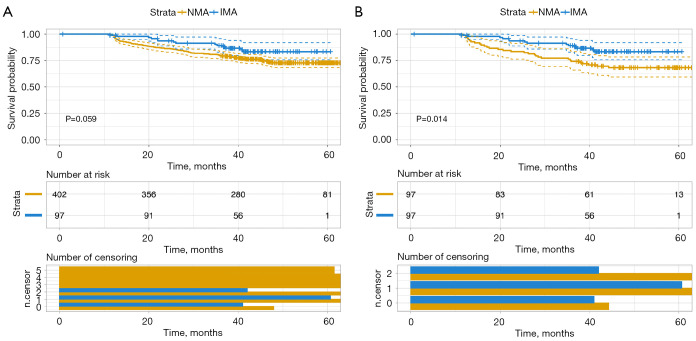
Prior to PSM, there was no significant difference in OS time between pulmonary IMA (3-year OS rate =89.2%; 5-year OS rate =83.4%) and NMA (3-year OS rate =79.6%; 5-year OS rate =72.8%) (HR =0.59; 95% CI: 0.34–1.03; P=0.059) (Figure 3A), and the median survival was not reached at the last follow-up. Notably, after PSM, we found that patients with pulmonary IMA (3-year OS rate =89.2%; 5-year OS rate =83.4%) had a better OS than patients with NMA (3-year OS rate =74.2%; 5-year OS rate =68.3%) (HR =0.46; 95% CI: 0.24–0.87; P=0.014) (Figure 3B).
Figure 3.
Overall survival (OS) in non-mucinous adenocarcinoma (IMA) and non-mucinous adenocarcinoma (NMA) group. Shown are Kaplan-Meier curves estimates of DFS between patients with IMA and NMA before (A) and after (B) propensity score matching (PSM).
In addition, we further investigated the correlation between OS and clinical variables in the matched study, as shown in Table 2. Age (P=0.024), N stage (P=0.022), and location of the lesion (P=0.046) served as prognostic factors by univariate analysis. After multivariate analysis, we identified age (P=0.005) and location of the lesion (P=0.003) as independent prognostic factors for OS.
Table 2. Results of univariate and multivariate analyses of survival after PSM.
| Characteristics | Univariable | Multivariable | |||
|---|---|---|---|---|---|
| HR (95% CI) | P value | HR (95% CI) | P value | ||
| Gender | |||||
| Female | Reference | ||||
| Male | 0.51 (0.17–1.52) | 0.226 | |||
| Age | |||||
| ≤60 years | Reference | Reference | |||
| >60 years | 4.33 (1.21–15.53) | 0.024* | 9.12 (1.96–42.53) | 0.005* | |
| Smoke | |||||
| No | Reference | ||||
| Yes | 1.06 (0.37–3.06) | 0.914 | |||
| T | |||||
| T1 | Reference | ||||
| T2 | 1.18 (0.36–3.94) | 0.782 | |||
| T3 | 2.18 (0.46–10.28) | 0.325 | |||
| N | |||||
| N0 | Reference | Reference | |||
| N1 | 3.98 (1.23–12.94) | 0.022* | 2.73 (0.82–9.04) | 0.101 | |
| N2 | 0 (0–Inf) | 0.998 | 0 (0–Inf) | 0.998 | |
| N3 | 1.32 (0.17–10.46) | 0.791 | 0.43 (0.05–3.80) | 0.444 | |
| Stage | |||||
| I | Reference | ||||
| II | 2.09 (0.45–9.69) | 0.345 | |||
| III | 0.80 (0.22–2.95) | 0.734 | |||
| Lymphovascular invasion | |||||
| No | Reference | ||||
| Yes | 1.32 (0.30–5.91) | 0.716 | |||
| Perineural invasion | |||||
| No | Reference | ||||
| Yes | 1.12 (0.15–8.57) | 0.914 | |||
| Pleural involvement | |||||
| No | Reference | ||||
| Yes | 1.19 (0.41–3.44) | 0.743 | |||
| Laterality | |||||
| Left | Reference | ||||
| Right | 1.15 (0.40–3.30) | 0.801 | |||
| Location | |||||
| Lower | Reference | Reference | |||
| Middle | 4.10 (1.02–16.45) | 0.046* | 13.60 (2.49–74.13) | 0.003* | |
| Upper | 1.62 (0.46–5.74) | 0.455 | 1.62 (0.45–5.82) | 0.462 | |
| Operation | |||||
| Sublobar resection | Reference | Reference | |||
| Lobectomy | 29,347,409.79 (0–Inf) | 0.998 | |||
| Pneumonectomy | 384,226,247.17 (0–Inf) | 0.998 | |||
*, P value is less than 0.05. PSM, propensity score matching; IMA, pulmonary invasive mucinous adenocarcinoma group; NMA, pulmonary non-invasive mucinous adenocarcinoma group; HR, hazard ratio; CI, confidence interval.
Subgroup analysis
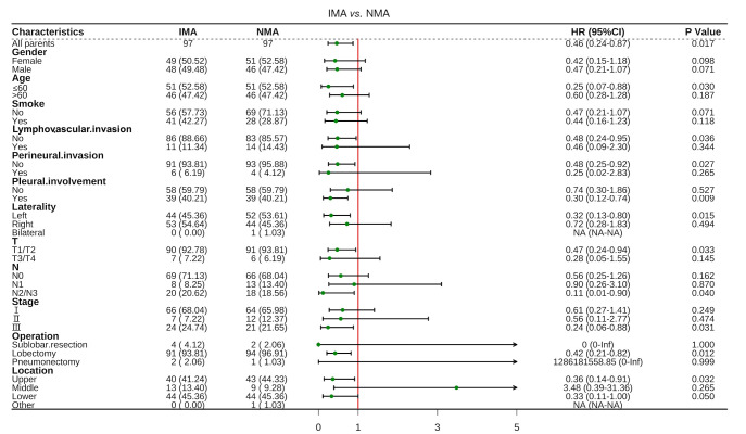
Subgroup analysis of OS was conducted according to the baseline characteristics (Figure 4). Improved OS in the IMA cohort versus NMA cohort was observed across cases ≤60 years old (HR =0.25; 95% CI: 0.07–0.88; P=0.030), absence of lymphovascular invasion (HR =0.48; 95% CI: 0.24–0.95; P=0.036) or perineural invasion (HR: 0.48; 95% CI: 0.25–0.92; P=0.027), and pleural invasion (HR =0.30; 95% CI: 0.12–0.74; P=0.009). A similar benefit was seen in the IMA cohort in cases with a tumor location on the left side (HR =0.32; 95% CI: 0.13–0.8; P=0.015) or in the upper lobe (HR =0.36; 95% CI: 0.14–0.91; P=0.032). In addition, in the group with early T stage (HR =0.47; 95% CI: 0.24–0.94; P=0.033), advanced N stage (HR =0.11; 95% CI: 0.01–0.90; P=0.04), and advanced pathological stage (HR =0.24; 95% CI: 0.06–0.88; P=0.031), patients with IMA showed prolonged OS compared with those with NMA. In terms of the choice of surgical approach, IMA patients who underwent lobectomy (HR =0.42; 95% CI: 0.21–0.82; P=0.012) had a longer OS than NMA patients.
Figure 4.
Forest plot for subgroup analysis for overall survival according to the baseline characteristics. OS, overall survival; NMA, non-mucinous adenocarcinoma; IMA, invasive mucinous adenocarcinoma; HR, hazard ratio; CI, confidence interval.
Discussion
As the predominant type of NSCLC, the incidence of LUAD is increasing (15). According to the latest WHO classification of pulmonary tumors (updated in 2015), primary pulmonary IMA is a unique variant of pulmonary adenocarcinoma. Previously, IMA was classified as a mucinous BAC, as its tumor cells were in a cup or columnar shape, and there was a large amount of mucin in the cytoplasm. The nuclei are usually not obvious or absent, and the surrounding alveolar spaces is often filled with viscin (16).
IMA is regarded as a distinct pathological type of LUAD based on its unique clinical and genetic features (17). However, while there have been many studies on the prognosis of pulmonary IMA, the conclusions among these studies have been highly contradictory. In this study, we sought to clarify the prognostic significance of IMA through an analysis of IMA and NMA patients while limiting the influence of data biases and confounding variables as much as possible through use of PSM. Then, we explored the prognosis of these patients using univariate and multivariate Cox regression analyses. We also conducted subgroup analysis, which can be helpful to verify the prognostic factors of IMA. The results of our study suggested that some IMA patients may have a better OS than NMA patients.
According to a previous report, the epidemiology of IMA is similar to that of other LUADs (18). In contrast to prior research (5), in our study, IMA was less common than NMA among women. This may have been due to differences between the eastern and western populations. The age distribution was similar between IMA and NMA (5), which is in accordance with our findings. Smoking, as an essential influencing factor affecting lung cancer, did not show differences between IMA and NMA cases in our study. In our cohort, the pathological tumor stage of IMA cases was earlier, and the tumor lesions were more commonly located in the lower lobes. These findings are consistent with those previously reported (5,19).
Due to their low incidence, the clinicopathological features and prognosis of IMAs have remained unclear and controversial. After PSM was performed, multivariate Cox analyses revealed that age >60 years (HR =9.12, P=0.005) and a middle location of the lesion (HR =13.6, P=0.003) were independent risk predictors for OS (Table 2). These results are consistent with previous studies (5,20). In addition, few studies have shown that tumor size and extent of invasion may be independent factors affecting the prognosis of IMAs (20-22). However, T stage did not reach statistical significance in univariate stratified Cox proportional hazards analyses. This may suggest that the prognostic impact of T stage is not significantly different for IMA patients. After excluding confounding factors in eastern populations, additional studies with larger samples are needed to verify this finding.
Some research has reported that compared to patients with other LUAD subtypes, IMA patients have poor OS (9,10,23,24). However, other previous studies have shown that the OS of IMA patients is comparable to that of NMA patients (13,21). Warth et al. showed that IMA patients have a better prognosis than other adenocarcinoma patients (14). In our study, after excluding confounding factors, IMA had a better prognosis in terms of OS than other LUADs. In the subgroup analysis of IMA patients, the overall prognostic characteristics of the IMA patients were significantly better than those of the NMA patients.
However, our study failed to demonstrate significant differences in DFS between IMA and NMA patients. This may be because OS may be influenced by non-first line therapy. Due to the differences in the gene mutation rates (such as EGFR and ALK) between the IMA and NMA groups (17,25), patients with IMA may have a greater chance of benefiting from targeted therapies.
To our knowledge, this was the first study to focus on the prognostic characteristics of lung IMA via PSM and subgroup analysis. In clinical practice, PSM plays an important role. Through appropriate randomization in retrospective studies to reduce selection bias, we can find the source of heterogeneity by using subgroup analysis. Therefore, by using both statistical tools simultaneously, we can provide a more reliable real world event analysis of the prognosis of primary IMA.
There were several limitations that need to be considered in our study. First, this was a retrospective study with a comparatively small sample size. Due to the low incidence of IMA, we could not gather sufficient samples to obtain the most accurate OS results. Thus, we used the PSM tool to reduce this weakness, and subgroup analysis was applied to further validate the outcomes of the prognostic analysis. Second, since we enrolled patients who were surgical candidates, the median survival of the patients could not be observed during the 3-year follow-up period. Additionally, there was a lack of complete non-first line treatment information and gene expression information that may affect OS. For these issues, more studies based on larger sample sizes and longer follow-up periods are necessary.
In summary, we conducted a real-world, large population-based analysis of the prognosis of IMA. We used PSM to reduce possible confounding factors in the statistical process, including gender, age, smoking, T stage, N stage, and operation. Then, subgroup analysis was applied to verify the overall prognostic characteristics and sources of heterogeneity of IMA. The prognostic features of the IMA patients were studied in-depth in this project.
Supplementary
The article’s supplementary files as
Acknowledgments
The authors appreciate the academic support from the AME Lung Cancer Collaborative Group.
Funding: This work was supported by grants from the National Natural Science Foundation of China (Nos. 81802995 and 81672315) and Key R&D Program Projects in Zhejiang Province (No. 2017C04G1360498).
Ethical Statement: The authors are accountable for all aspects of the work in ensuring that questions related to the accuracy or integrity of any part of the work are appropriately investigated and resolved. The study was conducted in accordance with the Declaration of Helsinki (as revised in 2013). The study was approved by the Ethics Committee of Zhejiang Cancer Hospital (No. IRB-2021-14). In accordance with national legislation and institutional requirements, written informed consent was not required for this study.
Footnotes
Reporting Checklist: The authors have completed the STROBE reporting checklist. Available at https://tlcr.amegroups.com/article/view/10.21037/tlcr-22-190/rc
Data Sharing Statement: Available at https://tlcr.amegroups.com/article/view/10.21037/tlcr-22-190/dss
Conflicts of Interest: All authors have completed the ICMJE uniform disclosure form (available at https://tlcr.amegroups.com/article/view/10.21037/tlcr-22-190/coif). The authors have no conflicts of interest to declare.
References
- 1.Siegel RL, Miller KD, Jemal A. Cancer statistics, 2019. CA Cancer J Clin 2019;69:7-34. 10.3322/caac.21551 [DOI] [PubMed] [Google Scholar]
- 2.Travis WD, Brambilla E, Burke AP, et al. Introduction to The 2015 World Health Organization Classification of Tumors of the Lung, Pleura, Thymus, and Heart. J Thorac Oncol 2015;10:1240-2. 10.1097/JTO.0000000000000663 [DOI] [PubMed] [Google Scholar]
- 3.Travis WD, Brambilla E, Noguchi M, et al. International association for the study of lung cancer/american thoracic society/european respiratory society international multidisciplinary classification of lung adenocarcinoma. J Thorac Oncol 2011;6:244-85. 10.1097/JTO.0b013e318206a221 [DOI] [PMC free article] [PubMed] [Google Scholar]
- 4.Travis WD, Brambilla E, Noguchi M, et al. International Association for the Study of Lung Cancer/American Thoracic Society/European Respiratory Society: international multidisciplinary classification of lung adenocarcinoma: executive summary. Proc Am Thorac Soc 2011;8:381-5. 10.1513/pats.201107-042ST [DOI] [PubMed] [Google Scholar]
- 5.Moon SW, Choi SY, Moon MH. Effect of invasive mucinous adenocarcinoma on lung cancer-specific survival after surgical resection: a population-based study. J Thorac Dis 2018;10:3595-608. 10.21037/jtd.2018.06.09 [DOI] [PMC free article] [PubMed] [Google Scholar]
- 6.Travis WD, Brambilla E, Riely GJ. New pathologic classification of lung cancer: relevance for clinical practice and clinical trials. J Clin Oncol 2013;31:992-1001. 10.1200/JCO.2012.46.9270 [DOI] [PubMed] [Google Scholar]
- 7.Xu L, Li C, Lu H. Invasive mucinous adenocarcinoma of the lung. Transl Cancer Res 2019;8:2924-32. 10.21037/tcr.2019.11.02 [DOI] [PMC free article] [PubMed] [Google Scholar]
- 8.Schneider BJ, Daly ME, Kennedy EB, et al. Stereotactic Body Radiotherapy for Early-Stage Non-Small-Cell Lung Cancer: American Society of Clinical Oncology Endorsement of the American Society for Radiation Oncology Evidence-Based Guideline. J Clin Oncol 2018;36:710-9. 10.1200/JCO.2017.74.9671 [DOI] [PubMed] [Google Scholar]
- 9.Casali C, Rossi G, Marchioni A, et al. A single institution-based retrospective study of surgically treated bronchioloalveolar adenocarcinoma of the lung: clinicopathologic analysis, molecular features, and possible pitfalls in routine practice. J Thorac Oncol 2010;5:830-6. 10.1097/JTO.0b013e3181d60ff5 [DOI] [PubMed] [Google Scholar]
- 10.Russell PA, Wainer Z, Wright GM, et al. Does lung adenocarcinoma subtype predict patient survival?: A clinicopathologic study based on the new International Association for the Study of Lung Cancer/American Thoracic Society/European Respiratory Society international multidisciplinary lung adenocarcinoma classification. J Thorac Oncol 2011;6:1496-504. 10.1097/JTO.0b013e318221f701 [DOI] [PubMed] [Google Scholar]
- 11.Yoshizawa A, Sumiyoshi S, Sonobe M, et al. Validation of the IASLC/ATS/ERS lung adenocarcinoma classification for prognosis and association with EGFR and KRAS gene mutations: analysis of 440 Japanese patients. J Thorac Oncol 2013;8:52-61. 10.1097/JTO.0b013e3182769aa8 [DOI] [PubMed] [Google Scholar]
- 12.Cai D, Li H, Wang R, et al. Comparison of clinical features, molecular alterations, and prognosis in morphological subgroups of lung invasive mucinous adenocarcinoma. Onco Targets Ther 2014;7:2127-32. [DOI] [PMC free article] [PubMed] [Google Scholar]
- 13.Shim HS, Kenudson M, Zheng Z, et al. Unique Genetic and Survival Characteristics of Invasive Mucinous Adenocarcinoma of the Lung. J Thorac Oncol 2015;10:1156-62. 10.1097/JTO.0000000000000579 [DOI] [PubMed] [Google Scholar]
- 14.Warth A, Muley T, Meister M, et al. The novel histologic International Association for the Study of Lung Cancer/American Thoracic Society/European Respiratory Society classification system of lung adenocarcinoma is a stage-independent predictor of survival. J Clin Oncol 2012;30:1438-46. 10.1200/JCO.2011.37.2185 [DOI] [PubMed] [Google Scholar]
- 15.Huang C, Qu X, Du J. Proportion of lung adenocarcinoma in female never-smokers has increased dramatically over the past 28 years. J Thorac Dis 2019;11:2685-8. 10.21037/jtd.2019.07.08 [DOI] [PMC free article] [PubMed] [Google Scholar]
- 16.Travis WD, Brambilla E, Burke AP, et al. WHO classification of Tumours of the Lung, Pleura. Thymus and Heart 2015;4:78-9. [DOI] [PubMed] [Google Scholar]
- 17.Cai L, Wang J, Yan J, et al. Genomic Profiling and Prognostic Value Analysis of Genetic Alterations in Chinese Resected Lung Cancer With Invasive Mucinous Adenocarcinoma. Front Oncol 2021;10:603671. 10.3389/fonc.2020.603671 [DOI] [PMC free article] [PubMed] [Google Scholar]
- 18.WD T, E B, AP B, et al. WHO Classification of Tumours of the Lung, Pleura, Thymus and Heart. 1.
- 19.Cha YJ, Kim HR, Lee HJ, et al. Clinical course of stage IV invasive mucinous adenocarcinoma of the lung. Lung Cancer 2016;102:82-8. 10.1016/j.lungcan.2016.11.004 [DOI] [PubMed] [Google Scholar]
- 20.Wang Y, Liu J, Huang C, et al. Development and validation of a nomogram for predicting survival of pulmonary invasive mucinous adenocarcinoma based on surveillance, epidemiology, and end results (SEER) database. BMC Cancer 2021;21:148. 10.1186/s12885-021-07811-x [DOI] [PMC free article] [PubMed] [Google Scholar]
- 21.Lee HY, Cha MJ, Lee KS, et al. Prognosis in Resected Invasive Mucinous Adenocarcinomas of the Lung: Related Factors and Comparison with Resected Nonmucinous Adenocarcinomas. J Thorac Oncol 2016;11:1064-73. 10.1016/j.jtho.2016.03.011 [DOI] [PubMed] [Google Scholar]
- 22.Oki T, Aokage K, Nomura S, et al. Optimal method for measuring invasive size that predicts survival in invasive mucinous adenocarcinoma of the lung. J Cancer Res Clin Oncol 2020;146:1291-8. 10.1007/s00432-020-03158-1 [DOI] [PMC free article] [PubMed] [Google Scholar]
- 23.Dacic S. Pros: the present classification of mucinous adenocarcinomas of the lung. Transl Lung Cancer Res 2017;6:230-3. 10.21037/tlcr.2017.04.11 [DOI] [PMC free article] [PubMed] [Google Scholar]
- 24.Yoshizawa A, Motoi N, Riely GJ, et al. Impact of proposed IASLC/ATS/ERS classification of lung adenocarcinoma: prognostic subgroups and implications for further revision of staging based on analysis of 514 stage I cases. Mod Pathol 2011;24:653-64. 10.1038/modpathol.2010.232 [DOI] [PubMed] [Google Scholar]
- 25.Xu X, Li N, Wang D, Chen W, Fan Y. Clinical Relevance of PD-L1 Expression and CD8+ T Cells' Infiltration in Patients With Lung Invasive Mucinous Adenocarcinoma. Front Oncol 2021;11:683432. 10.3389/fonc.2021.683432 [DOI] [PMC free article] [PubMed] [Google Scholar]
Associated Data
This section collects any data citations, data availability statements, or supplementary materials included in this article.
Supplementary Materials
The article’s supplementary files as


