Abstract
Background/Aim
We previously presented the real-world treatment outcomes of the EXTREME regimen as a first-line therapy for recurrent/metastatic squamous cell carcinoma of the head and neck (R/M SCCHN). This study aimed to evaluate the prognostic significance of pretreatment inflammatory biomarkers in patients with R/M-SCCHN treated with the EXTREME regimen as first-line therapy as a supplementary study of our previous retrospective cohort study.
Patients and Methods
The treatment outcomes of 100 patients with R/M-SCCHN treated with the EXTREME regimen as first-line therapy were compared according to patient characteristics and pretreatment inflammatory biomarkers using a Cox proportional hazards regression model. Survival was evaluated using the Kaplan-Meier method.
Results
In multivariate analysis, a lymphocyte-to-monocyte ratio (LMR) of <1.944 and Eastern Cooperative Oncology Group (ECOG) performance status (PS) of 1 were independent risk factors for poor overall and progression-free survival. Furthermore, we found that the PS-LMR score based on the ECOG PS and LMR could stratify patients to extract the poor prognostic characteristics of R/M-SCCHN patients treated with the EXTREME regimen as first-line therapy.
Conclusion
Further evaluation is warranted to study the reliability and applicability of this novel scoring system in predicting the prognosis of R/M-SCCHN patients in the future.
Keywords: R/M-SCCHN, performance status, lymphocytemonocyte ratio, inflammation biomarkers
Squamous cell carcinoma of the head and neck (SCCHN) is often diagnosed as an advanced disease, with 20-30% of patients developing local recurrences and/or distant metastases (1). Generally, the prognosis of patients with recurrent and/or metastatic SCCHN (R/M-SCCHN) is poor, with a median overall survival (OS) of less than 1 year (1). Recently, the prognosis of these patients has improved with the introduction of immune checkpoint inhibitors (ICIs) such as an anti-programmed cell death-1 (PD-1) antibody for the management of patients with R/M-SCCHN. Both nivolumab and pembrolizumab or pembrolizumab combined with platinum-fluorouracil chemotherapy resulted in prolonged survival with favorable safety and stable quality of life in previous CheckMate 141 and KEYNOTE-048 trials (2,3); however, the response rates of these ICI agents remain unsatisfactory. Thus, the development of biomarkers that can predict the efficacy of treatment and prognosis of R/M-SCCHN is eagerly awaited.
The significance of inflammatory biomarkers in predicting the prognosis of patients with various malignancies has been previously reported. In particular, blood cell-based biomarkers such as the neutrophil-to-lymphocyte ratio (NLR), platelet-to-lymphocyte ratio (PLR), and lymphocyte-to-monocyte ratio (LMR) reflecting tumor aggressiveness and immunity, or systemic inflammatory response have been reported as valuable prognostic makers in many cancers (4-6). Neutrophils and platelets are associated with tumor invasion and angiogenesis in malignancies. In addition, lymphocytes are known to contribute to immune defense for the elimination of cancer cells. Monocytes are also known to promote tumorigenesis and angiogenesis by stimulating several proinflammatory cytokines. Thus, a high NLR, high PLR, and low LMR could have a higher likelihood of increased mortality and recurrence rates of malignancies. We previously demonstrated the significance of the combined F-NLR classification score using NLR and fibrinogen as predictive prognostic markers for patients with advanced hypopharyngeal cancer (7). However, the prognostic significance of blood cell-based inflammatory biomarkers in patients with R/M-SCCHN is still unclear, while other prognostic markers reflecting cachexia and nutritional status have been widely reported.
Therefore, we evaluated the values of these inflammatory biomarkers to predict the prognosis of patients with R/M-SCCHN as a supplementary study of our previous multi-institutional retrospective cohort study (8). In the present study, we retrospectively reviewed the institutional records of 100 patients with R/M-SCCHN treated with the EXTREME regimen as first-line therapy to assess the relationship between pretreatment inflammatory markers and prognosis in these patients.
Patients and Methods
Study design. The present study was conducted as a supplementary analysis of our previous multi-institutional retrospective cohort study examining the treatment outcomes of the EXTREME regimen as first-line therapy for patients with R/M-SCCHN (8). Briefly, a retrospective chart review of 100 patients with R/M-SCCHN diagnosed and treated at Yokohama City University Hospital and Kansai Medical University Hospital between 2013 and 2018 was conducted. The primary endpoint of our previous study was OS. Details of the treatment regimens have been described previously (8). The protocol was approved by the review board of each institution (Yokohama City University Hospital and Kansai Medical University Hospital: approval IDs 2018249 and B1811200001, respectively).
Data collection. The following clinical features were obtained retrospectively from patients’ medical records as described previously: age, sex, tumor site, TNM classification based on the 7th edition of the Union for International Cancer Control, locoregional recurrence, multiple metastases (metastatic sites are more than two) after initial treatment, cisplatin concurrent chemoradiotherapy as initial treatment, selection of platinum-containing drugs in the first-line treatment, and Eastern Cooperative Oncology Group performance status (ECOG PS).
Blood samples were obtained within 2 weeks before the first-line treatment. NLR was calculated as the ratio of the peripheral neutrophil count to lymphocyte count (4), while PLR was calculated as the ratio of the peripheral platelet count to lymphocyte count (5). LMR was calculated as the ratio of peripheral lymphocyte count to monocyte count (6). In this study, we used the modified Glasgow Prognostic Score (mGPS) to assess pretreatment cachexia in patients. mGPS was classified into three groups: mGPS score of 2 (CRP >0.5 mg/dl and Alb <3.5 g/dl), score of 1 (CRP >0.5 mg/dl or Alb <3.5 g/dl), and score of 0 (CRP ≤0.5 mg/dl and Alb ≥3.5 g/dl) (9,10).
Statistical analysis. A receiver operating characteristic (ROC) curve was used to identify the cutoff point as the point nearest the upper-left corner of the chart with the highest sensitivity and specificity for the NLR, LMR, and PLR for OS. The chi-square test was used to evaluate correlations between categorical variables. Progression-free survival (PFS) was defined as the time of the first relapse of the disease or death from any cause. A Cox proportional hazard regression analysis was used to perform multivariate comparisons for categorical variables with p-values <0.05 in the chi-square analysis. OS and PFS were analyzed using the Kaplan-Meier method with the log-rank test. Bonferroni correction was also used for multiple comparisons. Statistical analyses were performed using JMP software version 12.2.0 (SAS Institute Inc., Cary, NC, USA); EZR version 1.27 (Saitama Medical Center, Jichi Medical University, Saitama, Japan), which is a graphical user interface for R version 3.1.1 (The R Foundation for Statistical Computing, Vienna, Austria); and GraphPad Prism version 6.05 (GraphPad Software, San Diego, CA). p≤0.05 was considered statistically significant.
Results
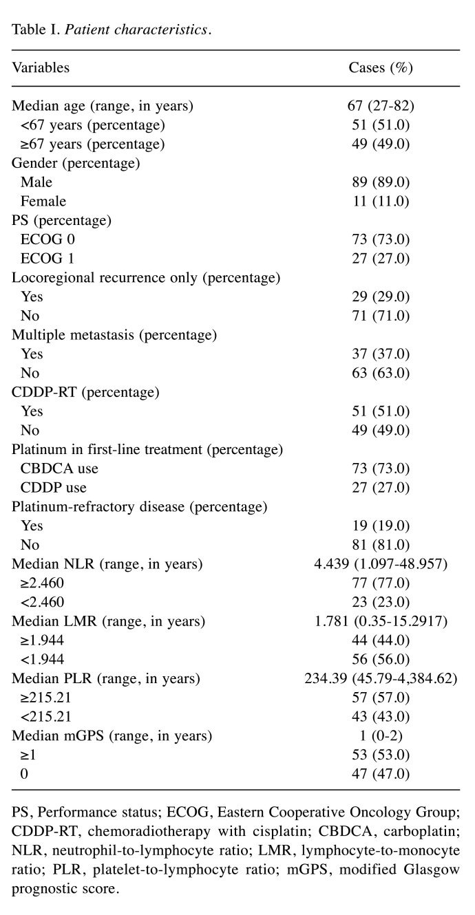
Patient characteristics. The clinical characteristics of the 100 R/M-SCCHN patients analyzed in this study are summarized in Table I. The median follow-up period was 9 months (range=1-65 months). The number of patients who died within the observation period was 73 (73.0%). The median age at diagnosis was 67 years (range=27-82 years). Most patients were male (89.0%) and had ECOG PS of 0 (73.0%). The number of patients with advanced-stage disease (stages III and IV) was 87 (87.0%). In addition, 29 (29.0%) patients had locoregional recurrence only, and 37 (37.0%) patients had multiple metastases. There were also 51 patients who underwent curative or postoperative chemoradiotherapy with cisplatin (CDDP-RT), including 19 patients with platinum-refractory disease. Furthermore, 73 and 27 patients underwent the carboplatin-based and cisplatin-based EXTREME regimens, respectively. The other clinical characteristics of the 100 R/M-SCCHN patients, such as treatment characteristics, have been described previously (8). The median NLR, LMR, PLR, and mGPS were 4.439 (range=1.097-48.96), 1.781 (range=0.350-15.29), 234.4 (range=45.79-4384), and 1 (range=0-2), respectively.
Table I. Patient characteristics.
PS, Performance status; ECOG, Eastern Cooperative Oncology Group; CDDP-RT, chemoradiotherapy with cisplatin; CBDCA, carboplatin; NLR, neutrophil-to-lymphocyte ratio; LMR, lymphocyte-to-monocyte ratio; PLR, platelet-to-lymphocyte ratio; mGPS, modified Glasgow prognostic score.
Cutoff values for inflammatory biomarkers. Optimal cutoff values for the NLR, LMR, and PLR were determined using ROC curves for OS, as shown in Figure 1A-C. The cutoff points of the NLR, LMR, and PLR were 2.460 with an area under the curve (AUC) of 0.613 (95% CI=0.484-0.742), 1.944 with an AUC of 0.669 (95% CI=0.557-0.781), and 215.2 with an AUC of 0.670 (95% CI=0.544-0.797), respectively.
Figure 1. Receiver operating characteristics curves for overall survival. (A) NLR, cutoff value=2.460, AUC=0.613 (95% CI=0.484-0.742). (B) LMR, cutoff value=1.944, AUC=0.669 (95% CI=0.557-0.781). (C) PLR, cutoff value=215.210, AUC=0.670 (95% CI=0.544-0.797).

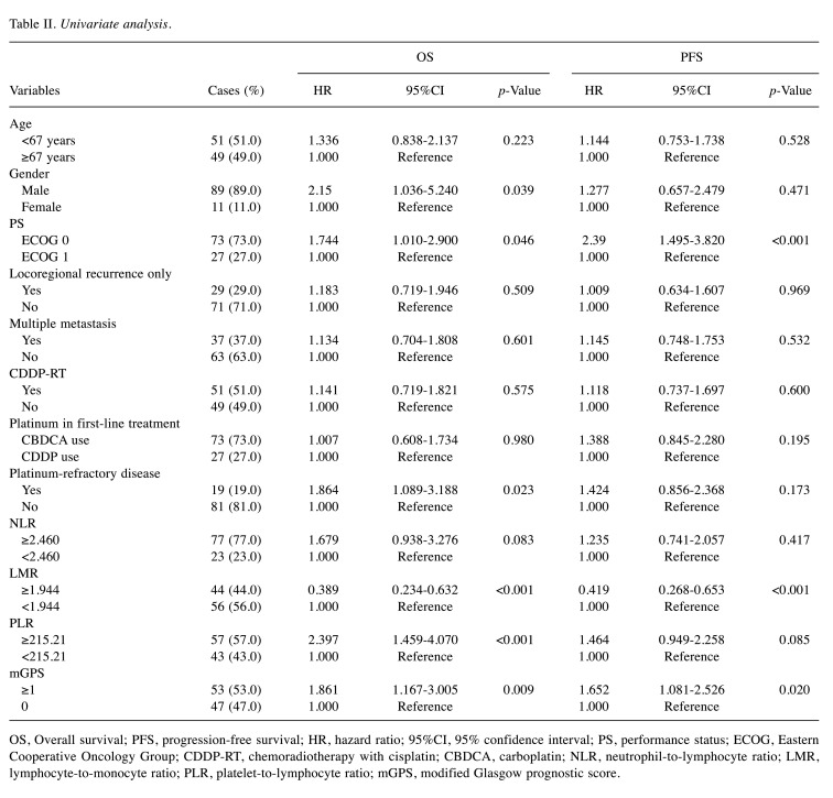
Univariate and multivariate analyses of prognostic biomarkers. Univariate analysis was performed to evaluate the prognostic significance of the NLR, PLR, LMR, and clinical characteristics for OS and PFS (Table II). We found that patients who were male, with ECOG PS of 1, platinum-refractory disease, LMR <1.944, PLR ≥215.2, and mGPS ≥1 had a significantly shorter OS (p=0.039, p=0.046, p=0.023, p<0.001, p<0.001, and p=0.009, respectively). In addition, we found that patients with ECOG PS of 1, LMR <1.944, and mGPS ≥1 had significantly shorter PFS (p<0.001, p<0.001, and p=0.020, respectively). In contrast, patients who are male, with platinum-refractory disease and PLR ≥215.2 were not significantly correlated with PFS. In the present study, other parameters such as age, the presence of locoregional recurrence and multiple metastatic lesions, CDDP-RT as previous treatment, platinum agent used in the EXTREME regimen, and NLR were significantly correlated with neither OS nor PFS.
Table II. Univariate analysis.
OS, Overall survival; PFS, progression-free survival; HR, hazard ratio; 95%CI, 95% confidence interval; PS, performance status; ECOG, Eastern Cooperative Oncology Group; CDDP-RT, chemoradiotherapy with cisplatin; CBDCA, carboplatin; NLR, neutrophil-to-lymphocyte ratio; LMR, lymphocyte-to-monocyte ratio; PLR, platelet-to-lymphocyte ratio; mGPS, modified Glasgow prognostic score.
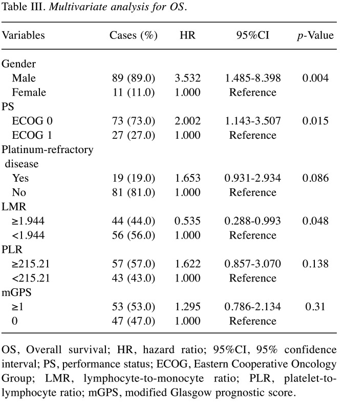
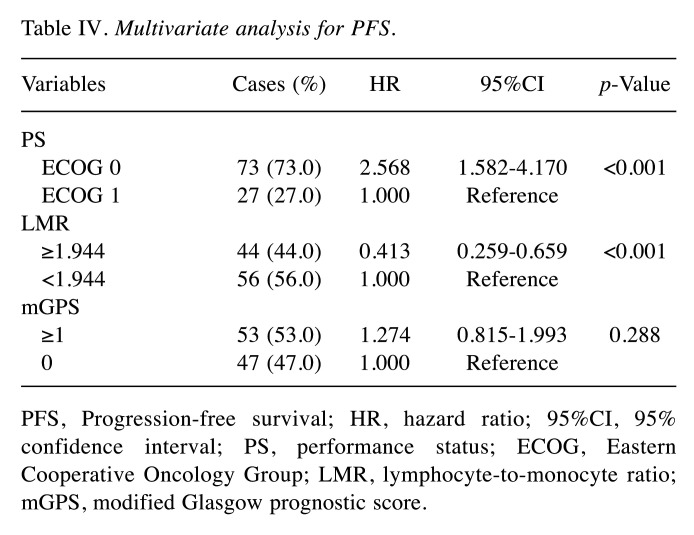
In addition, Cox multivariate analysis was performed to identify independent prognostic factors for survival in patients with R/M-SCCHN (Table III and Table IV). We found that patients who are male, with PS of 1, and LMR <1.944 were independently associated with poor OS (p=0.004, p=0.015, and p=0.048, respectively). In addition, we found that patients with LMR <1.944 and ECOG PS of 1 were independently associated with poor PFS (p<0.001 and p<0.001, respectively).
Table III. Multivariate analysis for OS.
OS, Overall survival; HR, hazard ratio; 95%CI, 95% confidence interval; PS, performance status; ECOG, Eastern Cooperative Oncology Group; LMR, lymphocyte-to-monocyte ratio; PLR, platelet-tolymphocyte ratio; mGPS, modified Glasgow prognostic score.
Table IV. Multivariate analysis for PFS.
PFS, Progression-free survival; HR, hazard ratio; 95%CI, 95% confidence interval; PS, performance status; ECOG, Eastern Cooperative Oncology Group; LMR, lymphocyte-to-monocyte ratio; mGPS, modified Glasgow prognostic score.
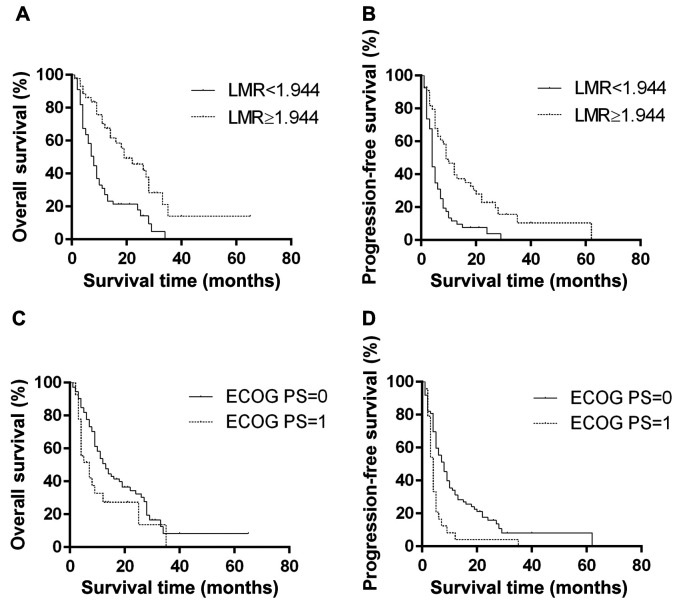
Furthermore, we observed that patients with an LMR of <1.944 showed significantly shorter OS and PFS than those with an LMR of ≥1.944 (HR=0.411; 95% CI=0.233-0.588; p<0.001; and HR=0.471; 95% CI=0.252-0.597; p<0.001, respectively; Figure 2A and B). Patients with a PS of 1 also had poorer OS and PFS than those with a PS of 0 (HR=1.712; 95% CI=1.097-3.616; p=0.030, and HR=2.074; 95% CI=1.637-5.225; p<0.001, respectively, Figure 2C and D). Thus, these results suggest that both an LMR <1.944 and a PS of 1 were independent prognostic factors for poor OS and PFS in this study.
Figure 2. Kaplan-Meier curves for overall survival (OS) and progression-free survival (PFS). (A) Kaplan-Meier curves for overall survival (OS) according to the lymphocyte-to-monocyte ratio (LMR). Black line, LMR <1.944; dashed line, LMR ≥1.944; p<0.001. (B) Kaplan-Meier curves for progression-free survival (PFS) according to the LMR. Black line, LMR <1.944; dashed line, LMR ≥1.944; p<0.001. (C) Kaplan-Meier curves for OS according to the Eastern Cooperative Oncology Group performance status (ECOG PS). Black line, ECOG PS of 0; dashed line, ECOG PS of 1; p=0.030. (D) Kaplan-Meier curves for PFS according to the ECOG PS. Black line, ECOG PS of 0; dashed line, ECOG PS of 1; p<0.001.
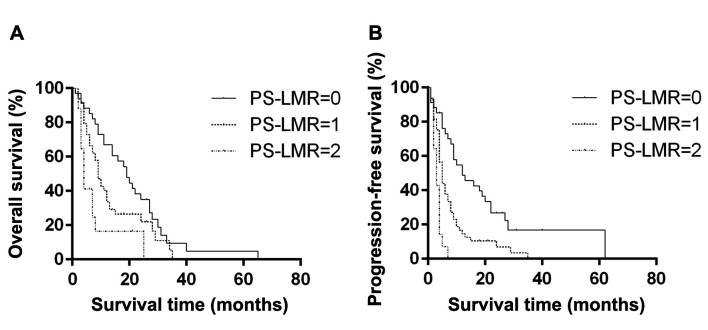
PS-LMR as a new prognostic score. In the present study, we assessed the clinical significance of the combined PS and LMR scores (PS-LMR score) as prognostic predictors for patients with R/M-SCCHN. PS-LMR scores were classified into three groups: PS-LMR score of 0 (PS of 0, LMR ≥1.944), PS-LMR score of 1 (PS of 0 or LMR ≥1.944), and PS-LMR score of 2 (ECOG PS of 1, LMR <1.944). We found that patients with a PS-LMR score of 2 had significantly shorter OS than those with PS-LMR scores of 0 and 1 (HR=2.762; 95% CI=2.275-12.20; p=0.001; and HR=2.021; 95% CI=1.296-5.863; p=0.041, respectively; Figure 3A). However, there was no significant difference between patients with PS-LMR scores of 0 and 1 (p=0.102). Furthermore, we found that a PS-LMR score of 2 was associated with significantly shorter PFS than PS-LMR scores of 0 and 1 (HR=4.065; 95% CI=6.945-47.98; p<0.001; and HR=2.361; 95% CI=2.095-10.29; p=0.003, respectively; Figure 3B). In addition, there was a significant difference in PFS between patients with PS-LMR scores of 0 and 1 (p=0.002). Thus, a high PS-LMR score was significantly associated with poor PFS. These results suggest that the PS-LMR score can be used as a pretreatment prognostic predictor of survival in R/M-SCCHN patients.
Figure 3. Kaplan-Meier curves of PS-LMR for overall (OS) and progression-free survival (PFS). Curves were evaluated using the log-rank test with Bonferroni’s adjustment to compare the three groups. (A) There was a significant difference in OS between PS-LMR scores of 0 and 1 (p<0.001) and between scores of 0 and 2 (p<0.002), but with no significant difference was noted between patients with PS-LMR scores of 1 and 2 (p=0.089). (B) There was a significant difference in PFS between PS-LMR scores of 0 and 1 (p<0.001), 0 and 2 (p<0.001), and 1 and 2 (p<0.001).
Discussion
In this study, we demonstrated that an ECOG PS of 1 and LMR <1.944 were independent prognostic factors for OS and PFS in patients with R/M-SCCHN treated with the EXTREME regimen as first-line therapy. Furthermore, we proposed the clinical significance of our novel combined PS and LMR scores (PS-LMR score) to extract poor prognosis in these patients. Our results on the association between low LMR and poor prognosis of patients with R/M-SCCHN were consistent with those of previous studies on other malignancies, including lung, pancreas, colorectum, ovarian, kidney, liver, esophagus, breast, stomach, and head and neck cancers (11). LMR is a blood cell-based inflammatory biomarker simply calculated by dividing the lymphocyte count by the monocyte count. As previously mentioned, lymphocytes are well known to play crucial roles in tumor immunity; therefore, high levels of T cells in the tumor microenvironment have been associated with improving the survival of cancer patients (12), while a low lymphocyte count could reflect immune deficiency (13). In particular, tumor-infiltrating lymphocytes (TILs), including infiltrating CD8+ T lymphocytes, have been reported as potent mediators of antitumor immunity. In the present study, median lymphocyte counts in patients with LMR <1.944 were lower than those with LMR ≥1.944 (data not shown), indicating that many patients with low LMR were associated with immune deficiency in our dataset.
Monocytes have also been reported to be associated with tumor progression and metastasis, as monocytes have the potential to differentiate into tissue macrophages, thereby accelerating angiogenesis and tumor cell motility. Macrophages can be divided into M1 and M2 phenotypes; the M1 phenotype is responsible for antitumor immunity, while the M2 phenotype is responsible for tumor growth and metastasis (14,15). Tumor-associated macrophages (TAMs) (16,17), derived from peripheral monocytes and located in or around cancer cells, normally represent M2 protumoral macrophages (16). Indeed, high TAM levels in the tumor have been reported to be associated with poor prognosis in several malignancies (12,17,18). Thus, LMR could reflect the balance between immune deficiency in tumor cells and tumor progression.
While the present study was conducted for patients with R/M-SCCHN treated with the EXTREME regimen as first-line treatment, ICIs are now being widely used as first-line treatment for these patients. Our data set included 30 patients treated with nivolumab as subsequent therapy, showing improved survival in these patients (8). Anti-PD-1 antibodies such as nivolumab and pembrolizumab have been reported to potentially suppress the activation of TILs and TAMs (19,20). Sekine et al. reported that a rapid increase in the LMR was significantly associated with the effects of nivolumab, and early changes in the LMR may be used as a novel effective surrogate marker to decide whether to continue anti-PD-1 therapy (21). This might be because LMR was the only independent prognostic predictor among blood cell-based biomarkers for patients with R/M-SCCHN in this study, as the treatment outcomes of ICI could be associated with TILs and TAMs, and LMR may be a useful biomarker for any treatment in patients with R/M-SCCHN. The prognostic significance of the LMR in patients with R/M-SCCHN treated with ICIs needs to be determined in the future.
In this study, ECOG PS was also an independent prognostic factor in patients with R/M-SCCHN, which is consistent with previous results indicating the prognostic significance of PS in many malignancies, including R/M-SCCHN (22,23), as ECOG PS is well known to reflect body composition and physical function in patients with advanced cancer. Furthermore, we report PS-LMR, the grading system that combines ECOG PS with LMR, as a novel prognostic biomarker for patients with R/M-SCCHN that reflects the potential of tumor progression and immunity as well as cachexia by combining two pretreatment markers more comprehensively.
In contrast, mGPS, an inflammation and nutrition-based score that consists of serum C-reactive protein and albumin levels, was not an independent prognostic factor in this study. This might be because our data set had only 25 of 100 R/M-SCCHN patients with an mGPS of 2 (data not shown), suggesting that our study included a small number of patients with cancer cachexia. Furthermore, other pretreatment parameters related to cancer cachexia, such as C-reactive protein-albumin ratio (24) and prognostic nutritional index (25), were not evaluated in the present study, as these parameters showed strong correlation among inflammatory prognostic predictive biomarkers.
This study has several limitations. First, the present study comprised only 100 R/M-SCCHN patients recruited from only two institutions in a retrospective manner. Second, consequent therapy using miscellaneous agents, including nivolumab, could affect the survival outcomes of patients in this study. As described above, the LMR could affect the levels of TILs and TAMs in tumors; however, actual counts of TILs and/or TAMs in tumors were not evaluated in this study. The heterogeneous background of patients with R/M disease may also have some impact on the results. Despite these limitations, our novel PS-LMR score could be a useful pretreatment biomarker for predicting the prognosis of patients with R/M-SCCHN cost-effectively. Further multi-institutional studies are necessary to confirm the reliability and applicability of this novel scoring system in predicting the prognosis of R/M-SCCHN patients in the future.
In conclusion, this supplementary analysis from our retrospective cohort study of real-world treatment outcomes of the EXTREME regimen as first-line therapy for R/M-SCCHN patients revealed the prognostic significance of both ECOG PS and LMR in these patients. In addition, we determined that our novel combined PS-LMR score was useful for extracting poor prognosis in these patients. Further prospective studies should be conducted to validate the reliability and applicability of this novel scoring system in predicting the prognosis of patients with R/M-SCCHN, particularly those treated with ICI in a prospective manner in the future.
Conflicts of Interest
The Authors declare that they have no competing interests in relation to this study.
Authors’ Contributions
JA and DS conceived the study. JA, TK and DS wrote the main manuscript and prepared the figures. JA, TK, DS, TF, MT, MS, TS, YI, HI, and NO were involved with data collection. DS and NO performed the analysis. All Authors discussed the results of the study, made comments on the manuscript, and approved the final manuscript.
References
- 1.Vermorken JB, Specenier P. Optimal treatment for recurrent/metastatic head and neck cancer. Ann Oncol. 2010;21(Suppl 7):vii252–vii261. doi: 10.1093/annonc/mdq453. [DOI] [PubMed] [Google Scholar]
- 2.Ferris RL, Blumenschein G Jr, Fayette J, Guigay J, Colevas AD, Licitra L, Harrington K, Kasper S, Vokes EE, Even C, Worden F, Saba NF, Iglesias Docampo LC, Haddad R, Rordorf T, Kiyota N, Tahara M, Monga M, Lynch M, Geese WJ, Kopit J, Shaw JW, Gillison ML. Nivolumab for recurrent squamous-cell carcinoma of the head and neck. N Engl J Med. 2016;375(19):1856–1867. doi: 10.1056/NEJMoa1602252. [DOI] [PMC free article] [PubMed] [Google Scholar]
- 3.Burtness B, Harrington KJ, Greil R, Soulières D, Tahara M, de Castro G Jr, Psyrri A, Basté N, Neupane P, Bratland Å, Fuereder T, Hughes BGM, Mesía R, Ngamphaiboon N, Rordorf T, Wan Ishak WZ, Hong RL, González Mendoza R, Roy A, Zhang Y, Gumuscu B, Cheng JD, Jin F, Rischin D, KEYNOTE-048 Investigators Pembrolizumab alone or with chemotherapy versus cetuximab with chemotherapy for recurrent or metastatic squamous cell carcinoma of the head and neck (KEYNOTE-048): a randomised, open-label, phase 3 study. Lancet. 2019;394(10212):1915–1928. doi: 10.1016/S0140-6736(19)32591-7. [DOI] [PubMed] [Google Scholar]
- 4.Templeton AJ, McNamara MG, Šeruga B, Vera-Badillo FE, Aneja P, Ocaña A, Leibowitz-Amit R, Sonpavde G, Knox JJ, Tran B, Tannock IF, Amir E. Prognostic role of neutrophil-to-lymphocyte ratio in solid tumors: a systematic review and meta-analysis. J Natl Cancer Inst. 2014;106(6):dju124. doi: 10.1093/jnci/dju124. [DOI] [PubMed] [Google Scholar]
- 5.Mao Y, Fu Y, Gao Y, Yang A, Zhang Q. Platelet-to-lymphocyte ratio predicts long-term survival in laryngeal cancer. Eur Arch Otorhinolaryngol. 2018;275(2):553–559. doi: 10.1007/s00405-017-4849-4. [DOI] [PubMed] [Google Scholar]
- 6.Nishijima TF, Muss HB, Shachar SS, Tamura K, Takamatsu Y. Prognostic value of lymphocyte-to-monocyte ratio in patients with solid tumors: A systematic review and meta-analysis. Cancer Treat Rev. 2015;41(10):971–978. doi: 10.1016/j.ctrv.2015.10.003. [DOI] [PubMed] [Google Scholar]
- 7.Kuwahara T, Takahashi H, Sano D, Matsuoka M, Hyakusoku H, Hatano T, Hiiragi Y, Oridate N. Fibrinogen and neutrophil-to-lymphocyte ratio predicts survival in patients with advanced hypopharyngeal squamous cell carcinoma. Anticancer Res. 2018;38(9):5321–5330. doi: 10.21873/anticanres.12859. [DOI] [PubMed] [Google Scholar]
- 8.Sano D, Fujisawa T, Tokuhisa M, Shimizu M, Sakagami T, Hatano T, Nishimura G, Ichikawa Y, Iwai H, Oridate N. Real-world treatment outcomes of the EXTREME regimen as first-line therapy for recurrent/metastatic squamous cell carcinoma of the head and neck: A multi-center retrospective cohort study in Japan. Anticancer Res. 2019;39(12):6819–6827. doi: 10.21873/anticanres.13898. [DOI] [PubMed] [Google Scholar]
- 9.Chang PH, Yeh KY, Wang CH, Chen EY, Yang SW, Huang JS, Chou WC, Hsieh JC. Impact of the pretreatment Glasgow prognostic score on treatment tolerance, toxicities, and survival in patients with advanced head and neck cancer undergoing concurrent chemoradiotherapy. Head Neck. 2017;39(10):1990–1996. doi: 10.1002/hed.24853. [DOI] [PubMed] [Google Scholar]
- 10.Toiyama Y, Miki C, Inoue Y, Tanaka K, Mohri Y, Kusunoki M. Evaluation of an inflammation-based prognostic score for the identification of patients requiring postoperative adjuvant chemotherapy for stage II colorectal cancer. Exp Ther Med. 2011;2(1):95–101. doi: 10.3892/etm.2010.175. [DOI] [PMC free article] [PubMed] [Google Scholar]
- 11.Mao Y, Chen D, Duan S, Zhao Y, Wu C, Zhu F, Chen C, Chen Y. Prognostic impact of pretreatment lymphocyte-to-monocyte ratio in advanced epithelial cancers: a meta-analysis. Cancer Cell Int. 2018;18:201. doi: 10.1186/s12935-018-0698-5. [DOI] [PMC free article] [PubMed] [Google Scholar]
- 12.Soo RA, Chen Z, Yan Teng RS, Tan HL, Iacopetta B, Tai BC, Soong R. Prognostic significance of immune cells in non-small cell lung cancer: meta-analysis. Oncotarget. 2018;9(37):24801–24820. doi: 10.18632/oncotarget.24835. [DOI] [PMC free article] [PubMed] [Google Scholar]
- 13.Dunn GP, Old LJ, Schreiber RD. The immunobiology of cancer immunosurveillance and immunoediting. Immunity. 2004;21(2):137–148. doi: 10.1016/j.immuni.2004.07.017. [DOI] [PubMed] [Google Scholar]
- 14.Condeelis J, Pollard JW. Macrophages: obligate partners for tumor cell migration, invasion, and metastasis. Cell. 2006;124(2):263–266. doi: 10.1016/j.cell.2006.01.007. [DOI] [PubMed] [Google Scholar]
- 15.Schoppmann SF, Birner P, Stöckl J, Kalt R, Ullrich R, Caucig C, Kriehuber E, Nagy K, Alitalo K, Kerjaschki D. Tumor-associated macrophages express lymphatic endothelial growth factors and are related to peritumoral lymphangiogenesis. Am J Pathol. 2002;161(3):947–956. doi: 10.1016/S0002-9440(10)64255-1. [DOI] [PMC free article] [PubMed] [Google Scholar]
- 16.Evrard D, Szturz P, Tijeras-Raballand A, Astorgues-Xerri L, Abitbol C, Paradis V, Raymond E, Albert S, Barry B, Faivre S. Macrophages in the microenvironment of head and neck cancer: potential targets for cancer therapy. Oral Oncol. 2019;88:29–38. doi: 10.1016/j.oraloncology.2018.10.040. [DOI] [PubMed] [Google Scholar]
- 17.Feng Q, Chang W, Mao Y, He G, Zheng P, Tang W, Wei Y, Ren L, Zhu D, Ji M, Tu Y, Qin X, Xu J. Tumor-associated macrophages as prognostic and predictive biomarkers for postoperative adjuvant chemotherapy in patients with stage II colon cancer. Clin Cancer Res. 2019;25(13):3896–3907. doi: 10.1158/1078-0432.CCR-18-2076. [DOI] [PubMed] [Google Scholar]
- 18.Zhang J, Chang L, Zhang X, Zhou Z, Gao Y. Meta-analysis of the prognostic and clinical value of tumor-associated macrophages in hepatocellular carcinoma. J Invest Surg. 2021;34(3):297–306. doi: 10.1080/08941939.2019.1631411. [DOI] [PubMed] [Google Scholar]
- 19.Fritz JM, Lenardo MJ. Development of immune checkpoint therapy for cancer. J Exp Med. 2019;216(6):1244–1254. doi: 10.1084/jem.20182395. [DOI] [PMC free article] [PubMed] [Google Scholar]
- 20.Ruan J, Ouyang M, Zhang W, Luo Y, Zhou D. The effect of PD-1 expression on tumor-associated macrophage in T cell lymphoma. Clin Transl Oncol. 2021;23(6):1134–1141. doi: 10.1007/s12094-020-02499-0. [DOI] [PubMed] [Google Scholar]
- 21.Sekine K, Kanda S, Goto Y, Horinouchi H, Fujiwara Y, Yamamoto N, Motoi N, Ohe Y. Change in the lymphocyte-to-monocyte ratio is an early surrogate marker of the efficacy of nivolumab monotherapy in advanced non-small-cell lung cancer. Lung Cancer. 2018;124:179–188. doi: 10.1016/j.lungcan.2018.08.012. [DOI] [PubMed] [Google Scholar]
- 22.Corrêa GT, Bandeira GA, Cavalcanti BG, Santos FB, Rodrigues Neto JF, Guimarães AL, Haikal DS, De Paula AM. Analysis of ECOG performance status in head and neck squamous cell carcinoma patients: association with sociodemographical and clinical factors, and overall survival. Support Care Cancer. 2012;20(11):2679–2685. doi: 10.1007/s00520-012-1386-y. [DOI] [PubMed] [Google Scholar]
- 23.Ueki Y, Takahashi T, Ota H, Shodo R, Yamazaki K, Horii A. Predicting the treatment outcome of nivolumab in recurrent or metastatic head and neck squamous cell carcinoma: prognostic value of combined performance status and modified Glasgow prognostic score. Eur Arch Otorhinolaryngol. 2020;277(8):2341–2347. doi: 10.1007/s00405-020-05945-5. [DOI] [PubMed] [Google Scholar]
- 24.Yu ST, Zhou Z, Cai Q, Liang F, Han P, Chen R, Huang XM. Prognostic value of the C-reactive protein/albumin ratio in patients with laryngeal squamous cell carcinoma. Onco Targets Ther. 2017;10:879–884. doi: 10.2147/OTT.S128391. [DOI] [PMC free article] [PubMed] [Google Scholar]
- 25.Onodera T, Goseki N, Kosaki G. Prognostic nutritional index in gastrointestinal surgery of malnourished cancer patients. Nihon Geka Gakkai Zasshi. 1984;85(9):1001–1005. [PubMed] [Google Scholar]


