Abstract
Background and Objective
Point-of-care type molecular diagnostic tests have been used for detecting SARS-CoV-2, although their clinical utility with nasal samples has yet to be established. This study evaluated the clinical performance of the cobas Liat SARS-CoV-2 & Influenza A/B (Liat) assay in nasal samples.
Methods
Nasal and nasopharyngeal samples were collected and were tested using the Liat, the cobas 6800 system and the cobas SARS-CoV-2 & Influenza A/B (cobas), and a method developed by National Institute of Infectious Diseases, Japan (NIID).
Results
A total of 814 nasal samples were collected. The Liat assay was positive for SARS-CoV-2 in 113 (13.9%). The total, positive, and negative concordance rate between the Liat and cobas/NIID assays were 99.3%/98.4%, 99.1%/100%, and 99.3%/98.2%, respectively. Five samples were positive only using the Liat assay. Their Ct values ranged from 31.9 to 37.2. The Ct values of the Liat assay were significantly lower (p < 0.001) but were correlated (p < 0.001) with those of other molecular assays. In the participants who tested positive for SARS-CoV-2 on the Liat assay using nasopharyngeal samples, 88.2% of their nasal samples also tested positive using the Liat assay.
Conclusion
The Liat assay showed high concordance with other molecular assays in nasal samples. Some discordance occurred in samples with Ct values > 30 on the Liat assay.
Supplementary Information
The online version contains supplementary material available at 10.1007/s40291-022-00580-8.
Key Points
| The cobas Liat SARS-CoV-2 & Influenza A/B assay showed high concordance with other molecular assays in nasal and nasopharyngeal samples. |
| Some discordance occurred in samples with Ct values > 30 on the Liat assay. |
| The Liat assay may be suitable for use in a variety of clinical situations, primarily where accurate detection of SARS-CoV-2 is necessary. |
Introduction
The severe acute coronavirus 2 (SARS-CoV-2) pandemic has had a detrimental effect on society globally [1]. The introduction of effective vaccines [2] and treatment [3] were expected to effectively control the pandemic; however, the numbers of new infections and deaths have increased in some countries [1]. Currently, population-based screening and early detection and isolation of infected individuals are still key to effective infection control [4].
The standard method for detecting SARS-CoV-2 is molecular testing because of its high diagnostic performance [5]. However, molecular diagnostics are less widely available and less convenient than antigen testing, and the turnaround time is longer [6]. Several molecular point-of-care tests (POCTs) have been developed [7] and applied in clinical settings to overcome the disadvantages of traditional molecular tests.
The cobas Liat system (Roche Molecular Systems, Inc., Pleasanton, CA, USA) is a small analyzer that automatically performs real-time polymerase chain reaction (PCR) testing using simple procedures and provides results with a short turnaround time [8]. The system has been used for point-of-care testing, and Liat assays are available for influenza virus [9], respiratory syncytial virus [10], Clostridioides difficile [11], group A Streptococcus [12], and SARS-CoV-2 [8]. The Liat system and cobas Liat SARS-CoV-2 & Influenza A/B (Liat) has been shown to have high sensitivity for detecting SARS-CoV-2 in nasopharyngeal samples [8], but the diagnostic performance with nasal samples has not been reported. Nasal sample collection is easier and less invasive than the nasopharyngeal sample collection [13]; therefore, the clinical utility of Liat would be greater if it could accurately detect SARS-CoV-2 in nasal samples.
This study aimed to clarify the diagnostic performance of the Liat SARS-CoV-2 assay with nasal samples. The results of Liat assays with nasal samples were prospectively compared with those of other assays with both nasal and nasopharyngeal samples.
Methods and Materials
This study was conducted in a PCR center located in Tsukuba Medical Center Hospital (TMCH) in Japan between 7 and 29 July 2021. The participants were outpatients suspected of having SARS-CoV-2 infection due to their symptoms or close contact history, all of whom underwent in-house PCR testing at the center [14]. They included individuals referred from 51 healthcare facilities and a local healthcare center, and healthcare workers working at TMCH. Clinical information of the participants was routinely recorded along with the in-house PCR. Verbal informed consent was obtained from all participants, and the ethics board of the University of Tsukuba approved the study (approval number: R03-41).
Sample Collection
One nasopharyngeal and two nasal samples were collected from each participant. The nasopharyngeal samples were collected as described previously [15]. Nasal samples were collected from the anterior nostrils according to recommended procedures [16]. Briefly, the swab was inserted to a depth of approximately 2 cm and rotated four times against the nasal mucosa. The procedure was repeated in the other nostril using the same swab. One nasal swab sample was used for molecular testing using Liat, and the other nasal swab sample was used for the evaluation of antigen testing in another study. All nasal sample collections for molecular testing followed those for antigen testing.
Both the nasal and the nasopharyngeal swab samples were diluted in Universal Transport Medium (UTMTM; Copan Diagnostics Inc., Murrieta, CA, USA) and stored at – 80 °C. The nasopharyngeal samples were frozen after being used for an in-house PCR test.
PCR Testing Procedures
After being thawed, all nasal samples underwent PCR testing according to three methods: (i) cobas Liat system and cobas Liat SARS-CoV-2 & Influenza A/B (Liat), (ii) cobas 6800 system and cobas SARS-CoV-2 & Influenza A/B (cobas; Roche Molecular Systems, Branchburg, NJ, USA) [17], and (iii) a national standard method developed by the National Institute of Infectious Diseases (NIID), Japan [18]. If a sample showed positivity on one of the methods and negativity on the other two, they were tested using the Gene Xpert system and the Xpert Xpress SARS-CoV-2 (Xpert Xpress; Cepheid, Sunnyvale, CA, USA) [19].
The nasopharyngeal samples were tested using Liat and the NIID test for the evaluation, and were not tested using the cobas system due to an insufficient amount of residual UTM sample. If the Liat and NIID results were discordant, the samples were tested using Xpert Xpress, in a similar manner to the nasal samples.
Liat, cobas, and Xpert Xpress perform sample preparation (purification and the extraction of RNA), real-time PCR, and the detection of the viruses using a fully automated process. For the Liat assay, a total of 200 μL of UTMTM sample was loaded into the test cartridge, which was then inserted into the system. The Liat test targets the ORF1a/b and N gene and shows positive results if one or both genes are detected. We re-tested samples if invalid results occurred. For the cobas assay, we used 1 mL of UTMTM sample (400 μL for the analysis and 600 μL for the dead space). The targeted regions were ORF1a/b and E gene, and the cobas test provided separate results for each target. The results of cobas were considered positive for SARS-CoV-2 when samples tested positive for one of the two targets. The Xpert Xpress used 300 μL of UTMTM sample and targeted the E and N2 genes. Similar to cobas, Xpert Xpress showed separate results for each target. All three assays were conducted according to the manufacturer’s instructions.
For the NIID test, purification and RNA extraction were performed using MagNA Pure 96 total NA Isolation Kit and the MagNA Pure 96 Instrument (Roche Molecular Systems, Branchburg, NJ, USA) from 140-µL aliquots of UTMTM sample. The NIID test targets the N2 region, and the equipment used for the RT-PCR included the PCR LightCycler®480 Instrument II (Roche Diagnostics International Ltd, Rotkreuz, Switzerland), the QuantiTect® Probe RT-PCR Kit (QIAGEN, Hilden, Germany), and a SARS-CoV-2 positive control (Nihon Gene Research Laboratories, Sendai, Japan). The RT-PCR was performed in duplicate.
The Ct values were automatically calculated after the detection of SARS-CoV-2 for all test methods used in this study.
Apart from detecting SARS-CoV-2, the Liat assay can also detect influenza virus; however, the results were not re-evaluated and validated by confirmatory molecular assays in this study.
Statistical Analyses
The results of the Liat assay were compared with those of the other molecular assays, and the concordance rate was calculated with 95% confident intervals (CIs), using the Clopper and Pearson method. The sensitivity, specificity, positive predictive value (PPV), and negative predictive value (NPV) of the Liat assay with nasal samples were also calculated. SARS-CoV-2 were considered positive when one of molecular assays showed positive on corresponding nasopharyngeal samples.
The Wilcoxon signed-rank test was used to compare the median Ct values. The correlations of the Ct values between two molecular assays were assessed using Pearson's product-moment correlation coefficient. The statistical analyses were performed using R version 3.14.1 (R Foundation for Statistical Computing, Vienna, Austria), and the figures were created using Python version 3.8.12 (Python Software Foundation, Wilmington, DE, USA). All codes and dataset used for the analyses are available in the Online Supplementary Material (OSM). p values < 0.05 were considered statistically significant.
Results
The study included a total of 843 participants, of whom 29 provided only nasopharyngeal samples. Therefore, we obtained 814 nasal and 843 nasopharyngeal samples. The Liat assay could not be performed on one nasopharyngeal sample due to an insufficient amount of residual sample (case ID: T0725). Supplementary Table 1 (OSM) presents data on the symptoms of those who were tested with the Liat assay of nasal samples.
Four nasal samples (0.49%) and 23 nasopharyngeal samples (2.7%) exhibited invalid results on the first test of the Liat assay. In the second (repeat) test, all four nasal samples tested negative, one nasopharyngeal sample tested positive, and 13 nasopharyngeal samples tested negative. Due to insufficient residual sample volume, the second test could not be performed on one of the nasopharyngeal samples. The nasopharyngeal sample was excluded from the comparative analysis of the Liat and the NIID assay (case ID: T0613). The remaining eight nasopharyngeal samples tested negative following the third test after being diluted threefold with UTMTM.
Finally, we compared the results of 814 nasal and 841 nasopharyngeal samples obtained in the Liat assay to those of the other molecular assays. The Liat assay was positive for SARS-CoV-2 in 113 (13.9%) of the nasal samples and 151 (18.0%) of the nasopharyngeal samples. A nasal sample was positive for influenza virus as detected by the Liat assay. No co-infection cases were identified in this study.
The Liat assay with nasal samples showed sensitivity, specificity, PPV, and NPV of 88.6%, 99.4%, 96.5%, and 98.0%, respectively, when compared to the results of molecular assays with their responding nasopharyngeal samples.
Comparison of the Results of the Liat Test With the Results of Other Molecular Assays Using Nasal Samples
The results of the Liat assay are compared with the results of the cobas and NIID assays performed using nasal samples in Tables 1 and 2, respectively.
Table 1.
Comparison of the results of the Liat and cobas SARS-CoV-2 assays performed using nasal samples
| All cases | Symptomatic cases | Asymptomatic cases | ||||
|---|---|---|---|---|---|---|
| Liat | Liat | Liat | ||||
| Positive | Negative | Positive | Negative | Positive | Negative | |
| cobas | ||||||
| Positive | 108 | 1 | 61 | 0 | 47 | 1 |
| Negative | 5 | 700 | 2 | 280 | 3 | 420 |
| Total concordance | 99.3 (98.4–99.7) | 99.4 (97.9–99.9) | 99.2 (97.8–99.8) | |||
| Positive concordance | 99.1 (94.5–100) | 100 (94.1–100) | 97.9 (88.9–99.9) | |||
| Negative concordance | 99.3 (98.4–99.8) | 99.3 (97.4–99.9) | 99.3 (97.9–99.9) | |||
cobas, cobas 6800 system and cobas SARS-CoV-2 & Influenza AB; Liat, cobas Liat system and cobas Liat SARS-CoV-2 & Influenza AB; SARS-CoV-2, severe acute coronavirus 2
The 95% confidence intervals are given in parentheses
Table 2.
Comparison of the results of the Liat and NIID SARS-CoV-2 assays performed using nasal samples
| All cases | Symptomatic cases | Asymptomatic cases | ||||
|---|---|---|---|---|---|---|
| Liat | Liat | Liat | ||||
| Positive | Negative | Positive | Negative | Positive | Negative | |
| NIID | ||||||
| Positive | 100 | 0 | 59 | 0 | 41 | 0 |
| Negative | 13 | 701 | 4 | 280 | 9 | 421 |
| Total concordance | 98.4 (97.3–99.1) | 98.8 (97.4–99.7) | 98.1 (96.4–99.1) | |||
| Positive concordance | 100 (96.4–100) | 100 (93.4–100) | 100 (91.4–100) | |||
| Negative concordance | 98.2(96.9–99.0) | 98.6 (96.4–99.6) | 97.9 (96.1–99.0) | |||
Liat, cobas Liat system and cobas Liat SARS-CoV-2 & Influenza AB; SARS-CoV-2, severe acute coronavirus 2; NIID, a national standard method developed by National Institute of Infectious Diseases (NIID), Japan
The 95% confidence intervals are given in parentheses
The total, positive, and negative concordance between the Liat and cobas assays were 99.3%, 99.1%, and 99.3%, respectively. The comparison was also performed stratified by the presence of relevant symptoms. In the symptomatic participants, the total, positive, and negative concordance was 99.4%, 100%, and 99.3%, respectively. In the asymptomatic participants, the total, positive, and negative concordance was 99.2%, 97.9%, and 99.3%, respectively.
Five nasal samples were positive only on the Liat assay, with Ct values ranging from 31.9 to 37.2. One of the five samples tested positive on additional analyses using Xpert Xpress with the Ct values of 39.6 for the E gene and 37.3 for the N2 gene.
When compared to the results of the NIID test, 13 sets of samples showed discordant results, all of which were Liat positive/NIID negative (Table 2). Eight of the 13 samples were positive on cobas assay. There were six sets of discordant results between the cobas and Liat assays, of which one sample was Liat-negative/cobas-positive and five samples were Liat positive/cobas negative.
Correlation of the Cycle Threshold Values of the Liat Assay with Those of the Other Assays
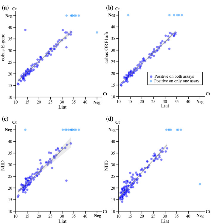
The correlation of the Ct values of the Liat assay and the other assays is shown in Fig. 1a–c. The median Ct values for each assay were as follows: Liat, 16.70; NIID, 23.7; cobas ORF1a/b gene, 24.4; cobas E gene, 23.6. The Ct values of the Liat assay were significantly lower (p < 0.001) but were correlated (p < 0.001) with those of the other assays.
Fig. 1.
Comparison of the cycle threshold values of Liat with those of other molecular assays using the same samples. a Liat vs. cobas E-gene in nasal samples; b Liat vs. cobas ORF1a/b in nasal samples; c Liat vs. NIID in nasal samples; d Liat vs. NIID in nasopharygeal samples. The lines and surrounding gray areas indicate linear regression lines with 95% confidence intervals. The blue circles are samples for which both molecular assays tested positive. The light blue circles are samples for which one assay was positive and the other was negative. cobas cobas 6800 system and cobas SARS-CoV-2 & Influenza A/B (cobas); Ct cycle threshold; Liat cobas Liat system and cobas Liat SARS-CoV-2 & Influenza A/B; NIID national standard method developed by the National Institute of Infectious Diseases of Japan
Analytical Performance of the Liat Assay Using Nasopharyngeal Samples
Of the 841 nasopharyngeal samples included in the final analysis, the results of the Liat and NIID assays were positive for SARS-CoV-2 in 151 and 144 samples, respectively, with a concordance of 99.2%. There were seven discordant samples, all of which were Liat positive/NIID negative. The Ct values of the two assays were significantly correlated (p < 0.001, Fig. 1d).
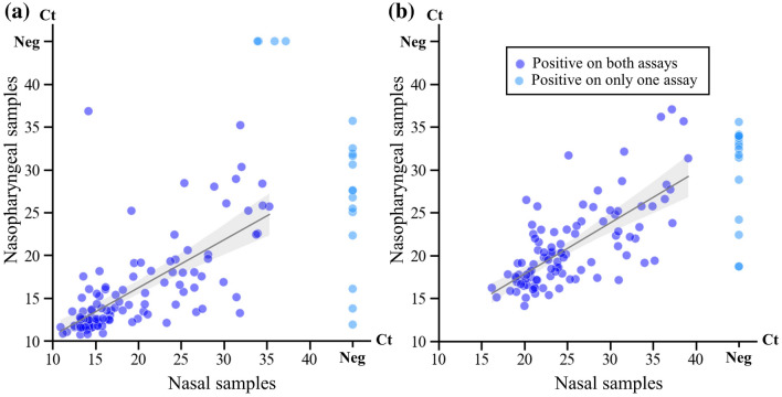
Both nasopharyngeal and nasal samples were obtained from 813 participants. The test results of the Liat assay performed on the two sample types were compared. Both sample types tested positive in 108 samples, 14 pairs of samples tested positive on the nasopharyngeal sample and negative on the nasal sample, and four pairs of samples tested positive on the nasal sample and negative on the nasopharyngeal sample. The correlation of the Ct values between the two sample types is shown in Fig. 2a, b.
Fig. 2.
Comparison of cycle threshold values between the nasal and nasopharyngeal samples collected from the same participants. a Liat; b NIID. cobas Liat system and cobas Liat SARS-CoV-2 & Influenza A/B; Ct cycle threshold; Liat cobas Liat system and cobas Liat SARS-CoV-2 & Influenza A/B; NIID national standard method developed by the National Institute of Infectious Diseases of Japan
Testing of Discordant Samples Using Different Molecular Assays
Table 3 summarizes the results of the discordant cases; the left panel shows nasal samples with positive results on only one of the three molecular assays (Liat, NIID, and cobas) and the right panel shows nasopharyngeal samples with discordant results of the Liat and NIID assay. Additional analyses using Xpert Xpress were performed for these cases, although one sample (case ID: T0198) could not be tested due to the lack of residual sample volume.
Table 3.
Cases with discordant results between molecular assyas
| Case number | Symptoms | Nasal samples | Nasopharyngeal samples | |||||
|---|---|---|---|---|---|---|---|---|
| Liat | NIID | cobas | Xpert Xpress | Liat | NIID | Xpert Xpress | ||
| T0161 | − | Neg | Neg | Neg | NA | Pos (35.7) | Neg | Pos (E: Neg, N2:42.0) |
| T0198 | − | Neg | Neg | Neg | NA | Pos (31.9) | Neg | NA |
| T0267 | − | Neg | Neg | Neg | NA | Pos (30.6) | Neg | Pos (E:34.8, N2:38.1) |
| T0339 | − | Pos (14.2) | Neg | Pos (38.8) | NA | Pos (36.8) | Neg | Pos (E: Neg, N2:41.6) |
| T0401 | − | Neg | Neg | Neg | NA | Pos (31.6) | Neg | Pos (E: Neg, N2:39.6) |
| T0438 | − | Neg | Neg | Neg | NA | Pos (32.5) | Neg | Pos (E:40.0, N2:42.1) |
| T0556 | − | Pos (35.3) | Neg | Neg | Neg | Pos (25.7) | Pos (34.1) | NA |
| T0625 | + | Pos (31.9) | Neg | Neg | Pos (E:39.6, N2:37.3) | Pos (35.2) | Neg | Pos (E: Neg, N2:41.7) |
| T0690 | + | Pos (37.2) | Neg | Neg | Neg | Neg | Neg | NA |
| T0735 | − | Neg | Neg | Pos (T2:37.8) | Neg | Pos (13.8) | Pos (18.7) | NA |
| T0760 | − | Pos (35.9) | Neg | Neg | Neg | Neg | Neg | NA |
| T0778 | − | Pos (33.9) | Neg | Neg | Neg | Neg | Neg | Neg |
Discordant cases in this table are defined as follows: for nasal samples, cases with positive results on only one of the three molecular assays (Liat, NIID, and cobas); for nasopharyngeal samples, cases with discordant results of the Liat and NIID assay.
Liat, cobas Liat system and cobas Liat SARS-CoV-2 & Influenza AB; NA not available, Neg negative, NIID a national standard method developed by National Institute of Infectious Diseases (NIID), Japan, Pos positive, SARS-CoV-2 severe acute coronavirus 2, Xpert Xpress Gene Xpert system and the Xpert Xpress SARS-CoV-2
In nasal samples, five samples were positive only using the Liat assay (Ct value range 31.9–37.2), and one sample was positive only using the cobas assay (Ct value 37.8). Of the five Liat-positive discordant samples, one sample tested positive using the Xpert Xpress assay, and another was positive on the corresponding nasopharyngeal sample from the same participant on both the Liat and the NIID assays.
In the nasopharyngeal samples, all seven discordant samples were Liat positive and NIID negative (Ct value range 30.6–36.8). The Xpert Xpress assay results were positive in six of the seven discordant samples.
Discussion
This prospective evaluation showed the high concordance in the nasal samples of the Liat assay results with those of other molecular assays. The Ct values of the Liat assay were significantly lower than those of other molecular assays, but were significantly correlated, although some discordant results were observed. Most of the cases of discordance occurred in samples with Ct values > 30 on the Liat assay.
In nasopharyngeal samples, the total, positive, and negative agreement of the results of the Liat and the cobas 6800/8800 assays were 98.6%, 100%, and 97.4%, respectively [8]. Similarly, this study showed high concordance between the results of the Liat assay and other molecular assays in nasal samples. Nevertheless, we observed some invalid results on the Liat assays, which may be caused by the presence of inhibitors in nasal mucosa or discharge. The Liat assays were successfully completed in all samples that initially provided invalid results after they were diluted. The dilution of samples reduces the influence of the inhibitors on molecular assays [20].
Some discordant results were observed in both nasal and nasopharyngeal samples, the majority of which were Liat positive and NIID/cobas negative. The discordant samples generally had Ct values > 30 on the Liat assay, indicating low viral concentrations. The Liat, cobas, and NIID assays have all been shown to have high analytical performance in previous studies [8, 17, 21]. However, the analytical performance on clinical specimens may be different due to the quality of the RNA extraction, the presence of inhibitors, genomic mutations, and stochasticity observed in samples with very low viral concentrations [22, 23]. A previous study comparing Liat and cobas reported that all discordant samples were Liat positive/cobas negative [8], which is consistent with the results of this study.
The Ct values of the Liat assay were strongly correlated with those of the cobas and NIID assays, but were significantly lower. Determining the Ct values is crucial to identify which patients with SARS-CoV-2 infection are most likely to transmit the virus, with higher Ct values indicating lower infectivity [24, 25]. However, Ct values can vary depending on the reagents and equipment used, even if the same samples are tested [25]; thus, the Ct values provided by each set of equipment should be carefully interpreted.
The nasal samples were more likely than nasopharyngeal samples to provide negative results. In this study, 88.2% of nasal samples tested positive on the Liat assay in participants whose nasopharyngeal samples tested positive using the Liat assay. A meta-analysis found that the sensitivity of RT-PCR of nasal samples was 82% compared to RT-PCR of nasopharyngeal samples [26], which is consistent with our study.
The Ct values between both nasal and nasopharyngeal samples were strongly correlated but varied (r = 0.68 and 0.70 for Liat and NIID assay, respectively), despite using the same collection media and assay procedures for both samples. The viral load is generally lower in the nostrils than in the nasopharynx [26], and previous studies found a similar variance of Ct values between those samples [27, 28]. The procedures for sample collection, the difference in viral dynamics between participants, and the conditions of samples may also have caused the fluctuation of their Ct values [25].
Our study has some limitations. First, we were unable to perform cobas testing on nasopharyngeal samples. The cobas assay has a high sensitivity and has been widely used worldwide [29, 30]. The level of discordance may vary depending on the equipment used for comparison. Second, we did not evaluate performance of the assays in samples from individuals infected with SARS-CoV-2 variants with gene mutations. The emergence of new variants could affect the diagnostic performance of the test. Third, the nasal samples used for molecular examination were collected after acquiring those used for antigen testing. The viral load in the nasal samples may have reduced due to the order of the procedure, which may have led to the variations in results obtained in the molecular examination. Finally, we did not use fresh samples for the evaluation of molecular examinations. Although to a miniscule degree, the storage process involving freezing and thawing also reportedly affects the viral load in samples [31].
In conclusion, the results of the Liat assay showed a high concordance with those of the other molecular assays in both nasal and nasopharyngeal samples. The findings of our study suggest that the Liat assay is suitable for use in a variety of clinical situations, primarily where accurate detection of SARS-CoV-2 is necessary.
Supplementary Information
Below is the link to the electronic supplementary material.
Supplementary Table 1. Symptom data of participants tested with Liat assays using nasal samples (XLSX 12 kb)
Supplementary File 1. Codes used for the analyses in this study (TXT 9 kb)
Supplementary File 2. Complete dataset analyzed in this study (XLSX 102 kb)
Acknowledgements
We thank Yoko Ueda, Mio Matsumoto, Masaomi Matsubayashi, Yumiko Tanaka, Mika Yaguchi, Naoko Minagawa, and the staff at the Tsukuba Medical Center Hospital for their intensive support in this study. The author Yuki Adachi and the technical support staff at Roche Diagnostics laboratory performed the reference RT-PCR testing. The authenticity of the data obtained can be guaranteed by the fact that all equipment used for PCR examinations were fully automated and that authors Yusaku Akashi and H. Suzuki individually validated appropriate data handling under the supervision of the University of Tsukuba. All dataset and codes used in this study are available in the Online Supplementary Material.
Declarations
Funding
This study was financially supported by Roche Diagnostics K.K.
Conflict of Interest
Roche Diagnostics K.K., provided support in the form of salaries to author Michiko Horie, Kenichi Togashi, and Yuki Adachi. Molecular examinations using Liat, cobas, NIID, and Xpert Xpress were performed with financial support from Roche Diagnostics K.K. H iromichi Suzuki was awarded lecture fee from Roche Diagnostics K.K.
Ethics Approval
The study was performed in accordance with the principles of the Declaration of Helsinki and with the STROBE guidelines and was approved by the ethics committee of the Uni versity of Tsukuba (approval number: R03 41).
Consent to Participate
Verbal informed consent was obtained from all participants.
Consent for Publication
Not applicable.
Availability of Data and Material
The dataset is appended as a supplementary file (Supplementary file 2)
Code Availability
All code used for the analysis is provided as a supplementary file (Supplementary file 1)
Author Contributions
All authors meet the authorship criteria set by the International Committee of Medical Journal Editors. Yusaku Akashi was the principal investigator, wrote the first draft of the manuscript, and performed the statistical analyses. Michiko Horie, Kenichi To gashi, and Hiromichi Suzuki designed this study. Yuki Adachi and Junichi Kiyotaki performed the molecular testing. Shigeyuki Notake, Atsuo Ueda, and Koji Nakamura collected the samples and performed the diagnostic testing. Hiromichi Suzuki supervised the project. All authors contributed to writing the final draft of the manuscript.
Contributor Information
Yusaku Akashi, Email: yusaku-akashi@umin.ac.jp.
Michiko Horie, Email: michiko.horie@roche.com.
Junichi Kiyotaki, Email: kiyo40324032@gmail.com.
Yuto Takeuchi, Email: yuto-takeuchi@umin.ac.jp.
Kenichi Togashi, Email: kenichi.togashi@roche.com.
Yuki Adachi, Email: yuki.adachi@roche.com.
Atsuo Ueda, Email: atsuo.ueda06090727@outlook.jp.
Shigeyuki Notake, Email: notake@tmch.or.jp.
Koji Nakamura, Email: koji-nakamura@tmch.or.jp.
Norihiko Terada, Email: terada.norihiko.ck@ms.hosp.tsukuba.ac.jp.
Yoko Kurihara, Email: kurihara.yoko.fu@un.tsukuba.ac.jp.
Yoshihiko Kiyasu, Email: kiyasu-tuk@umin.ac.jp.
Hiromichi Suzuki, Email: hsuzuki@md.tsukuba.ac.jp.
References
- 1.World Health Organization. Coronavirus disease (COVID-19) Weekly Epidemiological Update and Weekly Operational Update [Internet]. https://www.who.int/emergencies/diseases/novel-coronavirus-2019/situation-reports. . Available 17 Dec 2021
- 2.Pormohammad A, Zarei M, Ghorbani S, Mohammadi M, Razizadeh MH, Turner DL, et al. Efficacy and safety of COVID-19 vaccines: a systematic review and meta-analysis of randomized clinical trials. Vaccines (Basel). 2021;9:467. doi: 10.3390/vaccines9050467. [DOI] [PMC free article] [PubMed] [Google Scholar]
- 3.National Institutes of Health. Coronavirus Disease 2019 (COVID-19) Treatment Guidelines [Internet]. COVID-19 Treatment Guidelines. https://www.covid19treatmentguidelines.nih.gov/. Available 17 Dec 2021 [PubMed]
- 4.Mercer TR, Salit M. Testing at scale during the COVID-19 pandemic. Nat Rev Genet. 2021;22:415–426. doi: 10.1038/s41576-021-00360-w. [DOI] [PMC free article] [PubMed] [Google Scholar]
- 5.World Health Organization. Recommendations for national SARS-CoV-2 testing strategies and diagnostic capacities [Internet]. https://www.who.int/publications-detail-redirect/WHO-2019-nCoV-lab-testing-2021.1-eng. Available 17 Dec 2021
- 6.Infectious Diseases Society of America. COVID-19 Real-Time Learning Network. Rapid Testing [Internet]. https://www.idsociety.org/covid-19-real-time-learning-network/diagnostics/rapid-testing/. Available 17 Dec 2021
- 7.U.S. Food and Drug Administration. In Vitro Diagnostics EUAs - Molecular Diagnostic Tests for SARS-CoV-2. [Internet]. https://www.fda.gov/medical-devices/coronavirus-disease-2019-covid-19-emergency-use-authorizations-medical-devices/in-vitro-diagnostics-euas-molecular-diagnostic-tests-sars-cov-2. Available 17 Dec 2021
- 8.Hansen G, Marino J, Wang Z-X, Beavis KG, Rodrigo J, Labog K, et al. Clinical performance of the point-of-Care cobas Liat for detection of SARS-CoV-2 in 20 minutes: a multicenter study. J Clin Microbiol. 2021;59:e02811-20. doi: 10.1128/jcm.02811-20. [DOI] [PMC free article] [PubMed] [Google Scholar]
- 9.Akashi Y, Suzuki H, Ueda A, Hirose Y, Hayashi D, Imai H, et al. Analytical and clinical evaluation of a point-of-care molecular diagnostic system and its influenza A/B assay for rapid molecular detection of the influenza virus. J Infect Chemother. 2019;25:578–583. doi: 10.1016/j.jiac.2019.02.022. [DOI] [PubMed] [Google Scholar]
- 10.Goldstein EJ, Gunson RN. In-house validation of the cobas Liat influenza A/B and RSV assay for use with gargles, sputa and endotracheal secretions. J Hosp Infect. 2019;101:289–291. doi: 10.1016/j.jhin.2018.10.025. [DOI] [PubMed] [Google Scholar]
- 11.Hetem DJ, Bos-Sanders I, Nijhuis RHT, Tamminga S, Berlinger L, Kuijper EJ, et al. Evaluation of the Liat Cdiff assay for direct detection of clostridioides difficile toxin genes within 20 minutes. J Clin Microbiol. 2019;57:e00416–e419. doi: 10.1128/jcm.00416-19. [DOI] [PMC free article] [PubMed] [Google Scholar]
- 12.Wang F, Tian Y, Chen L, Luo R, Sickler J, Liesenfeld O, et al. Accurate detection of streptococcus pyogenes at the point of care using the cobas Liat Strep A nucleic acid test. Clin Pediatr (Phila) 2017;56:1128–1134. doi: 10.1177/0009922816684602. [DOI] [PubMed] [Google Scholar]
- 13.Takeuchi Y, Akashi Y, Kato D, Kuwahara M, Muramatsu S, Ueda A, et al. Diagnostic performance and characteristics of anterior nasal collection for the SARS-CoV-2 antigen test: a prospective study. Sci Rep. 2021;11:10519. doi: 10.1038/s41598-021-90026-8. [DOI] [PMC free article] [PubMed] [Google Scholar]
- 14.Kiyasu Y, Akashi Y, Sugiyama A, Takeuchi Y, Notake S, Naito A, et al. A prospective evaluation of the analytical performance of GENECUBE® HQ SARS-CoV-2 and GENECUBE® FLU A/B. Mol Diagn Ther. 2021;25:495–504. doi: 10.1007/s40291-021-00535-5. [DOI] [PMC free article] [PubMed] [Google Scholar]
- 15.Marty FM, Chen K, Verrill KA. How to Obtain a Nasopharyngeal Swab Specimen. N Engl J Med. 2020;382(22):e76. doi: 10.1056/nejmvcm2010260. [DOI] [PubMed] [Google Scholar]
- 16.Centers for Disease Control and Prevention. Interim Guidelines for Collecting and Handling of Clinical Specimens for COVID-19 Testing [Internet]. https://www.cdc.gov/coronavirus/2019-ncov/lab/guidelines-clinical-specimens.html. Available 18 Dec 2021
- 17.Craney AR, Velu PD, Satlin MJ, Fauntleroy KA, Callan K, Robertson A, et al. Comparison of two high-throughput reverse transcription-PCR systems for the detection of severe acute respiratory syndrome Coronavirus 2. J Clin Microbiol. 2020;58:e00890–e920. doi: 10.1128/jcm.00890-20. [DOI] [PMC free article] [PubMed] [Google Scholar]
- 18.Shirato K, Nao N, Katano H, Takayama I, Saito S, Kato F, et al. Development of genetic diagnostic methods for detection for novel Coronavirus 2019(nCoV-2019) in Japan. Jpn J Infect Dis. 2020;73:304–307. doi: 10.7883/yoken.jjid.2020.061. [DOI] [PubMed] [Google Scholar]
- 19.Loeffelholz MJ, Alland D, Butler-Wu SM, Pandey U, Perno CF, Nava A, et al. Multicenter evaluation of the Cepheid Xpert Xpress SARS-CoV-2 Test. J Clin Microbiol. 2021;59:e02955–e3020. doi: 10.1128/jcm.02955-20. [DOI] [PMC free article] [PubMed] [Google Scholar]
- 20.Schrader C, Schielke A, Ellerbroek L, Johne R. PCR inhibitors—occurrence, properties and removal. J Appl Microbiol. 2012;113:1014–1026. doi: 10.1111/j.1365-2672.2012.05384.x. [DOI] [PubMed] [Google Scholar]
- 21.Mizoguchi M, Harada S, Okamoto K, Higurashi Y, Ikeda M, Moriya K. Comparative performance and cycle threshold values of 10 nucleic acid amplification tests for SARS-CoV-2 on clinical samples. PLoS ONE. 2021;16:e0252757. doi: 10.1371/journal.pone.0252757. [DOI] [PMC free article] [PubMed] [Google Scholar]
- 22.Creager HM, Cabrera B, Schnaubelt A, Cox JL, Cushman-Vokoun AM, Shakir SM, et al. Clinical evaluation of the BioFire® Respiratory Panel 2.1 and detection of SARS-CoV-2. J Clin Virol. 2020;129:104538. doi: 10.1016/j.jcv.2020.104538. [DOI] [PMC free article] [PubMed] [Google Scholar]
- 23.Afzal A. Molecular diagnostic technologies for COVID-19: limitations and challenges. J Adv Res. 2020;26:149–159. doi: 10.1016/j.jare.2020.08.002. [DOI] [PMC free article] [PubMed] [Google Scholar]
- 24.Jefferson T, Spencer EA, Brassey J, Heneghan C. Viral cultures for COVID-19 infectious potential assessment—a systematic review. Clin Infect Dis. 2021;73:e3884–e3899. doi: 10.1093/cid/ciaa1764. [DOI] [PMC free article] [PubMed] [Google Scholar]
- 25.Rabaan AA, Tirupathi R, Sule AA, Aldali J, Mutair AA, Alhumaid S, et al. Viral dynamics and Real-Time RT-PCR Ct values correlation with disease severity in COVID-19. Diagnostics (Basel). 2021;11:1091. doi: 10.3390/diagnostics11061091. [DOI] [PMC free article] [PubMed] [Google Scholar]
- 26.Lee RA, Herigon JC, Benedetti A, Pollock NR, Denkinger CM. Performance of saliva, oropharyngeal swabs, and nasal swabs for SARS-CoV-2 molecular detection: a systematic review and meta-analysis. J Clin Microbiol. 2021;59:e02881–e2920. doi: 10.1128/jcm.02881-20. [DOI] [PMC free article] [PubMed] [Google Scholar]
- 27.Tu Y-P, Jennings R, Hart B, Cangelosi GA, Wood RC, Wehber K, et al. Swabs collected by patients or health care workers for SARS-CoV-2 testing. N Engl J Med. 2020;383:494–496. doi: 10.1056/NEJMc2016321. [DOI] [PMC free article] [PubMed] [Google Scholar]
- 28.Pinninti S, Trieu C, Pati SK, Latting M, Cooper J, Seleme MC, et al. Comparing nasopharyngeal and mid-turbinate nasal swab testing for the identification of SARS-CoV-2. Clin Infect Dis. 2021;72:1253–1255. doi: 10.1093/cid/ciaa882. [DOI] [PMC free article] [PubMed] [Google Scholar]
- 29.Poljak M, Korva M, Knap Gašper N, Fujs Komloš K, Sagadin M, Uršič T, et al. Clinical evaluation of the cobas SARS-CoV-2 test and a diagnostic platform switch during 48 hours in the midst of the COVID-19 pandemic. J Clin Microbiol. 2020;58:e00599–e620. doi: 10.1128/jcm.00599-20. [DOI] [PMC free article] [PubMed] [Google Scholar]
- 30.Roche’s cobas SARS-CoV-2 Test to detect novel coronavirus receives FDA Emergency Use Authorization and is available in markets accepting the CE mark [Internet]. https://www.roche.com/media/releases/med-cor-2020-03-13.htm. Available 18 Dec 2021.
- 31.Dzung A, Cheng PF, Stoffel C, Tastanova A, Turko P, Levesque MP, et al. Prolonged unfrozen storage and repeated freeze-thawing of SARS-CoV-2 Patient samples have minor effects on SARS-CoV-2 detectability by RT-PCR. J Mol Diagn. 2021;23:691–697. doi: 10.1016/j.jmoldx.2021.03.003. [DOI] [PMC free article] [PubMed] [Google Scholar]
Associated Data
This section collects any data citations, data availability statements, or supplementary materials included in this article.
Supplementary Materials
Supplementary Table 1. Symptom data of participants tested with Liat assays using nasal samples (XLSX 12 kb)
Supplementary File 1. Codes used for the analyses in this study (TXT 9 kb)
Supplementary File 2. Complete dataset analyzed in this study (XLSX 102 kb)


