



Abstract
Idiopathic retinal vasculitis is a chronic disease of unknown aetiology which results in ocular morbidity in patients of productive age group. Homocysteine has been implicated in various ocular conditions like age-related macular degeneration, retinal vein occlusions, diabetic retinopathy, and optic nerve diseases. We conducted a study to investigate the relation between serum homocysteine levels and retinal vasculitis at a tertiary care centre in North India. In this case-control study, 32 cases and 64 controls were included and the duration was from June 2017 to March 2019. Serum homocysteine of cases and controls was detected by reflectance photometry using VITROS Chemistry Products HCY 2 (Homocysteine) and the normal range of serum homocysteine as per this method was 6.60 to 14.80 micro mol per litre. Our study found that of the total 32 retinal vasculitis patients, serum homocysteine was raised in 65.62% (21 cases out of 32) and in 70.31% of control group (45 out of 64). Chi square test results showed that there was no significant association found between S. homocysteine levels and Reticular vasculitis (P: 0.64). The two groups were comparable in terms of the age with mean ± SD in cases being 33.47 ± 8.336 years and controls being 35.16 ± 8.568 years with a P value of 0.37 being statistically insignificant. The data collected was analysed using SSPS-16 (Statistical Package for Social Sciences Version 16) software and test of association was Odd’s ratio. Power of study was 80% and P < 0.05 is considered statistically significant. We found that there is no significant association between raised serum homocysteine and retinal vasculitis (with P < 0.64). Odds ratio was 0.80(0.33–1.99) implying that the cases and controls were statistically significantly different with respect to homocysteine levels.
Keywords: Retinal vasculitis, Homocysteine levels, Ocular involvement
Introduction
Retinal vasculitis is a term used to explain the spectrum of clinical manifestations which include perivascular sheathing or cuffing of retinal vessels, leakage, vascular occlusion, neovascularization and vitreous haemorrhage [1]. Along with sheathing and cuffing, features such as cotton-wool spots and intra-retinal hemorrhages can also be seen in most of the patients [2]. Inflammation of retinal veins is termed as phlebitis whereas the term arteriolitis is used when retinal arterioles are affected. Retinal vasculitis may manifest itself as an isolated condition or as complication of local or systemic disorders leading to inflammation of the retinal vessels [3]. Based on the clinical manifestations, systemic findings and laboratory investigations, the etiology may be infective, auto-immune, inflammatory or neoplastic disorders [4, 5]. It can be called an idiopathic condition, when all other etiology has been excluded. When multiple vessels are involved, the vascular inflammation can lead to ischemic changes, resulting in neovascularization elsewhere (NVE), neovascularization of the optic disc (NVD), and vitreous haemorrhage, eventually leading to neovascular glaucoma and loss of vision [6].
Idiopathic retinal vasculitis or Eales’ disease is prevalent in the Indian subcontinent, being rare in the other parts of the world. There are studies which show a positive correlation between retinal vasculitis and hypersensitivity reaction to the tubercular protein [7].
Homocysteine (HCYS) has been implicated in various ocular conditions like age related macular degeneration, retinal vein occlusions, diabetic retinopathy and optic nerve diseases [8]. There have been some studies showing a relationship between serum homocysteine and Behcet’s disease while serum homocysteine has been found to be elevated in retinal vascular occlusion and may be a contributory factor to atherosclerotic diseases [9–12]. But in case of retinal vasculitis per se there are not many studies to demonstrate the relationship between serum homocysteine and retinal vasculitis [13–15]. Also, a majority of the studies have been carried out in the western population. Hence, we decided to conduct a study to explore the role of raised serum homocysteine levels and retinal vasculitis. In this study, we assessed raised levels of serum homocysteine as an independent contributing factor to the development of retinal vasculitis in the Indian population.
Material and Methods
A case-control study was performed at a Government tertiary care center in North India, over a period of 22 months from June 2017 to March 2019. The inclusion criteria was based on diagnosis of Retinal vasculitis on the basis exudation and cuffing along the retinal vessels, multiple intra-retinal haemorrhages, and vitreous haemorrhage (Fig. 1). Subjects less than 18 years of age, patients with known diseases or co-morbidities like diabetes, hyperlipidemia, end stage renal failure, chronic hepatitis, cardiovascular disease, peripheral vascular diseases, inflammatory bowel disease, psoriasis, rheumatic disease, patients with any other ocular diseases and those taking methotrexate were excluded from the study. A total of 32 cases of idiopathic retinal vasculitis and 64 controls matched for age (± 2 years) in ratio of 1:2 was selected. All controls were hospital based randomly selected from the same ophthalmology OPD. Both cases and controls were non -smokers, and were not on any vitamin supplements.
Fig. 1.
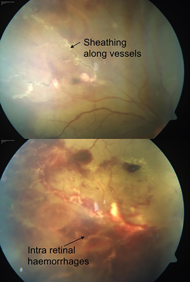
Retinal vasculitis with retinal haemorrhages, sheathing along vessels and hard exudates
Data Collection
Institute ethical clearance and written informed consent was taken from all participants. Fasting blood samples were collected from all individuals to determine serum homocysteine levels and estimated using reflectance photometry using VITROS Chemistry Products HCY 2 (Homocysteine) Reagent measured at 340 nm. Normal range of serum homocysteine as per this method was 6.60 to 14.80 μmol/L.
The data collected was analyzed using SSPS-16 (Statistical Package for Social Sciences Version 16) software for doing frequency analysis, percentage calculation, calculating mean and standard deviation. Odd’s ratio and confidence interval for odds ratio was calculated. The power of study was 80% and P < 0.05 is considered statistically significant.
Results
The age group of subjects ranged from 22 to 56 years with mean age in cases being 33.47 ± 8.3 and controls 35.16 ± 8.5 years. Among the cases, 13(40.62%) presented with diminution of vision, and 10 (31.25%) had floaters. 59.4% and 67.2% of the cases and controls had vision between 20/40–20/20. 25% and 5.6% of cases and controls had vision in the range of 20/60–20/200 (Fig. 2). Around 15% and 17% of cases and controls had vision less than 20/200. Chi square test results showed that there was no association between vision and the groups (Table 1). 19 (59.35%) cases had features of active vasculitis and 4(12.5%) patients were found to have fresh vitreous haemorrhage. 2(6.25%) individuals were detected to have macular edema and 2 (6.25%) had epiretinal membranes. The main cause of reduced vision in the controls was due to refractive errors [50/64(78.12%)]. The mean serum homocysteine value in cases was 20.35 ± 14.73 (Mean ± SD)and in control subjects was 20.92 ± 14.15 μmol/L (Mean ± SD) and was this difference was not statistically different (P value 0.74). Among the cases, 11(34.37%) had S. homocysteine levels < 14.8 μmol/L, and 21(65.62%) had S. homocysteine levels > 14.8 μmol/L (Fig. 3). In the control group, 19(29.68%) individuals had S. homocysteine levels less than 14.8 μmol/L, and 45 (70.31%) had greater than 14.8 μmol/L (Table 2). There was no significant association found between S. homocysteine levels and Retinal vasculitis (P: 0.64). The Odds ratio was 0.80 (0.33–1.99) implying that there is insufficient evidence to conclude that the cases and controls were statistically significantly different with respect to homocysteine levels.
Fig. 2.

Best corrected visual acuity distribution among cases and controls
Table 1.
Distribution of subjects based on visual acuity
| Vision | Cases | Controls |
|---|---|---|
| 20/20–20/40 | 19 (59.4) | 43 (67.2) |
| 20/60–20/200 | 8 (25.0) | 10 (15.6) |
| < 20/200 | 5 (15.6) | 11 (17.2) |
| Total | 32 | 64 |
Fig. 3.

Distribution of serum homocysteine among cases and controls
Table 2.
Comparison of cases and control with levels of serum homocysteine
| Serum homocysteine | Total | ||
|---|---|---|---|
| < 14.8 μmol/L | > 14.8 μmol/L | ||
| Cases | 11 | 21 | 32 |
| Controls | 19 | 45 | 64 |
| Total | 30 | 66 | 96 |
p: 0.64, OR − 0.80(0.33–1.99)
Discussion
Retinal vessels are unique in comparison to other vessels as far as microvascular changes are concerned in the body. Changes in vasculitis in different organs of the body like kidney, liver, heart or brain cannot be detected clinically whereas changes in retinal vessels can be seen on fundoscopy. These changes can be viewed and photographed for documentation purpose as well as for monitoring the changes in follow up. Retinal vasculitis patients mostly present with diminution of vision due to vitreous haemorrhage as found by a study by Saxena et al. [16]. The most common clinical sign of retinal vasculitis is perivascular sheathing [16]. The most common presentation in our study was found to be reduced vision, and sign was vascular sheathing. Other presentations of retinal vasculitis in our patients included intra retinal haemorrhages, sclerosis of the vessels, vitreitis, retinal ischemia, neovascularization elsewhere, cystoid macular edema, epiretinal membrane and vitreous haemorrhage.
Inflammation of vessels in other organs (CAD, stroke due to atherosclerosis etc.) have been found to have raised levels of serum homocysteine [13].What separates retinal vessel inflammation from other vascular inflammation is that changes can be seen in the retinal vasculature at an early stage which perhaps is not the case in other organs like of ischemic heart disease, atherosclerosis elsewhere, peripheral vascular diseases [16].One of the major causes of the loss of vision is the presence of inflammatory cells causing damage to the retina which is further enhanced by disruption of the blood retinal barrier leading to retinal and vitreous haemorrhage [17–19]. There have not been many studies, directly relating the association of serum homocysteine and retinal vasculitis. However, Muthuvel Bharathselvi et al. [20] studied the association of raised Serum homocysteine in 20 patients of Eales’ disease—and compared that to 20 healthy adult volunteers as control subjects. They stated that the homocysteine and homocysteine-thiolactone, which is formed by the error in homocysteine metabolism in hyperhomocysteinemia state, causes formation of the reactive oxygen species and degradation of glutathione (GSH), resulting in damage to the retinal vessels. Homocysteine concentration increases as result of deficiencies in cystathionine—synthetase, methylene-tetrahydrofolate reductase or other enzymes which are part of the methionine metabolism. Though the exact pathological mechanism is not understood, studies have shown that direct endothelial cells damage develop as a result of interaction between homocysteine and lipoprotein [21]. This leads to activation of the coagulation cascade by inactivating Protein C, upregulation of coagulation factor V, platelet adhesion, inhibition of thrombomodulin and increased thromboxane activity.
Present retrospective case–control study was conducted to determine whether there could be any relationship between serum homocysteine and retinal vasculitis. In retinal vasculitis, the laboratory investigations are an essential component to facilitate the detection of any underlying systemic disease and/or to establish a limited differential diagnosis. In our patients, a detailed history, clinical examination and laboratory investigations were carried out, none of the subjects had existing systemic illness. Three of the subjects were found to be diabetic during the study and were excluded from the study as per the exclusion criteria of the study. After subjecting the individuals to 14 h of fasting, the blood samples were collected for the assessment of serum homocysteine. The age group in our study was 22–56 years with an average of 33 years where as those in Bharathselvi et al. [20] were between 20 and 40 years.
Our study found that of the total 32 retinal vasculitis patients serum homocysteine was raised in 65.62% (21 cases) and in 70.31% of the control group (45 out of 64). Thus, in this study, we found that there was no significant association of raised serum homocysteine and retinal vasculitis (with P: 0.64). This was contrary to the result obtained in case of Eales’ disease by Bharathselvi M et al. [20, 22, 23]. They found that the plasma homocysteine levels were 18.6 (± 1.77) μmol/L in Eales’ disease cases and 11.2 (± 0.64) μmol/L in control subjects with P value less than 0.001 unlike our study with the mean serum homocysteine value in the cases 20.35 (SD 14.73) μmol/L and 20.92 in the control subjects (SD 14.15) μmol/L with P value 0.74. The Odds ratio was 0.80 (0.33–1.99) implying that there is no there is insufficient evidence to conclude that the cases and controls were statistically significantly different with respect to homocysteine levels.
Conclusion
In our study, we found that there is no statistically significant association between retinal vasculitis and raised levels of serum homocysteine.
Declarations
Conflict of interest
None.
Financial interest
None.
Footnotes
Publisher's Note
Springer Nature remains neutral with regard to jurisdictional claims in published maps and institutional affiliations.
Contributor Information
Angad Miskin, Email: angad.the.carpe.diem@gmail.com.
Raji Kurumkattil, Email: drrajisatyapal@yahoo.co.in.
Anurodh Gupta, Email: anurodhgoa@gmail.com.
Hemant Singh Trehan, Email: hstarti@gmail.com.
Puja Dudeja, Email: puja_dudeja@yahoo.com.
Sanjay Kumar Dhar, Email: csanjaydhar@yahoo.com.
Vijay Kumar Sharma, Email: vijayshrma007@gmail.com.
References
- 1.Sanders MD. Retinal vasculitis: a review. J R Soc Med. 1979;72:908–915. doi: 10.1177/014107687907201209. [DOI] [PMC free article] [PubMed] [Google Scholar]
- 2.Rosenbaum JT, Sibley CH, Lin P. Retinal vasculitis. CurrOpinRheumatol. 2016;28(3):228–235. doi: 10.1097/BOR.0000000000000271. [DOI] [PMC free article] [PubMed] [Google Scholar]
- 3.Walton RC, Ashmore ED. Retinal vasculitis. Curr Opin Ophthalmol. 2003;14:413–419. doi: 10.1097/00055735-200312000-00015. [DOI] [PubMed] [Google Scholar]
- 4.George RK, Walton RC, Whitcup SM, Nussenblatt RB. Primary retinal vasculitis. SystAassocDdiagnEvalOphthal. 1996;103(3):384–389. doi: 10.1016/s0161-6420(96)30681-7. [DOI] [PubMed] [Google Scholar]
- 5.Agarwal A, Afridi R, Agarwal R, Do VD, Gupta V, Nguyen QD. Multimodal imaging in retinal vasculitis. OculImmunolInflamm. 2017;25(3):424–433. doi: 10.1080/09273948.2017.1319494. [DOI] [PubMed] [Google Scholar]
- 6.Ali A, Ku JH, Suhler EB, Choi D, Rosenbaum JT. The course of retinal vasculitis. Br J Ophthalmol. 2014;98(6):785–789. doi: 10.1136/bjophthalmol-2013-303443. [DOI] [PubMed] [Google Scholar]
- 7.Biswas J, Ravi RK, Narayansamy A, Kulandai LT, Madhavan HN. Eales' disease - current concepts in diagnosis and management. J Ophthalmic Inflamm Infect. 2013;3(1):3–11. doi: 10.1186/1869-5760-3-11. [DOI] [PMC free article] [PubMed] [Google Scholar]
- 8.Nema N, Verma S, Kumar R. Investigation of methylenetetrahydrofolate reductase C677T and factor V Leiden mutation as a genetic marker for retinal vein occlusion. Tawian J Ophthal. 2018;8(2):99–103. doi: 10.4103/tjo.tjo_43_17. [DOI] [PMC free article] [PubMed] [Google Scholar]
- 9.Stanger O, Weger M, Obeid R. Impairment of homocysteine metabolism in patients with retinal vascular occlusion and non-arteritic ischemic optic neuropathy. Clin Chem Lab Med. 2005;43:1020–1025. doi: 10.1515/CCLM.2005.179. [DOI] [PubMed] [Google Scholar]
- 10.Narayanasamy A, Subramaniam B, Karunakaran C. Hyperhomocysteinemia and low methionine stress are risk factors for central retinal venous occlusion in an Indian population. Invest Ophthalmol Vis Sci. 2007;48:1441–1446. doi: 10.1167/iovs.06-0905. [DOI] [PubMed] [Google Scholar]
- 11.Ramakrishnan S, Sulochana KNL, Selvi S, Angayarkanni RN. Biochemistry of homocysteine in health and diseases. Indian J BiochemBiophys. 2006;43:275–283. [PubMed] [Google Scholar]
- 12.Cahill M, Karabatzaki M, Meleady R, Refsum H, Ueland P, Shields D, et al. Raised plasma homocysteine as a risk factor for retinal vascular occlusive disease. Br J Ophthalmol. 2000;84:154–157. doi: 10.1136/bjo.84.2.154. [DOI] [PMC free article] [PubMed] [Google Scholar]
- 13.Welch GN, Loscalzo J. Homocysteine and atherothrombosis. New Engl J Med. 1998;9:1042–1050. doi: 10.1056/NEJM199804093381507. [DOI] [PubMed] [Google Scholar]
- 14.Aflaki E, Mehryar M, Nazarinia M-A, Habibagahi Z, Rajaee A, Ranjbar-Omrani G. The relation between serum homocysteine level and eye involvement in Behcet’s disease. Arch Iran Med. 2008;11(96):625–628. [PubMed] [Google Scholar]
- 15.Paovic J, Paovic P, Sredovic V, Dimcic Z. Can Heterozygosity of MTHFR and hyperhomocysteinemia be risk factors for both retinal/vitreal hemorrhages in retinal vasculitis and retinal vein occlusion? Res Immunol Int J. 2015;2015:246396. [Google Scholar]
- 16.Saxena S, Kumar D. New classification system-based visual outcome in Eales' disease. Indian J Ophthalmol. 2007;55(4):267–269. doi: 10.4103/0301-4738.33038. [DOI] [PubMed] [Google Scholar]
- 17.Kumar S, Das RR, Biswas J, Kumar A. Profile of retinal vasculitis in a tertiary eye care centre in Eastern India. Indian J Ophthalmol. 2011;59(4):297–301. doi: 10.4103/0301-4738.82002. [DOI] [PMC free article] [PubMed] [Google Scholar]
- 18.Rosenbaum JT, Ku J, Ali A, Choi D, Suhler EB. Patients with retinal vasculitis rarely suffer from systemic vasculitis. Semin Arthritis Rheum. 2012;41(6):859–865. doi: 10.1016/j.semarthrit.2011.10.006. [DOI] [PMC free article] [PubMed] [Google Scholar]
- 19.Er H, Evereklioglu C, Cumurcu T, Türköz Y, Özerol E, Sahin K, et al. Serum homocysteine level is increased and correlated with endothelin-1 and nitric oxide in Behçet's disease. Br J Ophthalmol. 2002;86(6):653–657. doi: 10.1136/bjo.86.6.653. [DOI] [PMC free article] [PubMed] [Google Scholar]
- 20.Muthuvel B, Jyothirmay B, Selvi R, Coral K, Narayanaswamy A, Ramakrishnan S, et al. Increased homocysteine, homocysteine-thiolactone, protein homocysteinylation and oxidative stress in the circulation of patients with Eales’ disease. Ann ClinBiochem. 2013;50(4):330–338. doi: 10.1177/0004563213492146. [DOI] [PubMed] [Google Scholar]
- 21.Dudman NPH, Wilcker DEL, Wang JF, Lynch D, Lundberg P. Disordered methionine homocysteine metabolism in premature vascular disease. Arterioscler Thromb. 1993;13:1253–1260. doi: 10.1161/01.ATV.13.9.1253. [DOI] [PubMed] [Google Scholar]
- 22.Rao NA, Wu GS. Free radical mediated photoreceptor damage in uveitis. Prog Retin Eye Res. 2000;19:41–68. doi: 10.1016/S1350-9462(99)00003-8. [DOI] [PubMed] [Google Scholar]
- 23.Ramakrishnan S, Rajesh M, Sulochana KN. Eales’ disease: Oxidant stress and weak antioxidant defence. Indian J Ophthalmol. 2007;55(2):95–102. doi: 10.4103/0301-4738.30701. [DOI] [PubMed] [Google Scholar]