Abstract
A cluster of genes involved in antibiotic and heavy metal resistance has been characterized from a clinical isolate of the gram-negative bacterium Stenotrophomonas maltophilia. These genes include a macrolide phosphotransferase (mphBM) and a cadmium efflux determinant (cadA), together with the gene cadC coding for its transcriptional regulator. The cadC cadA region is flanked by a truncated IS257 sequence and a region coding for a bin3 invertase. Despite their presence in a gram-negative bacterium, these genetic elements share a common gram-positive origin. The possible origin of these determinants as a remnant composite transposon as well as the role of gene transfer between gram-positive and gram-negative bacteria for the acquisition of antibiotic resistance determinants in chronic, mixed infections is discussed.
Stenotrophomonas maltophilia has emerged in the last few years as an important nosocomial opportunistic pathogen. This bacterial species has been associated with different diseases, mainly in severely debilitated or immunosuppressed individuals (reviewed by Denton and Kerr [8]), as well as in the last stages of cystic fibrosis (12). Infections by S. maltophilia are difficult to treat (21, 23) due to the intrinsic antibiotic resistance of this bacterial species (2, 10). A combination of reduced permeability (31) and expression of efflux pump(s) (1, 33) might account at least in part for S. maltophilia intrinsic resistance to drugs. In addition to these mechanisms, antibiotic-inactivating enzymes such as metallo-beta-lactamases and cephalosporinases (19, 27, 29, 30) or, more recently, aminoglycoside-modifying enzymes (13), have been described to be encoded by S. maltophilia. Like other gram-negative bacilli, S. maltophilia is weakly susceptible to erythromycin. Besides a reduced permeability to the drug, S. maltophilia can pump out the antibiotic through a multidrug efflux determinant (A.A. and J.L.M., submitted for publication). In an attempt to further characterize the mechanisms involved in the reduced susceptibility to erythromycin in this bacterial species, we have cloned a DNA region capable of conferring erythromycin resistance to a hypersusceptible Escherichia coli strain. Sequencing of this region has demonstrated the presence of isoforms of genes previously found in Staphylococcus aureus and involved in resistance to erythromycin (mphBM) and cadmium (cadC and cadA). These genes are surrounded by a bin3 invertase (25) and a truncated IS257 sequence (20). The structure and G+C content of this DNA region suggests a gram-positive origin for these determinants. Gene transfer between gram-positive and gram-negative bacteria is well documented (7). We demonstrate here that the occurrence of such a transfer might be a powerful mechanism for acquiring antibiotic resistance genes in nosocomial pathogens such as S. maltophilia.
MATERIALS AND METHODS
Bacterial strains and growth conditions.
S. maltophilia D457R is a spontaneous multiresistant derivative of the clinical isolate S. maltophilia D457 (1). E. coli KZM120 (14) contains an acrAB null mutation (ΔacrAB::Tn903Kanr) that renders it drug hypersusceptible and was a kind gift from Dzwokai Ma. Bacterial strains were grown in Luria-Bertani medium (3) at 37°C with shaking, unless indicated otherwise. For selection purposes, medium was supplemented with ampicillin (200 μg/ml), kanamycin (25 μg/ml), and erythromycin (6 μg/ml).
Construction and screening of a DNA library.
Chromosomal DNA for library construction was extracted from S. maltophilia D457R as described previously (4). The obtained DNA was partially digested with Bsp1431 (MBI Fermentas, Vilnius, Lithuania), and fragments of 5 to 9 kb were isolated upon centrifugation on a 10 to 40% (wt/vol) sucrose gradient. DNA fragments were ligated to an alkaline phosphatase-treated BamHI-linearized plasmid pUC19 (26). E. coli KZM120 was electroporated with the ligation mixture, and transformants were selected on medium containing erythromycin, ampicillin, and kanamycin. Preparation and analysis of plasmid DNA was performed by standard methods as described previously (26).
Drug susceptibility measurements.
The MICs of erythromycin were determined in Mueller-Hinton medium (3) by E-Test (AB Biodisk, Solna, Sweden), according to the manufacturer's instructions.
DNA sequencing.
Automatic sequencing (Perkin-Elmer Gene Sequencer ABI310) of both strands of the DNA fragment contained in the plasmid pERY1 was carried out by primer walking. Analysis of the sequences was performed with the aid of Wisconsin Package version 9.1 (Genetics Computer Group, Madison, Wis.).
Southern blotting.
Chromosomal DNA from S. maltophilia D457 and D457R was treated with EcoRI (MBI Fermentas), electrophoresed on 0.7% agarose gel and transferred to Hybond-N (Amersham) as described earlier (26). λ DNA/HindIII (MBI Fermentas) was used as the molecular size marker. Membranes were subjected to overnight hybridization and subsequent washings under stringent conditions at 60°C with an mphBM probe obtained by PCR from pERY1 (see below). The obtained PCR product was purified with Micro Bio-Spin chromatography columns (Bio-Rad), labeled with [α-32P]dCTP using the DNA Labelling Kit–dCTP (Pharmacia Biotech), according to the manufacturer's instructions, and added to the hybridization buffer.
PCR.
An internal fragment of 140 bp from the mphBM gene was amplified by PCR using primer 1 (5′-CCAACCTCAAACAATCTCATTG-3′) and primer 2 (5′-GCTGCGGGTTTACCTGTAAG-3′). Reaction mixture (50 μl) contained 0.2 mM concentrations of each deoxynucleotide (dCTP, dTTP, dGTP, and dATP), 0.5 μM concentrations of each primer, 1.5 mM MgCl2, 10 mM Tris-HCl (pH 8.3), 50 mM KCl, 100 ng of template DNA, and 1.0 U of Taq DNA polymerase. The mixture was heated for 90 s at 94°C, followed by 35 cycles of 30 s at 94°C, 60 s at 60°C, and a 90-s extension step at 72°C and, finally, one 10-min extension cycle at 72°C before the end of the reaction. PCR products were analyzed by electrophoresis on an 1.6% agarose gel. A 100-bp DNA ladder (BioLabs) was used as the molecular size marker. Chromosomal DNAs from S. maltophilia D457R obtained with 1 year of difference were used as templates. The more recent DNA chromosomal preparation was obtained using the Genome DNA Kit (Bio 101).
Nucleotide sequence accession number.
The nucleotide sequence of the ERY1 region has been assigned GenBank accession number AJ251015.
RESULTS
Cloning of an erythromycin resistance gene from S. maltophilia D457R.
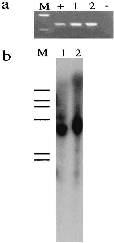
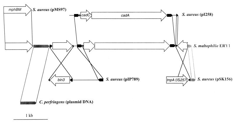
We have previously characterized an S. maltophilia spontaneous mutant (D457R) which shows an enhanced resistance to several different antibiotics (1), one of which is erythromycin. The MIC of erythromycin was 32 μg/ml for the wild-type strain S. maltophilia D457 and >256 μg/ml for the mutant strain S. maltophilia D457R. To clone the gene(s) responsible for erythromycin resistance in S. maltophilia D457R, we constructed a library in the plasmid pUC19 (see Materials and Methods) using as a receptor E. coli strain KZM120, which lacks the efflux pump determinant acrAB (14). Deletion of this multidrug resistance operon reduced the MIC of erythromycin from 16 to 2 μg/ml, making KZM120 a suitable strain for cloning macrolide resistance genes. The library was seeded onto plates containing erythromycin (6 μg/ml) as the selective agent. A single colony capable of growth under these conditions was isolated. Plasmid DNA (hereafter named pERY1) was obtained from such a clone, E. coli KZM120 was retransformed with this DNA preparation, and transformants were selected either in plates containing ampicillin at 200 μg/ml (the antibiotic selection marker of plasmid pUC19) or in plates containing erythromycin at 6 μg/ml. The number of transformants that grew under both selective conditions was the same. Thus, the 5,451-bp DNA fragment present in pERY1 carries a determinant for erythromycin resistance. Further confirmation was obtained from the analysis of susceptibility to erythromycin of strains either containing or not containing pERY1. As previously stated, the MIC of erythromycin for E. coli KZM120 is 2 μg/ml, and the same value was obtained for E. coli KZM120(pUC19). However, this value increased to reach 32 μg/ml for E. coli KZM120(pERY1), confirming that this plasmid contains an erythromycin resistance determinant. To assure that this DNA fragment is present in the genome of S. maltophilia D457R, PCR analysis was performed with chromosomal DNA obtained from S. maltophilia D457R by two different methods. As shown in Fig. 1a, a band with the predicted molecular size was amplified from both DNA preparations. Further confirmation of the presence of the ERY1 fragment in the genomes of S. maltophilia D457 and D457R was obtained by Southern blot analysis of restriction digests of chromosomal DNA from both strains using an internal probe from plasmid pERY1. The presence of hybridization signal bands with a molecular size of 4.4 kbp (Fig. 1b) indicated that the DNA fragment cloned in pERY1 is present in the genomes of both S. maltophilia D457 and S. maltophilia D457R. The genetic structure of this DNA region is shown in Fig. 2. The G+C content of this DNA region (35.1%) strongly suggests a gram-positive origin for this gene cluster.
FIG. 1.
Analysis of the presence of mphBM in the genome of S. maltophilia D457R. The presence of this gene in the genome of S. maltophilia was analyzed by two different methods. (a) Results of PCR amplification with primers specific for mphBM. M, molecular size markers. Top, 200 bp; bottom, 100 bp; +, positive control, with amplification using the plasmid pERY1 as the template; lanes 1 and 2, amplification with two different genomic DNA preparations from S. maltophilia D457 as templates. A band with the predicted molecular size (144 bp) was amplified from both DNAs. −, negative control. (b) Results of the hybridization of EcoRI-digested genomic DNAs from S. maltophilia D457 (lane 1) and D457R (lane 2) with an internal probe specific for the detection of mphBM. In both cases, a hybridization signal corresponding to a 4.2-kbp DNA fragment was detected. M, molecular size markers. Bars, from the top: 23, 9.4, 6.5, 4.4, 2.3, and 2.0 kbp.
FIG. 2.
Organization of the ERY1 region from S. maltophilia D457. The genetic structure of this region, as well as its relationship with some other previously analyzed sequences, is shown. The structure of ERY1 is shown in the middle of the figure. White arrows indicate the localization and orientation of the ORFs of the region. All of them present homologies of >90% with the previously characterized sequences shown in the figure. Black arrows indicate the localization and orientation of regions with homologies of >90% with sequences deposited at DNA data banks but which do not contain any ORFs. Gray arrows indicate the position and orientation of regions with homologies with sequences deposited at DNA data banks of <90%.
mphBM gene.
Sequencing of the DNA fragment and further analysis demonstrated the presence of a gene that is nearly identical to the previously described mphBM gene from S. aureus. mphBM encodes the synthesis of a macrolide phosphotranferase (15), and homologs for this gene have been described in E. coli (16, 17) and Streptomyces rochei (9). The homology of these genes ranges from 30 to 50%; however, in the case of S. maltophilia, the homology is 98.2% at the DNA level and 99.7% (with 98.3% identity) at the protein level compared with mphBM (Table 1). This extremely high homology indicates that the gene mphBM of S. maltophilia has been recently acquired from S. aureus and is just an isoform of the S. aureus gene. Erythromycin MICs were determined for E. coli KM120(pUC19) and E. coli KM120(pERY1). The MIC values were 2 and 32 μg/ml, respectively. The fact that the MIC of erythromycin increases in the presence of this gene in E. coli KZM120 indicates that it is functionally active in this bacterial species in spite of its possible gram-positive origin.
TABLE 1.
Homology of ERY1 with other DNA sequences
| ERY1 region (ORF)a | % G+C content | Homologous sequence (ORF)b | % Homology (% protein homology/ % aa identity)c | Function | Organism (plasmid) | Reference |
|---|---|---|---|---|---|---|
| 1–912 (13–912) | 36.8 | AB013298: 2284–319 (mphBM: 2296–3195) | 98.2 (99.7/98.3) | Macrolide 2′-phosphotransferase II | S. aureus (pMS97) | 15 |
| 950–1309 | 25.1 | X73562: 936–1285 | 56.9 | Unknown | C. perfringens (unknown plasmid) | 22 |
| 1309–2053 (1397–2005) | 31.8 | X16298: 961–1704 (inverted) (bin3: 1049–1657) | 86.3 (98/92.5) | Invertase | S. aureus (pI9789) | 25 |
| 2128–2247 | 36.1 | X16298: 1–120 (inverted) | 95.0 | Unknown | S. aureus (pI9789) | 25 |
| 2137–4903 (2277–2645) (2638–4821) | 37.1 | J04551: 563–3329 (cadC: 703–1071) (cadA: 1064–3247) | 99.1 (98.4/96.7) (99.5/98.9) | Cadmium efflux | S. aureus (pI258) | 18 |
| 4905–5013 | 36.1 | J04551: 3424–3533 | 99.1 | Unknown | S. aureus (pI258) | 18 |
| 4903–5318 (4989–5318 reverse strand) | 34.6 | AF053771: 3269–3624 (inverted) (tnpA [IS257]: 2945–3601) | 99.0 (100/99.1) | Transposase | S. aureus (pSK156) | 20 |
| 5318–5451 | 34.6 | AF053771: 3710–3840 | 58.8 | Unknown | S. aureus (pSK156) | 20 |
Region in ERY1 showing homology of >56% with an already known sequence. Localization of ORFs within this DNA region are is shown in parentheses.
The accession number of the homologous sequence and the region of homology within the sequence is given. The names and localization of the ORFs within the homologous sequence are indicated in parentheses.
The percentage of homology at DNA level is given. The percentage of homology at the amino acid level and the percentage of identical amino acids (aa) are indicated in parentheses.
bin3 gene.
Analysis of the sequence downstream from mphBM indicates the presence of a DNA region highly homologous (Table 1) to a central region of the transposon Tn552 from S. aureus. This region comprises the gene bin3, a divergent member of the resolvase-invertase family (25). The homologous region from S. maltophilia includes not only the bin3 isoform but also a palindromic sequence upstream from the open reading frame (ORF). A 107-bp sequence with an unknown function that is present 828 bp upstream from bin3 in Tn552 is also present, although it is inverted in this DNA region of S. maltophilia (Fig. 1).
cadC and cadA genes.
The 107-bp region, present in pERY1 and upstream from bin3 in Tn552 is also present upstream from the cadC gene in the plasmid pI258 (18) from S. aureus. cadC (32) is a regulator of the expression of cadA, a gene involved in the efflux of cadmium by S. aureus carrying the plasmid pI258 (18). Isoforms of both genes are also present, in the same order as in S. aureus in S. maltophilia (Fig. 1). Downstream from cadA, the homology between S. aureus and S. maltophilia is maintained to the end of the published S. aureus sequence, the only difference being a 103-bp internal region which is present in S. aureus and not in S. maltophilia (Fig. 1).
IS257.
The region downstream from the cadA ORF is highly homologous, not only to the surrounding cadA sequence from the S. aureus plasmid pI258 but also to the insertion sequence IS257. This indicates that an IS257 sequence is probably downstream from cadA in pI258. In the case of S. maltophilia D457, the homology includes one of the inverted repeats and part of the transposase gene. Only the half-carboxy-terminal part of the gene (amino acids 108 to 218) is present, and it is truncated by an additional 133-bp sequence (Fig. 1) which presents a 64.5% homology with the region from residue 3710 to residue 3840 from the IS257-containing plasmid pSK156 (20). The function of this region is unknown.
DISCUSSION
S. maltophilia is an opportunistic pathogen intrinsically resistant to several antibiotics. Some antibiotic resistance genes have been characterized from this bacterial species and, in most cases, they can be considered indigenous (and even housekeeping) genes more than acquired antibiotic resistance genes (13, 19, 27). In our work, we present evidence that S. maltophilia D457 has acquired a cluster of antibiotic and heavy metal resistance genes from gram-positive bacteria. Most of these genes are isoforms of genes previously found in S. aureus plasmids. Only, a 360-bp DNA region did not have an S. aureus counterpart in current DNA databases. This region was homologous with a sequence from Clostridium perfringens with unknown function. However, the fact that the homology of this region was <60%, indicates that it is not an isoform of a gene present in C. perfringens but only a homolog. Whether the organism from which the ERY1 DNA region has been transferred to S. maltophilia also contains the same homolog of this C. perfringens DNA is a matter of speculation.
The combination of ERY1 genes in the same DNA region has not yet been described. The genetic elements present in pERY1 were first characterized from S. aureus strains isolated at different geographic locations (in Japan and the United States) and in different years. The gram-positive origin of these genes is reinforced by the G+C content (Table 1). Overall, this value is 35.1%, a level closely similar to that for the genomes of gram-positive bacteria such as S. aureus and quite different from the 63 to 67.5% reported for S. maltophilia (8). IS257 is an insertion sequence ubiquitously found in the chromosome and plasmids of S. aureus (28), whereas its presence is uncommon in other bacterial species.
DNA exchange between gram-positive and gram-negative bacteria has been described; however, this is the first time in which this transfer has been documented for S. maltophilia. The organization of the sequenced region strongly suggests its origin as a transposon-like structure in which several insertion events might have occurred. In this way, the presence of a truncated IS257 sequence points to the possible insertion of another genetic element in this region. This complex structure resembles those found in the composite transposons from gram-positive bacteria (5, 6, 24). The strong similarities but also the differences (for instance, the deletion downstream of cadA from S. aureus) of these genetic elements with respect to their gram-positive counterparts indicate that several different recombination events have occurred to yield this genetic patchwork. Since the genetic elements of this region (Table 1) are characteristic of gram-positive bacteria, we think that these recombination events occurred before the acquisition of this DNA region by S. maltophilia.
For this transfer to occur, bacteria must share the same environment. This situation is common in the case of mixed infections and might be relevant in chronic infections such as cystic fibrosis. In fact, S. maltophilia D457 (the parental strain of D457R) is a clinical isolate from the sputum of a cystic fibrosis patient. Since S. aureus is frequently encountered in the lungs of cystic fibrosis patients (11), the DNA determinants present in the DNA region characterized in the present work might have been acquired from a strain of this bacterial species infecting the same individual as S. maltophilia D457. Alternatively, transfer of these determinants might have occurred in environmental conditions between S. maltophilia and gram-positive organisms such as Bacillus spp., which share the same environmental habitat.
ACKNOWLEDGMENTS
We thank Dzwokai Ma for the gift of E. coli KZM120 and A. Varas for technical assistance.
This research was supported in part by grant 08.2/022/98 from Comunidad Autónoma de Madrid. A. Alonso is a recipient of a fellowship from Gobierno Vasco. P. Sanchez is a recipient of a fellowship from Ministerio de Educación y Cultura.
REFERENCES
- 1.Alonso A, Martínez J L. Multiple antibiotic resistance in Stenotrophomonas maltophilia. Antimicrob Agents Chemother. 1997;41:1140–1142. doi: 10.1128/aac.41.5.1140. [DOI] [PMC free article] [PubMed] [Google Scholar]
- 2.Arpi M, Victor M A, Mortensen I, Gottschau A, Bruun B. In vitro susceptibility of 124 Xanthomonas maltophilia (Stenotrophomonas maltophilia) isolates: comparison of the agar dilution method with the E-test and two agar diffusion methods. APMIS. 1996;104:108–114. [PubMed] [Google Scholar]
- 3.Atlas R M. Handbook of microbiological media. London, England: CRC Press, Inc.; 1993. [Google Scholar]
- 4.Bagdasarian M, Bagdasarian M M. Gene cloning and expression. In: Gerhardt P, Murray R G E, Wood W A, Krieg N R, editors. Methods for general and molecular bacteriology. Washington, D.C.: American Society for Microbiology; 1994. pp. 406–417. [Google Scholar]
- 5.Bonafede M E, Carias L L, Rice L B. Enterococcal transposon Tn5384: evolution of a composite transposon through cointegration of enterococcal and staphylococcal plasmids. Antimicrob Agents Chemother. 1997;41:1854–1858. doi: 10.1128/aac.41.9.1854. [DOI] [PMC free article] [PubMed] [Google Scholar]
- 6.Byrne M E, Gillespie M T, Skurray R A. Molecular analysis of a gentamicin resistance transposonlike element on plasmids isolated from North American Staphylococcus aureus strains. Antimicrob Agents Chemother. 1990;34:2106–2113. doi: 10.1128/aac.34.11.2106. [DOI] [PMC free article] [PubMed] [Google Scholar]
- 7.Courvalin P. Transfer of antibiotic resistance genes between gram-positive and gram-negative bacteria. Antimicrob Agents Chemother. 1994;38:1447–1451. doi: 10.1128/aac.38.7.1447. [DOI] [PMC free article] [PubMed] [Google Scholar]
- 8.Denton M, Kerr K G. Microbiological and clinical aspects of infection associated with Stenotrophomonas maltophilia. Clin Microbiol Rev. 1998;11:57. doi: 10.1128/cmr.11.1.57. [DOI] [PMC free article] [PubMed] [Google Scholar]
- 9.Fernandez Moreno M A, Vallin C, Malpartida F. Streptothricin biosynthesis is catalyzed by enzymes related to nonribosomal peptide bond formation. J Bacteriol. 1997;179:6929–6936. doi: 10.1128/jb.179.22.6929-6936.1997. [DOI] [PMC free article] [PubMed] [Google Scholar]
- 10.Garrison M W, Anderson D E, Campbell D M, Carroll K C, Malone C L, Anderson J D, Hollis R J, Pfaller M A. Stenotrophomonas maltophilia: emergence of multidrug-resistant strains during therapy and in an in vitro pharmacodynamic chamber model. Antimicrob Agents Chemother. 1996;40:2859–2864. doi: 10.1128/aac.40.12.2859. [DOI] [PMC free article] [PubMed] [Google Scholar]
- 11.Gilligan P H. Microbiology of airway disease in patients with cystic fibrosis. Clin Microbiol Rev. 1991;4:35–51. doi: 10.1128/cmr.4.1.35. [DOI] [PMC free article] [PubMed] [Google Scholar]
- 12.Karpati F, Malmborg A S, Alfredsson H, Hjelte L, Strandvik B. Bacterial colonisation with Xanthomonas maltophilia—a retrospective study in a cystic fibrosis patient population. Infection. 1994;22:258–263. doi: 10.1007/BF01739911. [DOI] [PubMed] [Google Scholar]
- 13.Lambert T, Ploy M C, Denis F, Courvalin P. Characterization of the chromosomal aac(6′)-Iz gene of Stenotrophomonas maltophilia. Antimicrob Agents Chemother. 1999;43:2366–2371. doi: 10.1128/aac.43.10.2366. [DOI] [PMC free article] [PubMed] [Google Scholar]
- 14.Ma D, Cook D N, Alberti M, Pon N G, Nikaido H, Hearst J E. Genes acrA and acrB encode a stress-induced efflux system of Escherichia coli. Mol Microbiol. 1995;16:45–55. doi: 10.1111/j.1365-2958.1995.tb02390.x. [DOI] [PubMed] [Google Scholar]
- 15.Matsuoka M, Endou K, Kobayashi H, Inoue M, Nakajima Y. A plasmid that encodes three genes for resistance to macrolide antibiotics in Staphylococcus aureus. FEMS Microbiol Lett. 1998;167:221–227. doi: 10.1111/j.1574-6968.1998.tb13232.x. [DOI] [PubMed] [Google Scholar]
- 16.Noguchi N, Emura A, Matsuyama H, O'Hara K, Sasatsu M, Kono M. Nucleotide sequence and characterization of erythromycin resistance determinant that encodes macrolide 2′-phosphotransferase I in Escherichia coli. Antimicrob Agents Chemother. 1995;39:2359–2363. doi: 10.1128/aac.39.10.2359. [DOI] [PMC free article] [PubMed] [Google Scholar]
- 17.Noguchi N, Katayama J, O'Hara K. Cloning and nucleotide sequence of the mphB gene for macrolide 2′-phosphotransferase II in Escherichia coli. FEMS Microbiol Lett. 1996;144:197–202. doi: 10.1111/j.1574-6968.1996.tb08530.x. [DOI] [PubMed] [Google Scholar]
- 18.Nucifora G, Chu L, Misra T K, Silver S. Cadmium resistance from Staphylococcus aureus plasmid pI258 cadA gene results from a cadmium-efflux ATPase. Proc Natl Acad Sci USA. 1989;86:3544–3548. doi: 10.1073/pnas.86.10.3544. [DOI] [PMC free article] [PubMed] [Google Scholar]
- 19.Paton R, Miles R S, Amyes S G. Biochemical properties of inducible beta-lactamases produced from Xanthomonas maltophilia. Antimicrob Agents Chemother. 1994;38:2143–2149. doi: 10.1128/aac.38.9.2143. [DOI] [PMC free article] [PubMed] [Google Scholar]
- 20.Paulsen I T, Brown M H, Skurray R A. Characterization of the earliest known Staphylococcus aureus plasmid encoding a multidrug efflux system. J Bacteriol. 1998;180:3477–3479. doi: 10.1128/jb.180.13.3477-3479.1998. [DOI] [PMC free article] [PubMed] [Google Scholar]
- 21.Penzak S R, Abate B J. Stenotrophomonas (Xanthomonas) maltophilia: a multidrug-resistant nosocomial pathogen. Pharmacotherapy. 1997;17:293–301. [PubMed] [Google Scholar]
- 22.Perelle S, Gibert M, Boquet P, Popoff M R. Characterization of Clostridium perfringens iota-toxin genes and expression in Escherichia coli. Infect Immun. 1993;61:5147–5156. doi: 10.1128/iai.61.12.5147-5156.1993. [DOI] [PMC free article] [PubMed] [Google Scholar]
- 23.Quinn J P. Clinical problems posed by multiresistant nonfermenting gram-negative pathogens. Clin Infect Dis. 1998;27:S117–S124. doi: 10.1086/514912. [DOI] [PubMed] [Google Scholar]
- 24.Rice L B, Carias L L. Transfer of Tn5385, a composite, multiresistance chromosomal element from Enterococcus faecalis. J Bacteriol. 1998;180:714–721. doi: 10.1128/jb.180.3.714-721.1998. [DOI] [PMC free article] [PubMed] [Google Scholar]
- 25.Rowland S J, Dyke K G. Characterization of the staphylococcal beta-lactamase transposon Tn552. EMBO J. 1989;8:2761–2773. doi: 10.1002/j.1460-2075.1989.tb08418.x. [DOI] [PMC free article] [PubMed] [Google Scholar]
- 26.Sambrook J, Fritsch E F, Maniatis T. Molecular cloning: a laboratory manual. 2nd ed. Cold Spring Harbor, N.Y: Cold Spring Harbor Laboratory Press; 1989. [Google Scholar]
- 27.Sanschagrin F, Dufresne J, Levesque R C. Molecular heterogeneity of the L-1 metallo-beta-lactamase family from Stenotrophomonas maltophilia. Antimicrob Agents Chemother. 1998;42:1245–1248. doi: 10.1128/aac.42.5.1245. [DOI] [PMC free article] [PubMed] [Google Scholar]
- 28.Tenover F C, Arbeit R, Archer G, Biddle J, Byrne S, Goering R, Hancock G, Hebert G A, Hill B, Hollis R. Comparison of traditional and molecular methods of typing isolates of Staphylococcus aureus. J Clin Microbiol. 1994;32:407–415. doi: 10.1128/jcm.32.2.407-415.1994. [DOI] [PMC free article] [PubMed] [Google Scholar]
- 29.Walsh T R, MacGowan A P, Bennett P M. Sequence analysis and enzyme kinetics of the L2 serine beta-lactamase from Stenotrophomonas maltophilia. Antimicrob Agents Chemother. 1997;41:1460–1464. doi: 10.1128/aac.41.7.1460. [DOI] [PMC free article] [PubMed] [Google Scholar]
- 30.Walsh T R, Hall L, Assinder S J, Nichols W W, Cartwright S J, MacGowan A P, Bennett P M. Sequence analysis of the L1 metallo-beta-lactamase from Xanthomonas maltophilia. Biochim Biophys Acta. 1994;1218:199–201. doi: 10.1016/0167-4781(94)90011-6. [DOI] [PubMed] [Google Scholar]
- 31.Yamazaki E, Ishii J, Sato K, Nakae T. The barrier function of the outer membrane of Pseudomonas maltophilia in the diffusion of saccharides and beta-lactam antibiotics. FEMS Microbiol Lett. 1989;51:85–88. doi: 10.1016/0378-1097(89)90082-7. [DOI] [PubMed] [Google Scholar]
- 32.Yoon K P, Misra T K, Silver S. Regulation of the cadA cadmium resistance determinant of Staphylococcus aureus plasmid pI258. J Bacteriol. 1991;173:7643–7649. doi: 10.1128/jb.173.23.7643-7649.1991. [DOI] [PMC free article] [PubMed] [Google Scholar]
- 33.Zhang L, Li X Z, Poole K. Multiple antibiotic resistance in Stenotrophomonas maltophilia: involvement of a multidrug efflux system. Antimicrob Agents Chemother. 2000;44:287–293. doi: 10.1128/aac.44.2.287-293.2000. [DOI] [PMC free article] [PubMed] [Google Scholar]


