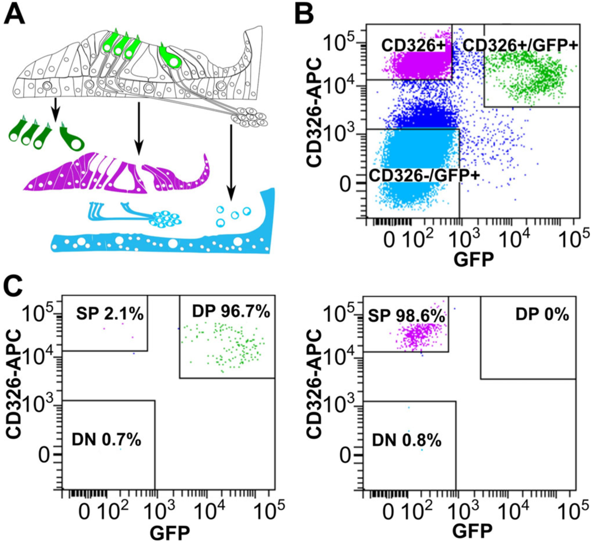
Fig. 5.

Cell sorting from a Math1-GFP mouse. (A) Schematic representing the organ of Corti and the three sorted cell populations. (B) Flow cytometry from cochlear tissue with HCs positive for CD326 and GFP (Double positive DP), supporting cells positive only for CD326 (Single Positive SP) and non-epithelial cells negative for both markers (Double negative DN). (C) Post-sort analyses of the cells sorted in (b) showing high purity for HCs (96.7%) and supporting cells (98.6%).
