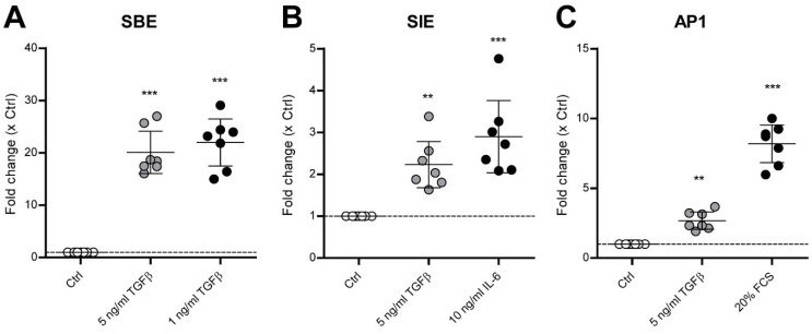
Figure 2.
TGF-β1 activates SMAD3:4, STAT3 and AP1 signaling in human OA chondrocytes. Primary human OA chondrocytes, cultured for a week to form a monolayer, were transduced for 6–8 h with 500 ng of lentivirus per 62,500 cells. After 48 h, the transduced cells were serum-starved overnight and then stimulated with a positive control (black dots) or with 5 ng/mL TGF-β1 (gray dots) for 6 h. Luminescence was determined at 470–480 nm, and fold change compared to medium-stimulated cells (Ctrl, white dots) was calculated. TGF-β1 (gray dots) activated cell signaling regulated by the transcription factors (A)SMAD3:4 (SBE: SMAD binding element), (B) STAT3 (SIE: Interleukin(IL)-6 sis-inducible element or STAT3 response element) and (C) AP1 (Activation Protein 1). Data are plotted as mean ± 95% CI, with each dot representing the average of a quadruple sample in one chondrocyte donor (total n = 7). Additionally, the induction of the pathways with positive controls is reported (black dots), which were 1 ng/mL TGF-β1 for the SBE pathway, 10 ng/mL IL-6 for the SIE pathway and 20% fetal calf serum (FCS) for the AP1 pathway. Statistical analysis was performed using one-way ANOVA with Bonferroni’s post hoc test: ** p ≤ 0.01; *** p < 0.001.

