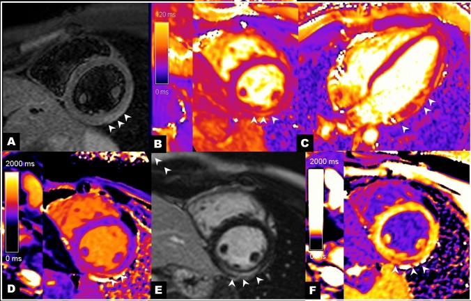
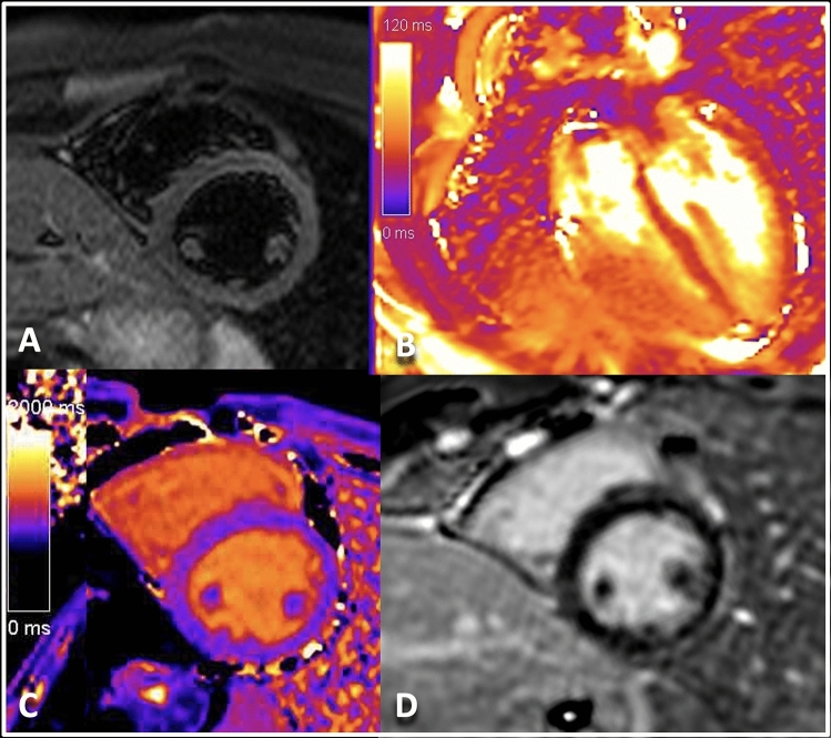
With the ongoing vaccination campaign against coronavirus disease-19 (COVID-19), cases of myocarditis associated with mRNA vaccination have been increasingly recognized. We describe here the case of a young male patient who developed chest pain and increased troponin 4 days after vaccination with Pfizer-BioNTech COVID-19 vaccine. Coronary angiography revealed patent coronary arteries and in the setting of clinically suspected myocarditis the patient was referred for cardiac magnetic resonance (CMR). CMR images showed normal biventricular function, edema in the inferior and infero-lateral left ventricular (LV) wall (Fig. 1, T2-STIR imaging in panel A, T2 mapping in panels B and C) with midwall fibrosis within the same region as indicate by increased signal in native T1 mapping (Fig. 1, panel D), late gadolinium enhancement (LGE) (Fig. 1, panel E) and reduced signal in post-contrast T1 mapping images (Fig. 1, panel F). All these findings were consistent with acute myocarditis. A further examination was scheduled at 6 months, to assess myocardial remodeling after the acute phase and evaluate the need for adjunctive treatments. The follow-up examination showed absence of edema and a significant reduction of LGE extension (Fig. 2). This result suggests the ongoing resolution of the disease and its benign course. Large cohorts of patients with myocarditis after mRNA vaccination for COVID-19 revealed the common occurrence of mild forms with benign outcome [1], albeit severe cases requiring intensive management might occur. Translating the approach applied to more classic forms of myocarditis [2], serial CMR might be useful to provide adjunctive clinical information and an improved risk-stratification.
Fig. 1.
CMR in the acute setting: A STIR short axis images of the left ventricle and B and C T2 mapping short axis and 4-chamber view showed edema in the inferior, infero-lateral and antero-lateral ventricular wall (white arrows head). Native T1 mapping (D) showed in the same area increased signal intensity. LGE (E) and T1 post-contrast (F) images showed midwall enhancement (white arrows head)
Fig. 2.
6-months follow-up CMR showed absence of edema and reduction of the extension of LGE in LV wall (T1-STIR image in A, T2 mapping in B, T1 mapping in C and LGE in D)
Author contributions
GC, FC and LA performed the cardiac magnetic resonance imaging examinations and wrote the draft of the manuscript, MPC and MPG were involved in the clinical management of the patient and collection of clinical data, all authors read and approved the final version of the manuscript.
Funding
The authors have not disclosed any funding.
Declarations
Competing interests
The authors declare no competing interests.
Footnotes
Publisher's Note
Springer Nature remains neutral with regard to jurisdictional claims in published maps and institutional affiliations.
References
- 1.Witberg G, Barda N, Hoss S, Richter I, Wiessman M, Aviv Y, et al. myocarditis after Covid-19 vaccination in a large health care organization. N Engl J Med. 2021;385:2132–2139. doi: 10.1056/NEJMOA2110737/SUPPL_FILE/NEJMOA2110737_DISCLOSURES.PDF. [DOI] [PMC free article] [PubMed] [Google Scholar]
- 2.Aquaro GD, GhebruHabtemicael Y, Camastra G, Monti L, Dellegrottaglie S, Moro C, et al. Prognostic value of repeating cardiac magnetic resonance in patients with acute myocarditis. J Am Coll Cardiol. 2019;74:2439–2448. doi: 10.1016/j.jacc.2019.08.1061. [DOI] [PubMed] [Google Scholar]


