

Abstract
Introduction:
Chest compression fraction (CCF) is the cumulative time spent providing chest compressions divided by the total time taken for the entire resuscitation. Targeting a CCF of at least 60% is intended to limit interruptions in compressions and maximize coronary perfusion during resuscitation. We aimed to identify the mean CCF and its relationship with various factors affecting it.
Methods:
Patients presenting to the emergency department in cardiac arrest at a single center were prospectively included in this study. Resuscitation was provided by trained health-care providers. The feedback device Cprmeter2™ was placed on the patient's sternum at the beginning of resuscitation. The total time taken for the entire resuscitation was noted by the device and CCF calculated.
Results:
The mean CCF was analyzed using descriptive statistics and was found to be 71.60% ± 7.52%. The total duration of resuscitation (R = −0.55, P = < 0.001, min-max, 2.02–34.31, mean 12.25 ± 6.54), number of people giving chest compressions (R = −0.48, P = < 0.001, min-max, 1–6, mean 4.04 ± 1.12), and total number of team members in resuscitation (R = −0.50, P = < 0.001, min-max, 4–10, mean 6.65 ± 1.32) had negative correlation with CCF. Diurnal variation (day, n = 35; mean 69.20% ± 7% and night, n = 20; mean 75.80% ± 5.6%, P = 0.001) and patients receiving defibrillation (receiving n = 10 mean 67.00% ± 4.11% and not receiving n = 45 mean 72.62 ± 7.42%, P = 0.005) were found to significantly affect CCF.
Conclusion:
The mean CCF for cardiac arrest patients was well within the targets of guideline recommendation. CCF decreased when resuscitation lasted longer, during daytime when the defibrillator was used, the total team members increased, and also when the number of people giving chest compressions increased. CCF during resuscitation may improve if there is a focus on improving these factors and requires validation in multicentric settings.
Keywords: Cardiac arrest, cardiopulmonary resuscitation, chest compression fraction, defibrillation, emergency department
INTRODUCTION
Improving survival from cardiac arrest is an important goal for the emergency medical team.[1,2] With an annual worldwide incidence of >6 million cardiac arrests the search continues for ways to improve survival.[3]
The delivery of high-quality chest compressions is one of the most essential components of cardiopulmonary resuscitation (CPR).[4] The American Heart Association (AHA) currently recommends minimizing the frequency and duration of interruptions in chest compressions to maximize the number of compressions delivered per minute.[5]
Chest compression fraction (CCF) is the cumulative time of chest compressions given during resuscitation divided by the total time taken for the entire resuscitation. It is a measure of time devoted exclusively to chest compressions. Rescuers should try to perform chest compressions at a rate of at least 100–120 per min and a depth of at least 2 inches, avoiding excessive depths >2.4 inches or 6 cm according to the new AHA guidelines.[6] The addition of a target compression fraction of at least 60% is intended to limit interruptions in compressions and to maximize blood flow and coronary perfusion during CPR.[6] Quantifying CPR by calculating the CCF helps in improving the quality of resuscitation.[6]
During resuscitation, providers may stop delivering compressions for reasons that are integral to patient care such as assessing rhythm and pulse, maintaining airway and ventilation, or defibrillation. Alternatively, they may pause compressions for reasons that are not related to patient care such as distraction, fatigue, confusion, or lack of knowledge and inability to perform lifesaving skills efficiently. Interruptions in chest compressions during CPR have a deleterious impact on cerebral and coronary perfusion during animal models of cardiac resuscitation.[3,7] The rescuers’ CPR performance, particularly the quality of chest compression, decreases rapidly over time.[8] Several devices providing audiovisual feedback during compressions (CPR feedback devices) have been introduced to improve basic life support (BLS) quality.[9,10] Nevertheless, the factors affecting CCF were proposed to vary across settings and we aimed to measure the average CCF achieved in the Indian academic emergency departments (EDs). In addition to CCF, we also sought to identify its relationship with factors such as total duration of resuscitation, the number of personnel involved, use of the defibrillator, airway management, and diurnal variation, in cardiac arrest cases presenting to our ED.
METHODS
Study design and setting
This was a prospective observational study done in the ED of a tertiary care teaching hospital in south India over 1 year. The study had independent approval from the Institutes Review Board, Institutes Ethical Committee (IEC no: 52/17/IEC/JMMC and RI). The study was also registered under the Clinical Trials Registry-India (CTRI/2018/05/013913).
The objective of the study
Primary objective: To estimate the CCF in cardiac arrest patients presenting to the ED in a tertiary care teaching hospital in Kerala.
Secondary objectives: To identify the relationship of chest compression fraction with
Total duration of resuscitation
Number of people giving chest compressions
Total number of team members
Usage of defibrillator
Definitive airway placement
Time when resuscitation was done.
Inclusion criteria
We included patients who were 18 years or older, who presented to the ED in cardiac arrest.
Exclusion criteria
Patients who presented in cardiac arrest following a trauma, pregnant ladies in cardiac arrest, with hard signs of death, and those in which resuscitation was terminated early were excluded.
Criteria for early termination of resuscitation involved a joint decision-making process, in the presence of preexisting chronic illness preventing meaningful recovery like disseminated malignancy, acute illness preventing recovery like 100% burns, catastrophic traumatic brain injury with no brain stem reflexes, and the time elapsed with no bystander CPR for more than 15 min or with bystander CPR more than 30 min.[11] The cases where bystander or patient denied consent or withdrew consent at any point of time were also not included.
Consent
Consent was sought at two points in the study. At admission consent was sought from the bystander or his/her legally authorized representative (LAR). Later in the event, if the individual had a neurologically intact outcome, consent was sought from the patients in the presence of their LAR, and they were allowed to withdraw their data from inclusion if they wished to do so. In the case of individuals who could not give informed consent or had no bystanders at the moment of admission, the data were still abstracted but included in the analysis only after procuring consent from the patient or his/her LAR.
Methods
All cardiac arrest patients brought to the ED who met the inclusion criteria were studied by an independent observer who was not part of the resuscitation. Resuscitation measures were initiated according to BLS and advanced cardiac life support–AHA-guidelines by trained health-care providers.
The feedback device CPRmeter2™ was attached to the body and switched on before compressions were started [Figure 1]. The device provides real-time auditory and visual feedback of the resuscitation measures with the help of two sensors – an accelerometer measuring depth, rate and a force sensor that gives information on compression/recoil. Data were extracted either directly from the device postresuscitation or retrieved through a Bluetooth® technology-enabled mobile device. The inactivity time between each cycle of chest compressions was measured by the device. The device was turned off at the end of the resuscitation, i.e., either return of spontaneous circulation (ROSC) or termination of resuscitation following the death of the patient. The total time taken for the entire resuscitation was noted, and the CCF was calculated by the device. Factors that affect the CCF were also looked into, using a predefined pro forma during the resuscitation, and consent was taken from bystanders at that time.
Figure 1.
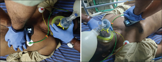
Real-time use of feedback device during resuscitation
Sample size calculation
Based on the mean (0.70) and standard deviation (0.17) of CCF observed in an earlier publication titled “chest compression rates and survival following out of hospital cardiac arrest,”[12] with 95% confidence interval and 5% relative allowable error, the minimum sample size was calculated as 50 using the formula (n) = ([Z1-α/2]2 S2)/d2, where Z1-α/2 was the value of normal deviate at the considered level of confidence, S was the expected standard deviation of variable in group, and d was the expected absolute allowable error in the mean.
Consecutive cases which presented to the ED during the study period that satisfied the inclusion and exclusion criteria were enrolled till the necessary sample size was met.
Data analysis and interpretation
Numerical variables are expressed as mean ± standard deviation (minimum-maximum) and categorical variables as frequency (percentage).
To compare the mean differences of CCF with the study variable, an independent two-sample t-test was applied. The relationship between numerical variables, which did not follow normality, was calculated using the Spearman rank correlation coefficient. Multivariate regression analysis was also done to adjust for confounders.
The statistical software, namely, Statistical Package for the Social Sciences, SPSS (International Business Machines Statistical Package for the Social Sciences, IBM Corp. Released 2013, version 22.0. Armonk, NY, USA: IBM Corp) was used for the analysis of the data. Microsoft Word and Microsoft Excel (2013 version) were used to enter data and generate tables and charts.
RESULTS
A total of 55 cardiac arrest victims who met the inclusion and exclusion criteria were studied.
Demographic data
In our study, the majority of the patients were males 41 (74.5%), and the remaining 14 (25.5%) were females.
Multiple comorbidities were present in most of the patients. Diabetes mellitus (63.63%) was the most common among them, followed by hypertension (47.27%), coronary heart disease (43.63%), and dyslipidemia (32.72%). The mean age of the study population was found to be 60.8 ± 12.4 years (min-max, 21–88). The mean CCF was 71.60 ± 7.25% (min-max, 47–90). The distribution of parameters across groups is shown in Table 1.
Table 1.
Distribution of parameters across groups
| With defibrillation (n=10) | Without defibrillation (n=45) | With advanced airway (n=26) | Without advanced airway (n=29) | Day time (n=35) | Night time (n=20) | |
|---|---|---|---|---|---|---|
| Age (years), mean±SD | 60±12.78 | 61.09±12.48 | 57.35±12.74 | 64.07±11.43 | 59.31±13.4 | 63.65±10.25 |
| Total number of people who gave chest compressions (n), mean±SD | 4.80±0.919 | 3.87±1.10 | 3.88±1.21 | 4.17±1.03 | 4.31±1.05 | 3.55±1.09 |
| Total number of team members (n), mean±SD | 7.60±1.17 | 6.27±1.45 | 6.77±1.45 | 6.28±1.50 | 6.91±1.61 | 5.80±0.89 |
| Total duration of resuscitation (min), mean±SD | 19.21±7.06 | 10.70±5.37 | 12.66±8.4 | 11.88±4.3 | 13.94±7.02 | 9.26±4.39 |
| Gender, n (%) | ||||||
| Male | 80 | 73.3 | 76.9 | 72.4 | 71.4 | 80 |
| Female | 20 | 26.7 | 23.1 | 27.6 | 28.6 | 20 |
| Diabetes mellitus, n (%) | ||||||
| Yes | 40 | 57.8 | 42.3 | 65.5 | 48.6 | 65 |
| No | 60 | 42.2 | 57.7 | 34.5 | 51.4 | 35 |
| Hypertension, n (%) | ||||||
| Yes | 60 | 44.4 | 38.5 | 55.2 | 48.6 | 45 |
| No | 40 | 56.6 | 61.5 | 44.8 | 51.4 | 55 |
| Dyslipidaemia, n (%) | ||||||
| Yes | 20 | 35.6 | 26.9 | 37.9 | 34.3 | 30 |
| No | 80 | 64.4 | 73.1 | 62.1 | 65.7 | 70 |
| CAD, n (%) | ||||||
| Yes | 50 | 42.2 | 42.3 | 44.8 | 40 | 505 |
| No | 50 | 57.8 | 57.7 | 56.2 | 60 | 50 |
CAD: Coronary artery disease, SD: Standard deviation
Comparison of various factors with chest compression fraction
Comparison of chest compression fraction with defibrillation
Defibrillation with biphasic defibrillator-200J-using paddles was done in 10 (18.18%) cases. Five had shockable opening rhythms, while five cases had shockable rhythms during the course of resuscitation. CCF was found to be low in patients with defibrillation (n = 10; CCF = 67 ± 4.11%; P = 0.005) when compared to patients without defibrillation (n = 45; CCF = 72.62 ± 7.42%, P = 0.005) [Table 2].
Table 2.
Comparison between chest compression fraction and factors analyzed
| Studied factor | n | CCF, mean±SD | P |
|---|---|---|---|
| Diurnal variation | |||
| Day | 35 | 69.20±7.06 | 0.001 |
| Night | 20 | 75.80±5.56 | |
| Definitive airway | |||
| Yes | 26 | 72.31±6.84 | 0.498 |
| No | 29 | 70.97±7.66 | |
| Defibrillation | |||
| Yes | 10 | 67.00±4.11 | 0.005 |
| No | 45 | 72.62±7.42 |
CCF: Chest compression fraction, SD: Standard deviation
Comparison of chest compression fraction with and without a definitive airway
A definitive airway was inserted as a part of their resuscitation in 47% (26/55) of patients. CCF was not significantly different in patients with a definitive airway (CCF = 72.31 ± 6.84%) compared to patients without a definitive airway (CCF = 70.97 ± 7.66%; P = 0.498) [Table 2].
Comparison of chest compression fraction during day and night time
Thirty-five patients that presented during the daytime (8 am–8 pm) had a mean CCF of 69.20 ± 7.06%, whereas twenty patients recruited at night time had a mean CCF of 75.80 ± 5.57% and the difference was significant (P = 0.001) [Table 2].
Correlation of chest compression fraction with variables studied
Correlation of chest compression fraction with total number of team members
The CCF was found to have a negative correlation with total team members involved in the resuscitation, R = −0.48 and P = < 0.001. CCF decreased when the number of team members increased. The minimum number of team members involved in a single resuscitation was 4 and maximum 10 (mean of 6.65 ± 1.32 and mode 7) [Table 3].
Table 3.
Correlation between studied variable and chest compression fraction
| Variable | CCF | ||
|---|---|---|---|
|
| |||
| n | Correlation coefficient | P | |
| Total number of people who gave chest compressions | 55 | −0.48 | <0.001 |
| Total duration of resuscitation | 55 | −0.51 | <0.001 |
| Total number of team members | 55 | −0.48 | <0.001 |
CCF: Chest compression fraction
Correlation of chest compression fraction with total duration of resuscitation
The CCF was found to have a negative correlation with the total duration of resuscitation with R = −0.51 and P = < 0.001. The mean duration of resuscitation was 12.25 ± 6.54 min (min-max, 2.02–34.31 min) [Table 3].
Correlation of chest compression fraction with total number of people giving chest compressions
The CCF was found to have a negative correlation with the total number of people giving chest compressions, with R = −0.48 and a P = < 0.001. When the number of people giving chest compressions increased, CCF decreased. The minimum number of people giving chest compressions in a single resuscitation was 1 and the maximum was 6 (mean n = 4.04 ± 1.12, mode = 4). Multivariate logistic regression analysis done showed that the relation between the total duration of resuscitation and CCF was significant (standardized coefficient beta [β*] = 0.44), whereas the other two variables; the number of team members involved (β* = 0.03) and total duration of resuscitation (β* = 0.11) were not [Table 3].
DISCUSSION
All cardiac arrest resuscitation in the ED was done with the feedback device in place and data collected. They were enrolled for the study after gaining consent from the patients’ bystanders after the resuscitation. The target CCF recommended by the AHA guideline is at least 60%. The results of this study showed a good mean CCF of 71.60% ± 7.25%.
Idris et al. in his study suggested that CCF is a major determinant of survival in out-of-hospital ventricular fibrillation (VF). They also concluded that it can be used as a single predictor of a better outcome in patients with shockable opening rhythms.[12] Edelson et al. proved that longer pauses between compressions before the first shock in VF patients was associated with poor outcomes.[13] We looked into various factors affecting CCF in our study and analyzed them statistically.
The duration of resuscitation had negatively affected CCF. Among 55 cases, 41 (74.5%) had a duration of fewer than 15 min. The mean duration of resuscitation was 12.25 ± 6.54 min. In the subset with resuscitation duration <15 min, they had a mean CCF greater than 70% ([0–5]-83.5%), [5–10]-73.66%, [10–15]-72.14%). When the duration of resuscitation increased, further CCF decreased. For a duration >15 min, even though the CCF was >60%, it was not as high as CCF for a lower duration of time. Ensuring CCF is a tedious process that merits constant attention and energy. People would get tired in the process of resuscitation, especially when the resuscitation lasts longer. Similar results were seen in a study conducted by Idris et al. where they had an inverse relation with the total duration of resuscitation and CCF.[12]
The CCF was found to be low in cases with defibrillation-mean CCF (67 ± 4.11%) when compared with cases with no defibrillation (72.4 ± 7.40%) and P = 0.005. The interruptions in between compressions for providing defibrillation have negatively impacted CCF. Analysis time during defibrillation and perishock pause are contributors to the delay. Newer methods such as artifact filtering technology[14] are introduced in practice which removes the arteiacts during compressions and allows for rhythm analysis during compressions. Another method to reduce interruption has been tried in studies conducted by Shannon M Fernando where hands-on defibrillation was provided to reduce the perishock pause.[14]
In our study, we found there was no significant difference in CCF between patients with insertion of definitive airway (n = 26, CCF = 72.31% ± 6.84%, P = 0.498) and without a definitive airway (n = 29, CCF = 70.97% ± 7.66%, P = 0.498). We attribute this to continuing compressions during definitive airway management. Definitive airway insertion minimizes interruptions during chest compressions. Apart from the time for analyzing rhythm, continuous chest compressions are provided for the entire duration of resuscitation.
The CCF had a negative correlation with the total number of people giving chest compressions. Two resuscitations required only a single compressor, as the victim achieved ROSC early. The majority of resuscitations had 3 and 4 people providing chest compressions and their mean CCF was 75.52% and 70.78%, respectively. The mean CCF progressively decreased when the number of people providing compressions increased. It was 66.3% with 5 people and 68.71% with 6 people involved in giving compressions. The decreasing trend of CCF, with an increase in the number of compressors, could be explained by the confusion and lack of synchrony while interchanging between many people.
The CCF had a negative correlation with the total number of team members involved in the resuscitation, (R = −0.48, P = < 0.001). When the total number of team members increased, CCF decreased. The majority of cases had 6 and 7 resuscitation team members and their mean CCF was 73.78% and 67.86%, respectively. The decrease in CCF associated with larger teams emphasizes the need for effective team dynamics and frequent simulation drills.
A diurnal variation in CCF was also observed (Day n = 35 CCF = 69.20% ± 7.06%, P = 0.001 and night n = 20 CCF = 75.80% ± 5.56%, P = 0.001). The higher CCF during the night could be explained by the overall lower number of cases during the night time in the ED and hence better resource utilization and more active involvement of the resuscitation team.
Limitations
This was an observational study in a tertiary care teaching hospital and the results may not be generalizable. It is a replicable model and needs validation in other larger settings.
The sample size in our study was relatively small and was calculated with a focus on the primary objective. Although it provided the preliminary data required for the secondary objective, it was not powered to test them. Hence, the secondary outcomes affecting CCF need further validation.
Even though the feedback device was used as part of the standard of care, being relatively newly implemented at the time, in the absence of blinding, a Hawthorne bias and novelty effect needs to be presumed in the setting.
CONCLUSION
The mean CCF for cardiac arrest patients presenting to our ED was 71.60% ± 7.52% which was found to be well within the guideline recommendation by AHA.
We have also found out that CCF decreased when resuscitation lasted longer, during the daytime, when a defibrillator was used when the total number of team members increased, and when a greater number of people gave chest compressions.
Although these parameters require further validation, it can be safely concluded that, with appropriate monitoring and interventions, we may improve CCF quality, thereby optimizing resuscitation and outcomes.
Quality metrics like CCF objectively evaluate CPR and hold the key to improvement of resuscitation quality and optimal outcomes for cardiac arrest resuscitation in the ED setting.
Research quality and ethics statement
This study was approved by the Institutional Review Board/Ethics Committee (IEC no: 52/17/IEC/JMMC and RI. Clinical Trials registry number: CTRI/2018/05/013913.). The authors followed applicable EQUATOR Network (http://www.equator-network.org/) guidelines during the conduct of this research project.
Financial support and sponsorship
Nil.
Conflicts of interest
There are no conflicts of interest.
REFERENCES
- 1.Becker LB, Ostrander MP, Barrett J, Kondos GT. Outcome of CPR in a large metropolitan area – Where are the survivors? Ann Emerg Med. 1991;20:355–61. doi: 10.1016/s0196-0644(05)81654-3. [DOI] [PubMed] [Google Scholar]
- 2.Sans S, Kesteloot H, Kromhout D. The burden of cardiovascular diseases mortality in Europe: Task Force of the European Society of Cardiology on Cardiovascular Mortality and Morbidity Statistics in Europe. Eur Heart J. 1997;18:1231–48. [PubMed] [Google Scholar]
- 3.Mehra R. Global public health problem of sudden cardiac death. J Electrocardiol. 2007;40:S118–22. doi: 10.1016/j.jelectrocard.2007.06.023. [DOI] [PubMed] [Google Scholar]
- 4.Berg RA, Hemphill R, Abella BS, Aufderheide TP, Cave DM, Hazinski MF, et al. Part 5: Adult basic life support: 2010 American Heart Association Guidelines for Cardiopulmonary Resuscitation and Emergency Cardiovascular Care. Circulation. 2010;122:S685–705. doi: 10.1161/CIRCULATIONAHA.110.970939. [DOI] [PubMed] [Google Scholar]
- 5.Sayre MR, Koster RW, Botha M, Cave DM, Cudnik MT, Handley AJ, et al. Part 5: Adult basic life support: 2010 international consensus on cardiopulmonary resuscitation and emergency cardiovascular care science with treatment recommendations. Circulation. 2010;122:S298–324. doi: 10.1161/CIRCULATIONAHA.110.970996. [DOI] [PubMed] [Google Scholar]
- 6.Kleinman ME, Brennan EE, Goldberger ZD, Swor RA, Terry M, Bobrow BJ, et al. Part 5: Adult basic life support and cardiopulmonary resuscitation quality: 2015 American Heart Association Guidelines update for cardiopulmonary resuscitation and emergency cardiovascular care. Circulation. 2015;132:S414–35. doi: 10.1161/CIR.0000000000000259. [DOI] [PubMed] [Google Scholar]
- 7.Berg RA, Sanders AB, Kern KB, Hilwig RW, Heidenreich JW, Porter ME, et al. Adverse hemodynamic effects of interrupting chest compressions for rescue breathing during cardiopulmonary resuscitation for ventricular fibrillation cardiac arrest. Circulation. 2001;104:2465–70. doi: 10.1161/hc4501.098926. [DOI] [PubMed] [Google Scholar]
- 8.Wik L, Kramer-Johansen J, Myklebust H, Sørebø H, Svensson L, Fellows B, et al. Quality of cardiopulmonary resuscitation during out-of-hospital cardiac arrest. JAMA. 2005;293:299–304. doi: 10.1001/jama.293.3.299. [DOI] [PubMed] [Google Scholar]
- 9.Abella BS, Edelson DP, Kim S, Retzer E, Myklebust H, Barry AM, et al. CPR quality improvement during in-hospital cardiac arrest using a real-time audiovisual feedback system. Resuscitation. 2007;73:54–61. doi: 10.1016/j.resuscitation.2006.10.027. [DOI] [PubMed] [Google Scholar]
- 10.Fischer H, Gruber J, Neuhold S, Frantal S, Hochbrugger E, Herkner H, et al. Effects and limitations of an AED with audiovisual feedback for cardiopulmonary resuscitation: A randomized manikin study. Resuscitation. 2011;82:902–7. doi: 10.1016/j.resuscitation.2011.02.023. [DOI] [PubMed] [Google Scholar]
- 11.Adnet F, Triba MN, Borron SW, Lapostolle F, Hubert H, Gueugniaud PY, et al. Cardiopulmonary resuscitation duration and survival in out-of-hospital cardiac arrest patients. Resuscitation. 2017;111:74–81. doi: 10.1016/j.resuscitation.2016.11.024. [DOI] [PubMed] [Google Scholar]
- 12.Idris AH, Guffey D, Pepe PE, Brown SP, Brooks SC, Callaway CW, et al. Chest compression rates and survival following out-of-hospital cardiac arrest. Crit Care Med. 2015;43:840–8. doi: 10.1097/CCM.0000000000000824. [DOI] [PubMed] [Google Scholar]
- 13.Edelson DP, Abella BS, Kramer-Johansen J, Wik L, Myklebust H, Barry AM, et al. Effects of compression depth and pre-shock pauses predict defibrillation failure during cardiac arrest. Resuscitation. 2006;71:137–45. doi: 10.1016/j.resuscitation.2006.04.008. [DOI] [PubMed] [Google Scholar]
- 14.Fernando SM, Cheskes S, Howes D. Hands-on defibrillation and electrocardiogram artefact filtering technology increases chest compression fraction and decreases peri-shock pause duration in a simulation model of cardiac arrest. CJEM. 2016;18:270–5. doi: 10.1017/cem.2015.103. [DOI] [PubMed] [Google Scholar]