Abstract
Schwannoma is a type of nerve tumor of the nerve sheath. They are preferentially localized on the head, neck, and flexor surfaces of the extremities. Retroperitoneal schwannoma is extremely rare. The diagnosis is uncommon and based on the anatomopathological and immunochemistry examination of the surgical specimen. We herein report an uncommon location of schwannoma treated with conventional surgery in a 53‐year‐old female patient admitted to our department for chronic abdominal pain. Retroperitoneal schwannoma is a rare disease that occurs in adult females. The histopathological examination is the only reliable examination for the diagnosis after total surgical resection.
Keywords: neurilemmoma, retroperitoneal schwannoma, retroperitoneal tumors, schwannoma
Retroperitoneal schwannoma is a rare disease. The clinical presentation is unspecific. The benign or malignant nature of the lesion is difficult to determine preoperatively. Surgical resection is, therefore, necessary and long‐term follow‐up is required, due to the potential risk of local recurrence or malignant transformation especially with incomplete excision.

1. INTRODUCTION
Schwannomas also called neurilemmoma are encapsulated nerve sheath tumors that correspond to a proliferation of Schwann cells derived from the neural crest. They are preferentially localized on the head, neck, and flexor surfaces of the extremities. 1 Retroperitoneal schwannoma is extremely rare. It represents only 4% of all retroperitoneal tumors and 3% of all schwannomas. 2 The diagnosis is uncommon and is based on the anatomopathological and immunochemistry examination of the surgical specimen. We herein report an uncommon location of schwannoma treated with conventional surgery in a 53‐year‐old female patient admitted to our department for chronic abdominal pain.
2. CASE PRESENTATION
We report the case of a 53‐year‐old female patient with hypertension admitted to our department for dull abdominal pain associated with right back pain evolving for a few months. Physical examination found a patient in a good general condition, with a depressible and painless abdomen without a palpable mass or visceromegaly. Abdominal computed tomography revealed a well‐capsulated retroperitoneal cystic mass. Subsequent abdominal MRI was done showing a poorly vascularized right retroperitoneal mass between the third portion of the duodenum and the inferior vena cava, with no specific communications, suggesting a primary retroperitoneal tumor [Figure 1].
FIGURE 1.

Radiological images of the retroperitoneal tumor. (A) axial view of the abdominal computed tomography scan showing a retroperitoneal cyst‐like mass. (B) MRI axial view
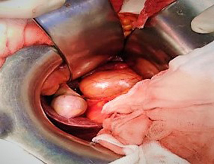
The patient was operated on by a subcostal approach. The posterior peritoneum was incised, and the duodenum was mobilized. A well‐limited retroperitoneal tumor of 6 cm was identified [Figure 2].
FIGURE 2.

Intraoperative findings
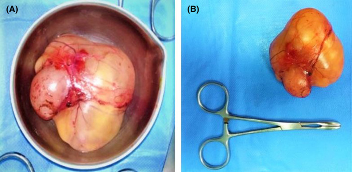
The tumor was easily cleavable from the surrounding structures and was completely excised [Figure 3].
FIGURE 3.

The operative specimen. (A) Posterior view, (B) Anterior view
The post‐operative course was uneventful. Pathological report of the operative specimen concluded in myxoid Schwannoma without histological signs of malignancies. Strong diffuse expression of the S100 protein was found on immunohistochemistry confirming the diagnosis. [Figure 4].
FIGURE 4.

(A) histology slide of this schwannoma showing Verocay body. (B) immunohistochemistry examination showing a diffuse expression of the S100 protein confirming the diagnosis of Schwannoma
The patient did not present any recurrence of pain or symptoms at 12 months of follow‐up.
3. DISCUSSION
Schwannomas are usually benign and solitary tumors. 1 Malignant transformation is exceptional except in cases of type 2 neurofibromatosis where it reaches 60% of cases. 3 Retroperitoneal involvement represents just 0.7% of benign schwannomas and 1.7% of malignant schwannomas. 4 , 5 , 6 They occur in patients of all ages but are most commonly found in women in the 2nd to the 5the decade. 1
The clinical presentation is unspecific. Masses less than 5 cm are often discovered incidentally. Sometimes, they can manifest themselves by low back pain, or by digestive and urinary disorders related to compression of the neighboring organs. 1 , 2 When the tumor is large, schwannoma may give rise to a palpable mass. CT and MRI are the two examinations of choice for exploring the retroperitoneum. The abdominal scan allows determining the location and the density of the tumor as well as its relationship with the neighboring organs. It generally finds a regular cystic mass. The homogeneous and well‐delimited character speaks in favor of a benign lesion. Abdominal MRI remains the gold standard because of its higher diagnostic predictability compared with ultrasound or CT. 2 , 3 It shows hypointense images of the tumor in T1 and hypersignal in T2. It allows a better exploration of a possible extension towards the intervertebral foramen. However, this extension remains exceptional. 7
Percutaneous biopsy is not recommended because of the risk of neoplastic dissemination in the event of a malignant tumor and the hemorrhagic risk. 4 , 8 Retroperitoneal schwannoma can have several differential diagnoses such as pheochromocytoma, paraganglioma and even liposarcoma, fibrosarcoma, and ganglioneuroma. 3
Complete surgical resection represents the cornerstone in therapeutic management. Both open and laparoscopic approaches seem to have good outcomes. Radical excision of retroperitoneal schwannoma is a challenging procedure given the bleeding risk according to the hyper‐vascularized nature of the tumor and the potential adhesion to retroperitoneal vessels. 2 , 8
Definitive diagnosis is based on histopathological examination of the surgical specimen. A strong diffuse expression of S100 protein on immunohistochemical is a distinct feature of schwannoma.
The prognosis of benign retroperitoneal schwannomas is good with a low recurrence rate after complete resection. Nevertheless, in the case of incomplete excision the incidence of tumor recurrence is 5% to 10%. 2 Therefore, long‐term follow‐up is required for these patients.
4. CONCLUSION
Retroperitoneal Schwannoma is a rare tumor whose diagnosis is often late due to unspecific symptoms. Complete surgical resection is the best treatment. The diagnosis is based on the pathological and immunohistochemical examination of the surgical specimen. The prognosis of benign retroperitoneal schwannomas is good. Long‐term follow‐up is required, due to the potential risk of local recurrence or malignant transformation especially with incomplete excision.
CONFLICT OF INTEREST
None.
AUTHOR CONTRIBUTION
Mehdi Debaibi conceived the idea for the document and contributed to the writing of the manuscript. Rime Essid contributed to the writing and editing of the manuscript. Asma Sghair and Rami Zourai reviewed and edited the manuscript. Moez Sahnoun reviewed and contributed to the to the literature review. Amen Dhaoui reviewed and supervised the manuscript. Adnen Chouchen edited, supervised, and approved the final manuscript. All authors read and approved the final manuscript.
ETHICAL APPROVAL
Personal data have been respected. Published with the consent of the patient.
CONSENT
Written informed consent was obtained from the patient to publish this report in accordance with the journal's patient consent policy.
ACKNOWLEDGMENTS
Published with the consent of the patient.
Debaibi M, Essid R, Sghair A, et al. Retroperitoneal schwannoma: Uncommon location of a benign tumor. Clin Case Rep. 2022;10:e05726. doi: 10.1002/ccr3.5726
Funding information
None
DATA AVAILABILITY STATEMENT
Personal data of the patient were respected. No data is available for this submission.
REFERENCES
- 1. Gao C, Zhu F‐C, Ma B, et al. A rare case of giant retroperitoneal neurilemmoma. J Int Med Res. 2020;48(9). [DOI] [PMC free article] [PubMed] [Google Scholar]
- 2. Radojkovic M, Mihailovic D, Stojanovic M, Radojković D. Large retroperitoneal schwannoma: a rare cause of chronic back pain. J Int Med Res. 2018;46(8):3404. [DOI] [PMC free article] [PubMed] [Google Scholar]
- 3. Harhar M, Ramdani A, Bouhout T, Serji B, El Harroudi T. Retroperitoneal schwannoma: two rare case reports. Cureus. 2021;13(2):e13456. [DOI] [PMC free article] [PubMed] [Google Scholar]
- 4. Beddouche A, Fahsi O, Kallat A, et al. Schwannome benin primitif retrovesical: une tumeur très rare à propos d'un cas. Pan Afr Med J. 2016;23:79. [DOI] [PMC free article] [PubMed] [Google Scholar]
- 5. Ben Moualli S, Hajri M, Ben Amna M, et al. Retroperitoneal schwannoma. Case report. Ann Urol. 2001;35(5):270‑272. [DOI] [PubMed] [Google Scholar]
- 6. Garcia G, Anfossi E, Prost J, Ragni E, Richaud C, Rossi D. Benign retroperitoneal schwannoma: report of three cases. Progres En Urol J Assoc Francaise Urol Soc Francaise Urol. 2002;12(3):450‑453. [PubMed] [Google Scholar]
- 7. Pollo C, Richard A, DE Preux J. Résection d'un schwannome rétropéritonéal en sablier par abord combiné. Neurochirurgie. 2004;50(1):53‐56. [DOI] [PubMed] [Google Scholar]
- 8. Skaini MSA, Haroon H, Sardar A, et al. Giant retroperitoneal ancient schwannoma: is preoperative biopsy always mandatory? Int J Surg Case Rep. 2015;6:233. [DOI] [PMC free article] [PubMed] [Google Scholar]
Associated Data
This section collects any data citations, data availability statements, or supplementary materials included in this article.
Data Availability Statement
Personal data of the patient were respected. No data is available for this submission.


