Abstract
The Rhesus D antigen (RhD) has been associated with susceptibility to several viral infections. Reports suggest that RhD-negative individuals are better protected against infectious diseases and have overall better health. However, potential mechanisms contributing to these associations have not yet been defined. Here, we used transcriptomic and genomic data from the Milieu Interieur cohort of 1000 healthy individuals to explore the effect of Rhesus status on the immune response. We used the rs590787 SNP in the RHD gene to classify the 1000 donors as either RhD-positive or -negative. Whole blood was stimulated with LPS, polyIC, and the live influenza A virus and the NanoString human immunology panel of 560 genes used to assess donor immune response and to investigate sex-specific effects. Using regression analysis, we observed no significant differences in responses to polyIC or LPS between RhD-positive and -negative individuals. However, upon sex-specific analysis, we observed over 40 differentially expressed genes (DEGs) between RhD-positive (n = 384) and RhD-negative males (n = 75) after influenza virus stimulation. Interestingly these Rhesus-associated differences were not seen in females. Further investigation, using gene set enrichment analysis, revealed enhanced IFNγ signalling in RhD-negative males. This amplified IFNγ signalling axis may explain the increased viral resistance previously described in RhD-negative individuals.
Subject terms: Innate immunity, Infection
Introduction
The Rhesus D blood group antigen (RhD) system is an important clinical factor in transfusion and obstetric medicine. RhD status is determined based on the presence or absence of the Rhesus antigen, a transmembrane protein found on the surface of red blood cells [1]. The function of the RhD antigen is largely unknown, although it may play a role in maintaining erythrocyte membrane integrity or transport of ammonium and carbon dioxide [2–4]. The Rhesus protein is highly immunogenic and resulting antibodies can induce severe adverse reactions in RhD-negative individuals should they encounter the D antigen following an unmatched blood transfusion. RhD-negative women can also be sensitised during pregnancy with an RhD-positive foetus or during delivery, often leading to haemolytic disease of the new born in subsequent pregnancies with RhD-positive foetuses [5].
Although less well studied, RhD status is also known to influence several other health outcomes [6]. An agnostic analysis of 1217 disease states found an association between RhD status and hypertension during pregnancy [6]. Several studies have demonstrated that RhD-positive and -negative subjects differ in resistance to the pathological effects of aging, fatigue, and smoking [7]. RhD-negative individuals also appear to be protected against certain infections, including latent toxoplasmosis [7]. RhD status also appears to affect susceptibility to SARS-CoV-2; in a study of 14,112 donors, RhD-negative individuals had a lower risk of initial infection, intubation and death- suggesting a protective role for RhD negativity [8]. RhD negativity varies substantially across different populations and may potentially confer an as yet unknown fitness advantage [9]. A major challenge in determining potential effects of RhD status on antiviral immunity is the lack of large, relevant human cohort studies with the power to detect potentially subtle immune differences.
The Milieu Interieur (MI) study is comprised of a cohort of 1000 healthy individuals, stratified by age (20–69, n = 200 per decade) and sex (500 males and 500 females). The overall aim of the MI study is greater understanding of the determinants of variation in the immune responsiveness of healthy adult humans [10]. Previous work from MI and others has shown that sex and cytomegalovirus (CMV) serostatus are key drivers of variation in the human immune response [11, 12]. Substantial genomic and transcriptomic data has been generated on the cohort to date and analysed using agnostic approaches [11, 13].
Here, we used genotype data on the rs590787 single nucleotide polymorphism (SNP) in the RHD gene to classify individuals from the MI study as either RhD-positive or -negative, an approach that has previously been used to determine RhD status in several studies [14–16]. Individuals who are RhD-negative are homozygous for the recessive alleles (CC), while RhD-positive individuals are heterozygous or homonymous dominant (CT, TT). Genomic data were integrated with transcriptomic data from whole blood stimulated with bacterial and viral ligands, LPS and polyIC, and the live influenza A virus, to investigate whether RhD status was associated with induced immune responses. We were particularly interested to compare RhD-positive and -negative males and females, as sex is emerging as a key factor in determining outcomes to viral infection, especially SARS-CoV-2 and influenza [17].
Materials and methods
Study population
1000 healthy individuals were included in this study, equally distributed across sex (500 males and 500 females) and age (20–69, with 200 individuals per decade). Only individuals of western European decent (i.e. French citizens for whom the last three generations of ancestors were from mainland France) were included [10]. The MI study was approved by the Comité de Protection des Personnes—Ouest 6 (Committee for the protection of persons) on June 13, 2012 and by French Agence nationale de sécurité du médicament (ANSM) on June 22, 2012. The study was sponsored by Institut Pasteur (Pasteur ID-RCB Number: 2012-A00238-35) and was conducted as a single centre interventional study without an investigational product. The original protocol was registered under ClinicalTrials.gov (study# NCT01699893). The samples and data used in this study were formally established as the MI biocollection (NCT03905993), with approvals by the Comité de Protection des Personnes—Sud Méditerranée and the Commission nationale de l’informatique et des libertés on April 11, 2018. The study was designed and conducted in accordance with the Declaration of Helsinki and good clinical practice, with all subjects giving informed consent.
SNP genotyping and determining RhD status
All participants were genotyped for the rs590787 SNP, through a genome-wide SNP array, using HumanOmniExpress and HumanExomeBeadChips [11]. Rhesus status (RhD-positive or -negative) was determined based upon the presence or absence of the rs590787 polymorphism in the gene sequence of each individual [14]. In total, 921 donors were retained for downstream analysis based on completeness of datasets available. For males, n = 384 RhD-positive and n = 75 RhD-negative. For females, n = 385 RhD-positive and n = 77 RhD-negative.
Whole blood stimulation
Whole blood was taken from each participant and 1 ml aliquoted into TruCulture tubes (Myriad Rules Based Medicine, USA) preloaded with the chosen stimuli: polyIC (20 μg/ml), LPS (10 ng/ml) and influenza A virus (H1N1 PR8; 100 HAU) as previously described [18] (Fig. 1). Tubes were incubated on a bench top heat block for 22 hours. Supernatants were harvested for proteomics. The cell pellet was resuspended in Trizol for later RNA extraction and transcriptomics.
Fig. 1. Schematic overview of the study.
Genomic variability was assessed through a genome-wide SNP array. Rhesus status was determined based on the rs590787 SNP. Whole blood samples were stimulated with a panel of ligands (polyIC and LPS) and a live influenza A virus. The expression of 560 immune-related genes was quantified using NanoString transcriptomics. R and gene set enrichment analysis (GSEA) were used to compare the response between RhD-positive and -negative individuals.
NanoString transcriptomics
Total RNA was extracted from the TruCulture cell pellets for the 1000 MI donors using NucleoSpin 96 miRNA kit (Macherey-Nagel). RNA concentrations were measured using Quantifluor RNA system kit (Promega) and RNA integrity numbers were determined using the Agilent RNA 6000 Nano kit (Agilent Technologies). The expression levels of 560 immune-related genes were quantified before and after stimulation using NanoString hybridisation arrays, producing highly reproducible transcriptomic data. Gene expression data were normalised as previously described [13].
CMV serology
CMV serostatus was assessed as previously described using a clinical-grade IgG assay according to the manufacturer’s instructions [19]. Anti-CMV IgG were measured using a chemiluminescence-based kit (CMV IgG kit, Beckman Coulter) on the Unicel Dxl 800 Access platform.
IFNγ protein quantification
IFNγ protein levels in the supernatants of influenza A virus stimulated whole blood were quantified using the Luminex xMAP system as previously described [18]. Samples were processed according to CLIA guidelines.
Statistical analysis
Transcriptomic data were analysed using R Studio (https://www.rstudio.com/.) In order to account for potential sex differences the cohort was subdivided into males and females. Normality of the data was assessed using Shapiro-Wilk tests and variance was assessed using Levene’s test. Regression analysis including CMV serostatus and age as covariates was used to assess differences between RhD-positive and -negative individuals. We included a quadratic term to account for the non-linear effects of age on influenza A virus stimulation [lm(counts ~ Rhesus + CMV + AGE + I(AGE^2), data = .)]. p values were adjusted for false discovery using the FDR correction to generate a q value. A q value of q < 0.1 was taken as significant. Additionally, models were run on log2 fold change data between unstimulated and stimulated conditions. Scatterplots were created using GraphPad Prism. Volcano plots were created in R studio using the EnhancedVolcano package. Heatmaps were generated in R using the package pheatmap.
Pathway analysis
Pathway analysis was carried out on genes with q values (<0.1), using gene set enrichment analysis (GSEA), found at https://www.gsea-msigdb.org/gsea/index.jsp. The genes were compared to panels of genes using the Hallmarks gene set.
Results
Rhesus status distribution is similar between males and females
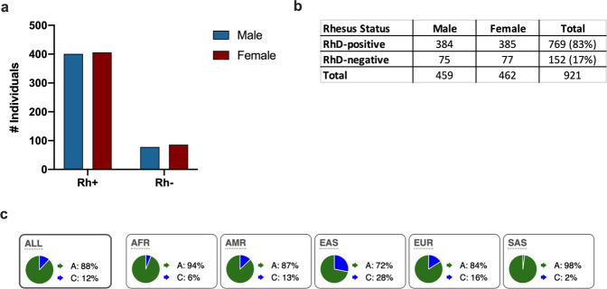
The presence or absence of rs590787, a SNP in the RhD gene, was used to determine the RhD status of donors in our cohort (Fig. 1) [14]. There was no difference in the distribution of RhD-positive or -negative individuals between males and females in our cohort (Fig. 2a). Eighty-three percent of donors in the MI cohort were RhD-positive (Fig. 2b). The genotype frequencies of the Rhesus SNP rs590787 were compared to those from the 1000 genomes cohort [20]. The genotype frequencies in the MI cohort were similar to those observed for Europeans in the 1000 Genome Study (Fig. 2c).
Fig. 2. Rhesus phenotype distribution of all individuals in the Milieu Interieur cohort.
RhD status was determined based on rs590787, a SNP in the RHD gene, using the Human Exome Bead Chip. a The frequencies of RhD-positive and -negative individuals was similar between males and females. b, c The genotype frequencies for the European cohort from the 1000 Genome Project were similar to those recorded for the MI cohort. The numbers in brackets are percentages.
Rhesus factor does not affect immune gene expression at baseline or in response to stimulation with pattern recognition receptors (PRR) ligands in whole blood
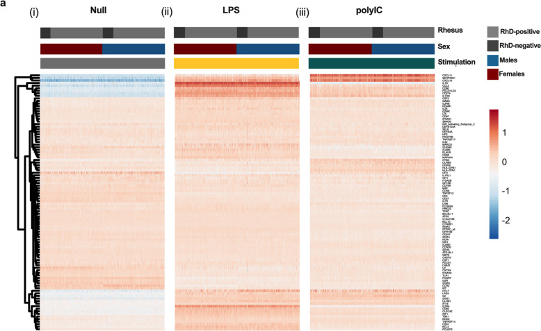
Whole blood was incubated for 22 hours and expression of 560 immune genes quantified. Gene expression in the unstimulated condition was similar between all RhD-positive and -negative individuals. Given the important sex-specific differences emerging regarding pathogen susceptibility, we also investigated sex-specific effects. The cohort was stratified by sex and assessed for differential gene expression between RhD-positive and -negative males and females. Baseline immune gene expression was similar between RhD-positive and -negative males and females (Fig. 3(i)).
Fig. 3. Rhesus antigenicity does not affect immune gene expression at baseline or in response to stimulation with PRR ligands in whole blood.
Immune gene expression in unstimulated and LPS and polyIC stimulated whole blood was assessed using NanoString transcriptomics. Comparisons between RhD-positive and -negative individuals in all donors, females only and males only showed no significant differences (q > 0.1, regression analysis with FDR correction) for (i) Null, (ii) LPS or (iii) polyIC. Shown in the heatmap are 100 representative genes chosen at random.
To assess specific TLR induced immunity, namely TLR3 and TLR4, we stimulated whole blood with the widely used ligands, polyIC and LPS. PolyIC is a dsRNA mimic that acts via TLR3 to upregulate a type I interferon response [21]. LPS is a bacterial ligand that can activate both the type I interferon response, as well as other pro-inflammatory pathways leading to upregulation of key mediators including IL6, TNFα and COX2 [22]. Using linear regression, we found the response to both polyIC and LPS to be similar in RhD-negative and -positive individuals whether male or female (Fig. 3(ii, iii)). Data are presented as a heatmap with 100 randomly selected genes shown. Differences in cell counts can have a major impact on the whole blood transcriptional response [13]. Therefore, we compared major circulating immune populations between RhD-positive and -negative individuals. Following FDR correction, we observed no significant differences in any cell population examined (Supplementary Table. 1).
Increased IFNγ mediated responses in RhD-negative males only
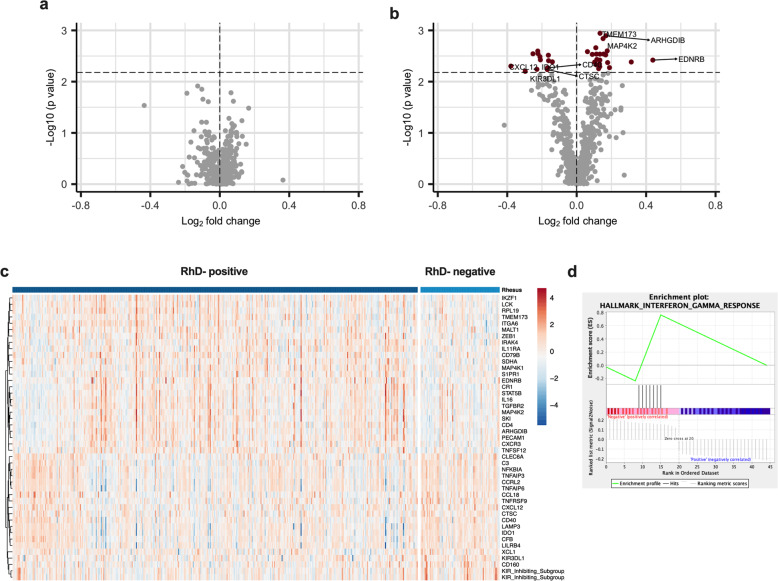
Infection with a live virus results in the activation of several PRRs and downstream pathways that upregulate pro-inflammatory cytokines and antiviral mechanisms, and help clear infection [23]. To determine whether the Rhesus factor has an impact on the antiviral response to a live virus, we stimulated whole blood with the live influenza A virus. No significant difference was observed between RhD-positive and -negative individuals when comparing the entire cohort, or when looking at females only (Fig. 4a). Interestingly, however, when examining the whole blood response to influenza A virus between RhD-positive and -negative males only, we observed differential expression of 45 immune-related genes (Fig. 4b, c; Supplementary List 1; q < 0.1, regression analysis with FDR correction). These differences were also seen when comparing log2 fold change data calculated on the unstimulated to influenza A virus conditions using the same regression model (Supplementary List 2). To further interrogate differences in the immune response to influenza A between RhD-positive and -negative individuals we used GSEA. Using this approach we found the IFNγ pathway to be significantly enriched in RhD-negative males (Fig. 4d).
Fig. 4. Following influenza A virus stimulation, 45 genes were differentially expressed between RhD-positive and -negative males.
Whole blood was stimulated with influenza A virus and gene expression assessed using NanoString. a Volcano plot showing no significant differences between RhD-positive and -negative females (q > 0.1, regression analysis with FDR adjustment). b Volcano plot of the differentially expressed genes between RhD-positive and -negative donors in the male group (q < 0.1, regression analysis with FDR adjustment). c Heatmap of differentially expressed genes between RhD-positive and negative males. d Enrichment plot indicating upregulation of the IFNγ pathway in the RhD-negative males (p < 0.01).
Discussion
In a French cohort of 1000 well-characterised healthy individuals, we investigated the impact of RhD on the innate immune response. Following stimulation with live influenza A virus, we found differential expression of 45 immune genes between RhD-positive and -negative males. In contrast, RhD-negative and -positive women had similar responses to influenza A virus. Further analysis of the differentially expressed genes revealed enrichment for IFNγ signalling in RhD-negative males. The increased IFNγ signalling found here in RhD-negative males may begin to explain their reduced susceptibility to infection.
Several association studies have found a relationship between RhD negativity and enhanced resistance to viral infection [6–8, 24]. However, systemic analysis of immune activity by RhD status had never been carried out. RhD-negative individuals have reduced risk of initial infection with SARS-CoV-2, as well as decreased risk of both intubation and death [8]. The increased IFNγ signalling that we find in RhD-negative donors could contribute to reduced susceptibility to SARS-CoV-2 infection and pathology.
This study analysed transcriptomic data from studies using well-described PRR ligands, LPS, and polyIC, which are mimics relevant to bacterial and viral infection. We also chose to include a more complex live stimulus, the influenza A virus. The responses to the bacterial and viral mimics in RhD-positive and -negative individuals were similar across all groups. Studies on other blood types including the Lewis (Le) group found no differences in expression in responses to LPS between Le+ and Le− individuals in either males or females [25]. Another study found associations between the inheritance of polymorphisms in genes that encode and regulate the expression of the Le blood antigens and protection against infections with Helicobacter pylori [26]. This suggests that analysis focused exclusively on specific PRR ligands may not be sufficient to identify differential effects of blood groups on the innate immune response to infectious agents. Studies using whole organisms may be more informative.
Here, using stimulation with the influenza A (but not the surrogate viral ligand polyIC) we observed significant differential expression of 45 immune genes between RhD-positive and -negative males. Influenza A viral pathogenesis has already been described as different between sexes, with males at risk of developing more severe disease compared with females of a similar age [27]. Recent studies have shown that females mount a more robust immune response and have increased production of several key inflammatory proteins known to be important in control of viral infection [28]. This more potent innate immune response in females could mask potential differences in RhD-positive and -negative females in our system.
The distribution of blood groups, including RhD, varies globally- this may explain, at least in part, the regional differences in disease occurrence. RhD-negativity is particularly enriched in East Asian populations (28% RhD-negative) compared with European or African populations (16% and 6%, respectively) [6]. This may reflect the different historic disease burdens on these populations. As the cohort used in our study includes only donors of Western European descent, more cohorts should be analysed to include individuals of different ethnicities to further probe the differences in the immune response between RhD-positive and -negative individuals.
While non-communicable disease states have been studied in the context of blood antigens, viral illness is often overlooked as it is typically transient and often underreported [29]. Further association studies exploring the relationships between blood types and viral infection are warranted to fully understand the contribution of blood type to risk of severe viral disease. Findings from these studies may inform clinical management of patients based on RhD status. Recently, Alsten et al. looked at differences in 96 circulating inflammatory proteins between several blood groups. While RhD was not among those examined, differences were found in the circulating inflammatory profiles of ABO and Duffy blood types [30].
In this study we found evidence of an enhanced IFNγ mediated immune response in RhD-negative males. IFNγ has been shown to be important in the control of several viral infections, including Ebola and SARS-CoV-2 [31, 32]. The enhanced signature observed in RhD-negative individuals in our cohort may contribute to the increased resistance to infection described in these individuals. The enhanced gene signature was not reflected at the protein level. However, this discrepancy could be explained by the relatively low induction of IFNγ by influenza in our system, or by differences in the kinetics of IFNγ protein and its downstream transcripts. Increased responsiveness to IFNγ by RhD-negative individuals could also explain the enhanced IFNγ response in the absence of an increase in IFNγ protein.
Although modest, the differences between RhD-positive and -negative males were widespread and may contribute to the increased viral resistance reported elsewhere. Several genes related to natural killer cells, including CD160 and KIRs, were among those that are upregulated in RhD-negative individuals. NK cells, potent IFNγ producers and cytotoxic lymphocytes, are key players in control of viral infection. While there was no difference in NK cell counts in our cohort, the differences observed in marker expression could be indicative of an increased activation state and IFNγ production [33]. These findings are the first to shed light on the potential immune differences between RhD-positive and -negative individuals and support the use of systemic investigations to understand the impact of blood types on the whole blood immune response.
Supplementary information
Acknowledgements
This work was funded by awards to COF through a Science Foundation Ireland Investigator Award (12/IA/1667) and under the Science Foundation Ireland Phase 2 COVID-19 Rapid Response Call (20/COV/8487). This work benefited from support of the French government’s Invest in the Future programme. This programme is managed by the Agence Nationale de la Recherche, reference ANR-10-LABX-69-01. We thank the Cytometry and Biomarkers UTechS for generation of Nanostring gene expression datasets.
Author contributions
JAS and MS analysed the data and wrote the manuscript. CP and BC generated the data and revised the manuscript. NMB, DD and COF supervised the study and edited the manuscript. All authors approved the final version of the manuscript.
Funding
Open Access funding provided by the IReL Consortium.
Competing interests
The authors declare no competing interests.
Footnotes
A list of authors and their affiliations appears at the end of the paper.
Publisher’s note Springer Nature remains neutral with regard to jurisdictional claims in published maps and institutional affiliations.
These authors contributed equally: Nollaig M. Bourke, Darragh Duffy, Cliona O’Farrelly.
Contributor Information
Jamie A. Sugrue, Email: sugruej@tcd.ie
Milieu Interieur Consortium:
Laurent Abel, Andres Alcover, Hugues Aschard, Philippe Bousso, Nollaig Bourke, Petter Brodin, Pierre Bruhns, Nadine Cerf-Bensussan, Ana Cumano, Christophe D’Enfert, Ludovic Deriano, Marie-Agnès Dillies, James Di Santo, Françoise Dromer, Gérard Eberl, Jost Enninga, Jacques Fellay, Ivo Gomperts-Boneca, Milena Hasan, Gunilla Karlsson Hedestam, Serge Hercberg, Molly A. Ingersoll, Olivier Lantz, Rose Anne Kenny, Mickaël Ménager, Hugo Mouquet, Cliona O’Farrelly, Etienne Patin, Sandra Pellegrini, Antonio Rausell, Frédéric Rieux-Laucat, Lars Rogge, Magnus Fontes, Anavaj Sakuntabhai, Olivier Schwartz, Benno Schwikowski, Spencer Shorte, Frédéric Tangy, Antoine Toubert, Mathilde Touvier, Marie-Noëlle Ungeheuer, Christophe Zimmer, Matthew L. Albert, Darragh Duffy, and Lluis Quintana-Murci
Supplementary information
The online version contains supplementary material available at 10.1038/s41435-022-00169-5.
References
- 1.Avent ND, Reid ME. The Rh blood group system: a review. Blood. 2000;95:375–87. doi: 10.1182/blood.V95.2.375. [DOI] [PubMed] [Google Scholar]
- 2.Westhoff CM. The Rh blood group system in review: a new face for the next decade. Transfusion. 2004;44:1663–73. doi: 10.1111/j.0041-1132.2004.04237.x. [DOI] [PubMed] [Google Scholar]
- 3.Marini AM, Matassi G, Raynal V, André B, Cartron JP, Chérif-Zahar B. The human Rhesus-associated RhAG protein and a kidney homologue promote ammonium transport in yeast. Nat Genet. 2000;26:341–4. doi: 10.1038/81656. [DOI] [PubMed] [Google Scholar]
- 4.Endeward V, Cartron J-P, Ripoche P, Gros G. RhAG protein of the Rhesus complex is a CO2 channel in the human red cell membrane. FASEB J. 2008;22:64–73. doi: 10.1096/fj.07-9097com. [DOI] [PubMed] [Google Scholar]
- 5.Pegoraro V, Urbinati D, Visser GHA, Di Renzo GC, Zipursky A, Stotler BA, et al. Hemolytic disease of the fetus and newborn due to Rh(D) incompatibility: a preventable disease that still produces significant morbidity and mortality in children. PLoS ONE. 2020;15:1–11.. doi: 10.1371/journal.pone.0235807. [DOI] [PMC free article] [PubMed] [Google Scholar]
- 6.Dahlén T, Clements M, Zhao J, Olsson ML, Edgren G. An agnostic study of associations between ABO and RhD blood group and phenome-wide disease risk. Elife. 2021;10:e65658. doi: 10.7554/eLife.65658. [DOI] [PMC free article] [PubMed] [Google Scholar]
- 7.Flegr J, Hoffmann R, Dammann M. Worse health status and higher incidence of health disorders in Rhesus negative subjects. PLoS ONE. 2015;10. 10.1371/journal.pone.0141362. [DOI] [PMC free article] [PubMed]
- 8.Zietz M, Zucker J, Tatonetti NP. Associations between blood type and COVID-19 infection, intubation, and death. Nat Commun. 2020;11. 10.1038/s41467-020-19623-x. [DOI] [PMC free article] [PubMed]
- 9.Perry GH, Xue Y, Smith RS, Meyer WK, Calışkan M, Yanez-Cuna O, et al. Evolutionary genetics of the human Rh blood group system. Hum Genet. 2012;131:1205–16. doi: 10.1007/s00439-012-1147-5. [DOI] [PMC free article] [PubMed] [Google Scholar]
- 10.Thomas S, Rouilly V, Patin E, Alanio C, Dubois A, Delval C, et al. The Milieu Intérieur study—an integrative approach for study of human immunological variance. Clin Immunol. 2015;157:277–93. doi: 10.1016/j.clim.2014.12.004. [DOI] [PubMed] [Google Scholar]
- 11.Patin E, Hasan M, Bergstedt J, Rouilly V, Libri V, Urrutia A, et al. Natural variation in the parameters of innate immune cells is preferentially driven by genetic factors. Nat Immunol. 2018;19:645. doi: 10.1038/s41590-018-0105-3. [DOI] [PubMed] [Google Scholar]
- 12.Picarda G, Benedict CA. Cytomegalovirus: shape-shifting the immune system. J Immunol. 2018;200:3881–9. doi: 10.4049/jimmunol.1800171. [DOI] [PMC free article] [PubMed] [Google Scholar]
- 13.Piasecka B, Duffy D, Urrutia A, Quach H, Patin E, Posseme C, et al. Distinctive roles of age, sex, and genetics in shaping transcriptional variation of human immune responses to microbial challenges. Proc Natl Acad Sci USA. 2018;115:E488–E497. doi: 10.1073/pnas.1714765115. [DOI] [PMC free article] [PubMed] [Google Scholar]
- 14.Shi J, Luo Y. Effects of RHD gene polymorphisms on distinguishing weak D or DEL from RhD− in blood donation in a Chinese population. Mol Genet\Genom Med. 2019;7:e00681. doi: 10.1002/mgg3.681. [DOI] [PMC free article] [PubMed] [Google Scholar]
- 15.Ali AA, Aalto M, Jonasson J, Osman A. Genome-wide analyses disclose the distinctive HLA architecture and the pharmacogenetic landscape of the Somali population. Sci Rep. 2020;10:5652. doi: 10.1038/s41598-020-62645-0. [DOI] [PMC free article] [PubMed] [Google Scholar]
- 16.Park YM, Cheong HS, Lee J-K. Genome-wide detection of allelic gene expression in hepatocellular carcinoma cells using a human exome SNP chip. Gene. 2014;551:236–42. doi: 10.1016/j.gene.2014.09.001. [DOI] [PubMed] [Google Scholar]
- 17.Klein SL, Dhakal S, Ursin RL, Deshpande S, Sandberg K, Mauvais-Jarvis F. Biological sex impacts COVID-19 outcomes. PLoS Pathog. 2020;16:e1008570. doi: 10.1371/journal.ppat.1008570. [DOI] [PMC free article] [PubMed] [Google Scholar]
- 18.Duffy D, Rouilly V, Libri V, Hasan M, Beitz B, David M, et al. Functional analysis via standardized whole-blood stimulation systems defines the boundaries of a healthy immune response to complex stimuli. Immunity. 2014;40:436–50. doi: 10.1016/j.immuni.2014.03.002. [DOI] [PubMed] [Google Scholar]
- 19.Scepanovic P, Alanio C, Hammer C, Hodel F, Bergstedt J, Patin E, et al. Human genetic variants and age are the strongest predictors of humoral immune responses to common pathogens and vaccines. Genome Med. 2018;10:59. doi: 10.1186/s13073-018-0568-8. [DOI] [PMC free article] [PubMed] [Google Scholar]
- 20.Auton A, Abecasis GR, Altshuler DM, Durbin RM, Abecasis GR, Bentley DR, et al. A global reference for human genetic variation. Nature. 2015;526:68–74. doi: 10.1038/nature15393. [DOI] [PMC free article] [PubMed] [Google Scholar]
- 21.Marshall-Clarke S, Downes JE, Haga IR, Bowie AG, Borrow P, Pennock JL, et al. Polyinosinic acid is a ligand for toll-like receptor 3. J Biol Chem. 2007;282:24759–66. doi: 10.1074/jbc.M700188200. [DOI] [PubMed] [Google Scholar]
- 22.Park BS, Lee J-O. Recognition of lipopolysaccharide pattern by TLR4 complexes. Exp Mol Med. 2013;45:e66. doi: 10.1038/emm.2013.97. [DOI] [PMC free article] [PubMed] [Google Scholar]
- 23.Chen X, Liu S, Goraya MU, Maarouf M, Huang S, Chen J-L. Host immune response to influenza A virus Infection. Front Immunol. 2018;9. 10.3389/fimmu.2018.00320. [DOI] [PMC free article] [PubMed]
- 24.Flegr J. REVIEW Influence of latent Toxoplasma infection on human personality, physiology and morphology: pros and cons of the Toxoplasma-human model in studying the manipulation hypothesis. J Exp Biol. 2013;216:127–33. doi: 10.1242/jeb.073635. [DOI] [PubMed] [Google Scholar]
- 25.Heneghan MA, McCarthy CF, Moran AP. Relationship of blood group determinants on Helicobacter pylori lipopolysaccharide with host lewis phenotype and inflammatory response. Infect Immun. 2000;68:937–41. doi: 10.1128/IAI.68.2.937-941.2000. [DOI] [PMC free article] [PubMed] [Google Scholar]
- 26.Björkholm B, Lundin A, Sillén A, Guillemin K, Salama N, Rubio C, et al. Comparison of genetic divergence and fitness between two subclones of Helicobacter pylori. Infect Immun. 2001;69:7832–8. doi: 10.1128/IAI.69.12.7832-7838.2001. [DOI] [PMC free article] [PubMed] [Google Scholar]
- 27.Klein SL, Hodgson A, Robinson DP. Mechanisms of sex disparities in influenza pathogenesis. J Leukoc Biol. 2012;92:67–73. doi: 10.1189/jlb.0811427. [DOI] [PMC free article] [PubMed] [Google Scholar]
- 28.Takahashi T, Ellingson MK, Wong P, Israelow B, Lucas C, Klein J, et al. Sex differences in immune responses that underlie COVID-19 disease outcomes. Nature. 2020;588:315–20. doi: 10.1038/s41586-020-2700-3. [DOI] [PMC free article] [PubMed] [Google Scholar]
- 29.Roberts H, Boktor SW, Waller K, Daar ZS, Boscarino JA, Dubin PH, et al. Underreporting of Hepatitis B and C virus infections - Pennsylvania, 2001-2015. PLoS ONE. 2019;14:e0217455. doi: 10.1371/journal.pone.0217455. [DOI] [PMC free article] [PubMed] [Google Scholar]
- 30.Van Alsten SC, Aversa JG, Santo L, Camargo MC, Kemp T, Liu J, et al. Association between ABO and Duffy blood types and circulating chemokines and cytokines. Genes Immun. 2021;22:161–71. doi: 10.1038/s41435-021-00137-5. [DOI] [PMC free article] [PubMed] [Google Scholar]
- 31.Rhein BA, Powers LS, Rogers K, Anantpadma M, Singh BK, Sakurai Y, et al. Interferon-γ Inhibits Ebola Virus Infection. PLOS Pathog. 2015;11:e1005263. doi: 10.1371/journal.ppat.1005263. [DOI] [PMC free article] [PubMed] [Google Scholar]
- 32.Ruetsch C, Brglez V, Crémoni M, Zorzi K, Fernandez C, Boyer-Suavet S, et al. Functional exhaustion of type I and II interferons production in severe COVID-19 patients. Front Med. 2021;7:1106. doi: 10.3389/fmed.2020.603961. [DOI] [PMC free article] [PubMed] [Google Scholar]
- 33.Vivier E, Tomasello E, Baratin M, Walzer T, Ugolini S. Functions of natural killer cells. Nat Immunol. 2008;9:503–10. doi: 10.1038/ni1582. [DOI] [PubMed] [Google Scholar]
Associated Data
This section collects any data citations, data availability statements, or supplementary materials included in this article.


