Abstract
Background
Few studies with large sample sizes are available regarding patients with Wellens’ syndrome. Therefore, we sought to assess the current incidence, risk factors, clinical presentation and long-term outcomes of this population.
Methods
Among a total of 3528 patients with ACS who underwent angioplasty from 2017 to 2019 in our centre, 2127 NSTE-ACS patients with culprit LAD vessels were enrolled in this study. According to electrocardiographic criteria, the patients were divided into a Wellens’ group (n = 200) and non-Wellens’ group (n = 1927). The primary endpoint was cardiac death; the secondary endpoint was MACCE, a composite of all-cause death, cardiac death, recurrent myocardial infarction, target lesion revascularization, heart failure and stroke.
Results
The incidence of Wellens’ syndrome was 5.7% (200 of 3528) of all ACS patients. Wellens’ syndrome more often manifested as NSTEMI (69% vs. 17.5%, P < 0.001). The percentages of preexisting coronary heart disease (39.6% vs. 23%) and previous PCI (19.5% vs. 9%) were significantly higher in the non-Wellens’ group than in the Wellens’ group (all P < 0.001). More importantly, the proportion of early PCI was higher in the Wellens’ group (68% vs. 59.3%, P = 0.017). At a median follow-up of 24 months, Wellens’ syndrome was not associated with an increased risk of MACCE (P = 0.05) or cardiac death (P = 0.188).
Conclusions
The presence of Wellens’ syndrome is not definitively associated with adverse prognosis in patients with NSTE-ACS. Age ≥ 65 years, diabetes, NSTEMI, eGFR < 60 ml/min and left main disease are associated with the incidence of cardiac death. Early recognition and aggressive intervention are critical, as they may help to attenuate adverse outcomes.
Keywords: Wellens’ syndrome, NSTE-ACS, MACCE, Cardiac death, Angioplasty
Introduction
Acute coronary syndrome (ACS) remains a leading cause of mortality and morbidity worldwide, need for emergency care and eventual hospitalization [1–3]. The diagnosis of ACS relies on clinical history, electrocardiographic (ECG) changes, and cardiac biomarkers; but within the spectrum of ACS, subtle presentations exist that cannot be overlooked. Wellens’ syndrome is one such example, in which a patient can present with ECG changes that are not classic for myocardial ischaemia and even with negative cardiac biomarkers. As a well-known high-risk ACS, Wellens’ syndrome, first described by de Zwaan and Wellens in 1982 [4], is the characteristic ST-T segment change in the precordial leads, indicating a critical stenosis high in the left anterior descending arterial (LAD). Identifying the syndrome carries significant diagnostic and prognostic value [5]. According to the Fourth Universal Definition of myocardial infarction [6], absence of ST-elevation in the precordial leads, the symmetrical and often deep (> 2 mm) T wave inversions in the anterior precordial leads are an early sign that may precede the elevation of the ST-segment. Thus, this syndrome has been considered an acute ST-elevation myocardial infarction (STEMI) equivalent [7].
Patients with Wellens’ syndrome have an increased risk for extensive anterior wall myocardial infarction, and early coronary revascularization is essential in the management of these cases. These individuals constitute a special cohort with their own clinical characteristics, which may affect the outcomes in this population. However, the limited literature on the syndrome consists of mostly sporadic case reports and clinical experience. There is still a paucity of recent data on patients with Wellens’ syndrome. Updated information on the incidence, risk factors, angiographic findings and prognosis of this subset of patients should be taken into consideration when taking care of these patients.
The aims of this retrospective control study were therefore to investigate the incidence and the risk factor profile in Wellens’ syndrome patients versus other ACS patients with culprit LAD vessels admitted to Beijing Friendship Hospital in China between 2017 and 2019 and to study the clinical presentation, treatment and long-term outcomes in these patients.
Study methods
Study design and participants
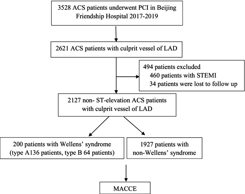
This retrospective study was based on data from the Cardiovascular Center of Beijing Friendship Hospital Data Bank. The protocol was approved by the ethics committee of Beijing Friendship Hospital (2021-P2-096-01). From January 2017 to December 2019, coronary angioplasty was performed in 3528 consecutive ACS patients at our centre, and a total of 2621 patients with culprit LAD vessels were enrolled in this study. To rule out pathological Q waves in the ECG, 460 patients with STEMI were excluded. In addition, 34 patients were lost to follow-up. Finally, a total of 2127 patients were included in the final analysis. Among the 2127 cases, 200 met the ECG criteria of Wellens’ syndrome, including 64 cases of type A, 136 cases of type B, all of which were shown angiographically to have significant LAD stenosis. Baseline characteristics, percutaneous coronary intervention (PCI) procedures, management, and long-term outcomes were collected from medical records and the data bank and then analysed. A flowchart of the patient enrolment is shown in Fig. 1.
Fig. 1.
Flow chart of patient enrollment. ACS, acute coronary syndromes; PCI, percutaneous coronary intervention; LAD, left anterior descending arterial; STEMI, ST-elevation myocardial infarction; MACCE, major adverse cardiac and cerebrovascular events
In all patients, 12-lead ECGs were routinely obtained once daily while they were in the hospital. Additional ECGs were checked during and after new attacks of chest pain. The criteria for Wellens’ syndrome are as follows [8, 9]: (a) prior history of chest pain, (b) minimal or no elevation of cardiac enzymes, (c) insignificant ST-segment elevation usually (< 1 mm), (d) no loss of precordial R waves, (e) absent precordial Q waves and (f) biphasic T waves in leads V2 and V3, or asymmetric, often deeply inverted T waves in leads V2 and V3. Wellens’ syndrome can be divided into two different types according to the precordial T wave pattern that is seen during the pain-free period. In type A, there are biphasic T waves typically observed in V2 and V3. In Type B, which is the most common form, there are deep, negative T waves in leads V2 and V3.
Data collection and clinical outcomes
The patients’ demographic information and cardiovascular risk factors, including hypertension, dyslipidaemia, diabetes mellitus, chronic kidney disease (CKD), peripheral arterial disease (PAD), heart failure (HF), smoking history, preexisting coronary heart disease (CHD) and previous PCI, were retrospectively collected from medical records. Body mass index (BMI) was calculated by dividing weight in kilograms by height in metres squared (kg/m2). Smoking history was defined as regularly smoking one or more cigarettes daily or smoking cessation within the past 12 months. Left ventricular ejection fraction (LVEF) and end diastolic dimension (EDD) were measured by transthoracic echocardiology before PCI. The angiogram data, including the number of stenosed coronary vessels, left main (LM) disease and PCI strategy, were obtained by reading the surgical report.
The primary endpoint for this analysis was cardiac death, defined as death caused by myocardial infarction, HF, or arrhythmia and unexplained sudden death [10]. The secondary endpoints were major adverse cardiovascular and cerebrovascular events (MACCEs), a composite of all-cause death, cardiac death, recurrent myocardial infarction, target lesion revascularization, HF, and stroke. Recurrent myocardial infarction was defined by the Fourth Universal Definition [6].
All MACCEs were thoroughly analysed and confirmed by two separate cardiologists simultaneously. Follow-up information after patient discharge from the hospital was obtained by clinic visits or phone interviews every 1–3 months, which were recorded in the data bank.
Statistical analysis
Continuous variables are expressed as the mean ± standard deviation or as the median with interquartile range; one-way analysis of variance was used to compare differences between continuous variables. Categorical variables are expressed as percentages and were analysed using Pearson’s χ2 test or Fisher’s exact test of variance. The cumulative incidence was estimated by the Kaplan–Meier method, and differences between groups were assessed by the log-rank test [11].
Cox regression was used to estimate relative risks among groups of patients. All factors showing significance in the univariate analysis (P < 0.05) or an indicator clinically considered to be important for the outcome were then examined by a multivariate analysis. The results are reported as adjusted hazard ratios (HRs) with associated 95% confidence intervals (CIs). All statistical tests were two-tailed, with statistical significance defined as a P value of < 0.05. All analyses were performed by using SPSS (version 25.0, Chicago, IL, USA); Kaplan–Meier survival curves were generated with GraphPad Prism software (version 5; GraphPad, Inc., San Diego, CA) [11].
Results
Baseline characteristics
Patients stratified by Wellens’ syndrome characteristics are summarized in Table 1. The study cohort included 2127 NSTE-ACS patients with culprit LAD vessels. Of these patients, 200 (9.4%) had Wellens’ syndrome. Wellens’ syndrome most often manifested as non-ST-elevation myocardial infarction (NSTEMI) (69% vs. 17.5%, P < 0.001). The percentages of preexisting coronary heart disease (39.6% vs. 23%) and previous PCI (19.5% vs. 9%) were significantly higher in the non-Wellens’ group than in the Wellens’ group (all P < 0.001). The two groups of patients had a similar prevalence of sex distribution, hypertension, diabetes mellitus and hyperlipemia. There were also no significant differences in the medical history of prior myocardial infarction, HF, CKD, PAD or stroke. Compared with non-Wellens’ patients, more Wellens’ patients had a lower BMI (25.21 ± 2.97 vs. 25.93 ± 3.45 kg/m2, P = 0.005), waist circumference (90.79 ± 9.84 vs. 92.5 ± 10.08 cm, P = 0.025) and LVEF (0.62 ± 0.09 vs. 0.65 ± 0.08, P = 0.002). However, the LDL-C and EDD were higher in the Wellens’ group.
Table 1.
Comparison of the baseline characteristics among patients
| Variables | Wellens’ (N = 200) | Non-Wellens’ (N = 1927) | P value |
|---|---|---|---|
| Age (years) | 63.3 ± 10.4 | 64.2 ± 9.7 | 0.158 |
| Male | 146 (73) | 1314 (68.2) | 0.163 |
| Days | 6 (5.8) | 6 (4.7) | < 0.001 |
| BMI (kg/m2) | 25.21 ± 2.97 | 25.93 ± 3.45 | 0.005 |
| WC (cm) | 90.79 ± 9.84 | 92.5 ± 10.08 | 0.025 |
| Smoking | 114 (57) | 1017 (55.8) | 0.255 |
| HT | 133 (66.5) | 1353 (70.2) | 0.276 |
| DM | 70 (35) | 810 (42) | 0.055 |
| Hyperlipemia | 100 (50) | 1030 (53.5) | 0.352 |
| History of CHD | 46 (23) | 763 (39.6) | < 0.001 |
| Prior MI | 15 (7.5) | 199 (10.3) | 0.206 |
| HF | 1 (0.5) | 13 (0.7) | 0.771 |
| CKD | 10 (5) | 71 (3.7) | 0.355 |
| PAD | 14 (7) | 202 (10.5) | 0.121 |
| Stroke | 39 (19.5) | 317 (16.5) | 0.271 |
| Previous PCI | 18 (9) | 376 (19.5) | < 0.001 |
| NSTEMI | 138 (69) | 338 (17.5) | < 0.001 |
| EDD (cm) | 5.19 ± 0.56 | 5.08 ± 0.52 | 0.006 |
| LVEF | 0.62 ± 0.09 | 0.65 ± 0.08 | 0.002 |
| LDL-C (mmol/L) | 2.5 ± 0.73 | 2.25 ± 0.75 | < 0.001 |
| eGFR (ml/min) | 86.69 ± 24.76 | 88.09 ± 22.2 | 0.408 |
| HBA1c (%) | 6.54 ± 1.46 | 6.7 ± 21.45 | 0.159 |
| Multivessel disease | 177 (88.5) | 1712 (88.8) | 0.884 |
| LM disease | 18 (9) | 229 (11.9) | 0.226 |
| Plain balloon angioplasty | 7 (3.5) | 279 (14.6) | < 0.001 |
| Drug-eluting stent | 193 (96.5) | 1635 (84.8) | < 0.001 |
| Stent length < 30 mm | 58 (29) | 542 (28.1) | 0.794 |
| Number of stents | 1.35 ± 0.63 | 1.14 ± 0.69 | < 0.001 |
| Early PCI (< 48 h after first presentation) | 136 (68) | 1143 (59.3) | 0.017 |
Bold values indicate that the difference is statistically significant (P < 0.05)
Data are presented as absolute numbers and percentages (for categorical variables) or mean value ± SD (for continuous variables) unless otherwise specified
BMI, body mass index; WC, waist circumference; HT, hypertension; DM, diabetes mellitus; CHD, coronary heart disease; MI, myocardial infarction; HF, heart failure; CKD, chronic kidney disease; PAD, peripheral arterial disease; PCI, percutaneous coronary intervention; NSTEMI, non-ST‑elevation myocardial infarction; EDD, left ventricular end diastolic dimension; LVEF, left ventricular ejection fraction; LDL-C, low-density lipoprotein cholesterol; eGFR, estimated glomerular filtration rate (calculated via Modification of Diet in Renal Disease equation); HbA1c, glycated hemoglobin, LM, left main
Table 1 also shows the differences in angiographic characteristics and treatment data between Wellens’ and non-Wellens’ patients. The two groups had a similar rate of LM disease and multivessel disease. Compared to the non-Wellens’ group, Wellens’ patients had more drug-eluting stents implanted (96.5% vs. 84.8%, P < 0.001) and greater stent numbers (1.35 ± 0.63 vs. 1.14 ± 0.69, P < 0.001) but less plain balloon angioplasty (3.5% vs. 14.6%, P < 0.001). More importantly, the proportion of early PCI (< 48 h after first presentation) was higher in Wellens’ group (68% vs. 59.3%, P = 0.017).
Clinical outcomes
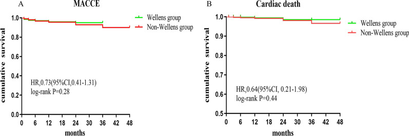
The median follow-up time was 24 months. The clinical outcomes in the Wellens’ and non-Wellens’ groups are shown in Table 2. Compared with Wellens’ group, the non-Wellens’ group had significantly higher rates of rehospitalization (19.3% vs. 10.5%, P = 0.002). There were no differences in MACCE (Wellens’ 4.5% vs. non-Wellens’ 6.5%, P = 0.26), all-cause death, cardiac death, HF, target vessel revascularization, recurrent myocardial infarction or stroke. Kaplan–Meier analysis also revealed no difference in MACCE (Plog-rank = 0.28) or cardiac death (Plog-rank = 0.44) between the Wellens’ and non-Wellens’ groups (Fig. 2). Univariate and multivariate Cox regression analysis results of the effect of Wellens' syndrome on clinical outcomes in NSTE-ACS patients are shown in Table 3. Wellens’ syndrome did not directly affect MACCEs, cardiac death or all-cause death.
Table 2.
Comparison of clinical outcome between Wellens’ and non-Wellens’ group
| Variables, n (%) | Wellens’(N = 200) | No-Wellens’ (N = 1927) | P value |
|---|---|---|---|
| MACCE | 9 (4.5) | 126 (6.5) | 0.26 |
| All-cause death | 4 (2) | 58 (3) | 0.419 |
| Cardiac death | 2 (1) | 34 (1.8) | 0.425 |
| Re-hospitalization | 21 (10.5) | 372 (19.3) | 0.002 |
| Heart failure | 1 (0.5) | 25 (1.3) | 0.329 |
| Target vessel revascularization | 0 | 18 (0.9) | 0.17 |
| Recurrent myocardial infarction | 5 (2.5) | 48 (2.5) | 0.994 |
| Stroke | 2 (1) | 21 (1.1) | 0.907 |
Values are n (%)
MACCE: major adverse cardiac and cerebrovascular events, a composite of all-cause death, cardiac death, recurrent myocardial infarction, target vessel revascularization, heart failure, and stroke
Fig. 2.
Kaplan–Meier analysis of MACCE (secondary endpoint, panel A) and cardiac death (panel B) for overall patients stratified by Wellens’ syndrome (green line) and non-Wellens’ syndrome (red line)
Table 3.
Univariate and multivariate Cox regression analysis results of Wellens' on Clinical outcome in NSTE-ACS patients
| Univariate | Multivariate | |||
|---|---|---|---|---|
| HR (95%CI) | P value | HR (95%CI) | P value | |
| MACCE | 0.7 (0.35–1.37) | 0.293 | 0.5 (0.25–1) | 0.05 |
| All-cause death | 0.68 (0.25–1.87) | 0.451 | 0.48 (0.17–1.37) | 0.171 |
| Cardiac death | 0.57 (0.14–2.39) | 0.444 | 0.38 (0.09–1.61) | 0.188 |
Compared with the non-Wellens’ group, Hazard ratios for events in Wellens’ group
Predictors of survival
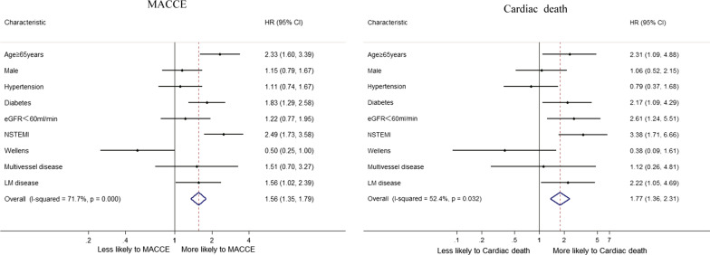
Considering all patients, a multivariable Cox regression analysis (Table 4) was used to identify clinical and angiography independent predictors of cardiac death and MACCE. For MACCEs, the final multivariable mode included age ≥ 65 years, diabetes mellitus, NSTEMI and LM disease. For cardiac death, the final multivariable mode included age ≥ 65 years, diabetes mellitus, NSTEMI, eGFR < 60 ml/min and LM disease. Overall, Wellens’ syndrome was not associated with an increased risk of MACCEs (P = 0.05) or cardiac death (P = 0.188). In Fig. 3, NSTEMI was the biggest influencing factor for poor MACCEs (HR: 2.49, 95% CI: 1.73–3.58) and cardiac death (HR: 3.38, 95% CI: 1.71–6.66).
Table 4.
Multivariate Cox regression analysis in the overall patients
| Predictor variable | HR (95% CI) | P value |
|---|---|---|
| MACCE | ||
| Age ≥ 65 years | 2.33 (1.6–3.39) | < 0.001 |
| Male | 1.15 (0.79–1.67) | 0.468 |
| HT | 1.11 (0.74–1.66) | 0.606 |
| DM | 1.83 (1.29–2.58) | 0.001 |
| eGFR < 60 ml/min | 1.22 (0.77–1.95) | 0.396 |
| NSTEMI | 2.49 (1.73–3.58) | < 0.001 |
| Wellens’ syndrome | 0.5 (0.25–1) | 0.05 |
| Multivessel disease | 1.5 (0.69–3.26) | 0.302 |
| LM disease | 1.56 (1.02–2.39) | 0.04 |
| Cardiac death | ||
| Age ≥ 65 years | 2.31 (1.09–4.88) | 0.029 |
| Male | 1.06 (0.52–2.15) | 0.883 |
| HT | 0.79 (0.37–1.68) | 0.534 |
| DM | 2.16 (1.09–4.29) | 0.027 |
| eGFR < 60 ml/min | 2.61 (1.24–5.51) | 0.012 |
| NSTEMI | 3.38 (1.71–6.66) | < 0.001 |
| Wellens’ syndrome | 0.38 (0.09–1.61) | 0.187 |
| Multivessel disease | 1.11 (0.26–4.8) | 0.884 |
| LM disease | 2.22 (1.05–4.69) | 0.037 |
HR, Hazard ratio; CI, Confidence interval; MACCE, major adverse cardiac and cerebrovascular events; HT, hypertension; DM, diabetes mellitus; NSTEMI, non-ST‑segment elevation myocardial infarction; LM, Left main
Fig. 3.
Factors independently associated with MACCE and cardiac death in overall patients in multivariable Cox regression analysis
Discussion
Wellens’ syndrome is a pattern of precordial T-wave abnormalities, first described by Wellens and his group, representing critical LAD coronary artery stenosis. In Wellens’ initial study group of 145 patients admitted for unstable angina, 9% had the typical pattern upon presentation, with a further 9% developing T-wave changes within 24 h. Seventy-five percent of these patients went on to suffer an anterior wall myocardial infarction despite the relief of symptoms with medical therapy. In a second prospective study, 14% of patients with unstable angina met the ECG criteria, all of whom were shown angiographically to have significant LAD stenosis [12].
In our study, patients with Wellens’ syndrome represented 5.7% (200 of 3528) of ACS patients hospitalized for three years. More specifically, the incidence of Wellens’ syndrome in NSTE-ACS patients with culprit LAD vessels was 9.4% (200 of 2127). Of these patients, 64 cases presented with Type A Wellens’ syndrome, which comprises 32% (64 of 200) of cases and shows biphasic T waves in leads V2 and V3. The remaining 68% (136 of 200) had Type B Wellens’ syndrome, which shows deeply inverted, symmetrical T waves in predominantly V2 and V3. This percentage is roughly in line with previous findings, which showed that type A Wellens’ constitutes roughly 24% of cases and that type B Wellens’ accounts for the remaining 76% of cases [9]. Thus, we confirmed that type B Wellens’ syndrome is more common.
The mechanism of Wellens’ syndrome remains unclear. It is considered a preinfarction stage of CHD, as the T-wave changes in the syndrome usually occur during the pain-free period [13, 14]. It is also postulated that the changes in the ECG account for reperfusion of the ischaemic myocardium due to alleviation of spasm of the proximal LAD artery [15–17]. There is also a view that the syndrome may be related to myocardial stunning or myocardial hibernation.
Multiple risk factors are common in patients with Wellens’ syndrome, and the majority of these patients are reported to have at least one traditional cardiovascular risk factor. In our study, patients with Wellens’ syndrome did not differ from the non-Wellens’ group in terms of factors such as hypertension, diabetes mellitus, and dyslipidaemia. More than half of the patients with Wellens’ syndrome were current smokers (57%). Compared with non-Wellens’ patients, they had higher LDL-C levels (2.5 ± 0.73 vs. 2.25 ± 0.75 mmol/L, P < 0.001) but were less likely to be diagnosed with hyperlipemia (50% vs. 53.5%, P = 0.352). Patients with Wellens’ syndrome were also less likely than non-Wellens’ patients to have a history of CHD and previous PCI at admission, which means that Wellens’ syndrome tends to occur in patients with new-onset cardiovascular disease. In addition, there were no significant differences between the two groups regarding comorbidities, such as HF, CKD, PAD, and stroke.
NSTEMI was the clinical presentation in 69% of Wellens’ patients, which was much higher than the 17.5% in the non-Wellens’ group. However, previous studies have suggested that unstable angina is the main clinical manifestation in Wellens’ patients [4, 12]. The increased incidence rate of NSTEMI should be due to the adoption of the Fourth Universal Definition of Myocardial Infarction defined by elevated cardiac troponin, especially hs-cTn, which indicates myonecrosis.
The angiographic characteristics of CHDin patients with Wellens’ syndrome may differ from the presentation in other ACS patients. Although there were no differences in LM disease and multivessel disease between the two groups, Wellens’ patients had more drug-eluting stents implanted and greater stent numbers than non-Wellens’ patients. This discrepancy may, to a large degree, be explained by a higher percentage of proximal and middle LAD lesions in patients with Wellens’ syndrome. Balloon angioplasty, including plain old balloon angioplasty and drug-coated balloon angioplasty, is not appropriate for these critical sites [18–20].
It is well known that patients with Wellens’ syndrome are at high risk of extensive anterior wall infarction, which might lead to serious left ventricular dysfunction, malignant arrhythmias, and sudden death. To our surprise, there was no statistically significant difference in MACCEs between the two groups during the follow-up period (mean 24 months), even though the Wellens’ group had a higher rate of myocardial infarction at admission. Furthermore, the 2-year incidence of cardiac death was similar in the two groups (Wellens’: 1% vs. non-Wellens’: 1.8%, P = 0.425). We believe that this low incidence was obtained by our approach, which was to treat these patients aggressively with urgent angiography and intervention. Data showed that the proportion of early PCI was higher in the Wellens’ group (68% vs. 59.3%, P = 0.017). This highlights the importance of timely identification of Wellens’ syndrome and appropriate management in this group of patients. Most patients, when identified early and taken for cardiac catheterization, do well after appropriate intervention. Regarding the strength of the improvement in medical procedures, a small number of Wellens’ patients (10.5%) were readmitted after PCI for 2 years, which was much lower than that of the non-Wellens’ group (19.3%, P = 0.002). We did not find a direct correlation between Wellens’ syndrome and adverse prognosis in patients with NSTE-ACS. NSTEMI was the biggest influencing factor for poor MACCEs and cardiac death. This suggests that the prognosis of CHD depends on its severity.
Early PCI is currently the preferred treatment for patients with high-risk NSTE-ACS [21, 22]. However, few patients in the real world can receive such treatment within the 24 h recommended by the guidelines, especially those patients who are pain-free at admission. In our study, we presumed that an aggressive invasive strategy (< 48 h after first presentation) could attenuate the risk of MACCE and avoid long-term adverse outcomes in patients with Wellens’ syndrome.
The main strengths of this study are the large and unselected population comprising nearly all NSTE-ACS patients with culprit LAD vessels treated at our hospital from 2017 to 2019 and the nearly complete follow-up. However, this was a single-centre, retrospective observational study. Therefore, the choice of therapeutic strategy reflected the convention and tendency of our single centre, which may affect the objectivity of the conclusions. Further prospective multicentre studies are needed to validate our findings.
Limitations
The following limitations were present in this study. (1) Lack of more information on angiographic and procedural characteristics of the study population. (2) Although we used multivariate Cox regression analysis to adjust for differences in baseline characteristics, there may still be unknown confounding factors. Therefore, the research results should be cautiously interpreted. (3) As the follow-up time was short, the long-term effect of Wellens’ syndrome has yet to be determined.
Conclusions
First, our study revealed that the incidence of Wellens’ syndrome can reach up to 5.7% as assessed by coronary arteriography in clinical practice. Second, Wellens’ patients have a lower prevalence of a history of CHD and previous PCI at admission; thus, Wellens’ syndrome is more common in populations with new-onset cardiovascular disease. Third, the presence of Wellens’ syndrome is not definitively associated with adverse prognosis in patients with NSTE-ACS. Early recognition and aggressive intervention are critical, as they may help to attenuate adverse outcomes.
Acknowledgements
The authors gratefully acknowledge the assistance with data acquisition by Guoliang Zhao.
Abbreviations
- ACS
Acute coronary syndromes
- MACCE
Major adverse cardiac and cerebrovascular events
- NSTEMI
Non-ST-elevation myocardial infarction
- PCI
Percutaneous coronary intervention
- CHD
Coronary heart disease
- LM
Left main disease
- LAD
Left anterior descending arterial
- LCX
Left circumflex artery
- RCA
Right coronary artery
- CKD
Chronic kidney disease
- PAD
Peripheral arterial disease
- HF
Heart failure
- EDD
Left ventricular end diastolic dimension
- LVEF
Left ventricular ejection fraction
- LDL-C
Low-density lipoprotein cholesterol
- eGFR
Estimated glomerular filtration rate
- BMI
Body mass index
- HR
Hazard ratio
- CI
Conference interval
Authors' contributions
All authors gave the final approval of the version published. LZ wrote the first draft of the manuscript. XhG did the statistics. TD and HhC collected data. HC and HwL made revisions to the manuscript. All authors read and approved the final manuscript.
Funding
This study was supported by Beijing Municipal Administration of Hospitals Incubating Program (code PX2020006), Beijing Municipal Administration of Hospitals' Youth Programme (QML20200106) and Beijing Key Clinical Subject Program.
Availability of data and materials
The datasets generated and/or analysed during the current study are not publicly available due to database principle but are available from the corresponding author on reasonable request.
Declarations
Ethics approval and consent to participate
This study was approved by the Institutional Review Board of Beijing Friendship Hospital (approval number 2021-P2-096-01). All procedures performed in studies involving human participants were in accordance with the ethical standards of the institution and with the 1964 Helsinki Declaration and its later amendments or comparable ethical standards. All participants gave informed consent.
Consent for publication
Not applicable.
Competing interests
The authors declare that they have no competing interests.
Footnotes
Publisher's Note
Springer Nature remains neutral with regard to jurisdictional claims in published maps and institutional affiliations.
Li Zhou and Xuhe Gong have contributed equally to this work and share first authorship
References
- 1.Roth GA, Forouzanfar MH, Moran AE, Barber R, Nguyen G, Feigin VL, et al. Demographic and epidemiologic drivers of global cardiovascular mortality. N Engl J Med. 2015;372:1333–1341. doi: 10.1056/NEJMoa1406656. [DOI] [PMC free article] [PubMed] [Google Scholar]
- 2.Collaborators GBDCoD. Global, regional, and national age-sex specific mortality for 264 causes of death, 1980–2016: a systematic analysis for the Global Burden of Disease Study 2016. Lancet. 2017;390:1151–210. 10.1016/S0140-6736(17)32152-9. [DOI] [PMC free article] [PubMed]
- 3.Roth GA, Johnson C, Abajobir A, Abd-Allah F, Abera SF, Abyu G, et al. Global, regional, and National Burden of cardiovascular diseases for 10 causes, 1990 to 2015. J Am Coll Cardiol. 2017;70:1–25. doi: 10.1016/j.jacc.2017.04.052. [DOI] [PMC free article] [PubMed] [Google Scholar]
- 4.De Zwaan C, Bär FW, Wellens HJ. Characteristic electrocardiographic pattern indicating a critical stenosis high in left anterior descending coronary artery in patients admitted because of impending myocardial infarction. Am Heart J. 1982;103:730–736. doi: 10.1016/0002-8703(82)90480-x. [DOI] [PubMed] [Google Scholar]
- 5.Nisbet BC, Zlupko G. Repeat Wellens' syndrome: case report of critical proximal left anterior descending artery restenosis. J Emerg Med. 2010;39:305–8. doi: 10.1016/j.jemermed.2007.10.043. [DOI] [PubMed] [Google Scholar]
- 6.Thygesen K, Alpert JS, Jaffe AS, Chaitman BR, Bax JJ, Morrow DA, et al. Fourth universal definition of myocardial infarction (2018) Circulation. 2018;138:e618–e651. doi: 10.1161/CIR.0000000000000617. [DOI] [PubMed] [Google Scholar]
- 7.de Winter RJ, Verouden NJ, Wellens HJ, Wilde AA. A new ECG sign of proximal LAD occlusion. N Engl J Med. 2008;359:2071–2073. doi: 10.1056/NEJMc0804737. [DOI] [PubMed] [Google Scholar]
- 8.Rhinehardt J, Brady WJ, Perron AD, Mattu A. Electrocardiographic manifestations of Wellens’ syndrome. Am J Emerg Med. 2002;20:638–643. doi: 10.1053/ajem.2002.34800. [DOI] [PubMed] [Google Scholar]
- 9.Tandy TK, Bottomy DP, Lewis JG. Wellens’ syndrome. Ann Emerg Med. 1999;33:347–351. doi: 10.1016/s0196-0644(99)70373-2. [DOI] [PubMed] [Google Scholar]
- 10.Gul M, Uyarel H, Ergelen M, Akgul O, Karaca G, Turen S, et al. Prognostic value of total bilirubin in patients with ST-segment elevation acute myocardial infarction undergoing primary coronary intervention. J Am Coll Cardiol. 2013;111:166–171. doi: 10.1016/j.amjcard.2012.09.011. [DOI] [PubMed] [Google Scholar]
- 11.Gong X, Zhou L, Ding X, Li H, Chen H. A sex-stratified long-term clinical outcome analysis in coronary chronic total occlusion patients. Biol Sex Differ. 2021;12:9. doi: 10.1186/s13293-020-00354-z. [DOI] [PMC free article] [PubMed] [Google Scholar]
- 12.de Zwaan C, Bär FW, Janssen JH, Cheriex EC, Dassen WR, Brugada P, et al. Angiographic and clinical characteristics of patients with unstable angina showing an ECG pattern of critical narrowing of the proximal LAD coronary artery. Am Heart J. 1989;117:657–665. doi: 10.1016/0002-8703(89)90742-4. [DOI] [PubMed] [Google Scholar]
- 13.Win Htut Oo SZ, Khalighi K, Kodali A, May C, Aung TT, Snyder R. Omnious T-wave inversions: Wellens' syndrome revisited. J Community Hosp Intern Med Perspect. 2016;6:32011. doi: 10.3402/jchimp.v6.32011. [DOI] [PMC free article] [PubMed] [Google Scholar]
- 14.Sakata K, Yoshino H, Houshaku H, Koide Y, Yotsukura M, Ishikawa K. Myocardial damage and left ventricular dysfunction in patients with and without persistent negative T waves after Q-wave anterior myocardial infarction. Am J Cardiol. 2001;87:510–515. doi: 10.1016/s0002-9149(00)01422-3. [DOI] [PubMed] [Google Scholar]
- 15.Sheng FQ, He MR, Zhang ML, Shen GY. Wellens’ syndrome caused by spasm of the proximal left anterior descending coronary artery. J Electrocardiol. 2015;48:423–425. doi: 10.1016/j.jelectrocard.2015.03.009. [DOI] [PubMed] [Google Scholar]
- 16.Maadarani O, Bitar Z, Shoeb S, Alsaddah J. From Wellens to Kounis syndrome: an unlucky patient. Eur J Case Rep Intern Med. 2020;7:001689. doi: 10.2890/2020_001689. [DOI] [PMC free article] [PubMed] [Google Scholar]
- 17.Ibarrola M. Wellens’ syndrome and finding of multiple coronary cameral fistulae: Is it time to discard this term? Ann Noninvasive Electrocardiol. 2020;25:e12693. doi: 10.1111/anec.12693. [DOI] [PMC free article] [PubMed] [Google Scholar]
- 18.Jeger RV, Eccleshall S, Wan Ahmad WA, Ge J, Poerner TC, Shin ES, et al. Drug-coated balloons for coronary artery disease: third report of the international DCB consensus group. JACC Cardiovasc Interv. 2020;13:1391–1402. doi: 10.1016/j.jcin.2020.02.043. [DOI] [PubMed] [Google Scholar]
- 19.Her AY, Shin ES, Bang LH, Nuruddin AA, Tang Q, Hsieh IC, et al. Drug-coated balloon treatment in coronary artery disease: Recommendations from an Asia-Pacific Consensus Group. Cardiol J. 2021;28:136–149. doi: 10.5603/CJ.a2019.0093. [DOI] [PMC free article] [PubMed] [Google Scholar]
- 20.Richelsen R, Overvad TF, Jensen SE. Drug-eluting balloons in the treatment of coronary de novo lesions: a comprehensive review. Cardiol Ther. 2016;5:133–160. doi: 10.1007/s40119-016-0064-4. [DOI] [PMC free article] [PubMed] [Google Scholar]
- 21.Collet JP, Thiele H, Barbato E, Barthélémy O, Bauersachs J, Bhatt DL, et al. 2020 ESC Guidelines for the management of acute coronary syndromes in patients presenting without persistent ST-segment elevation. Eur Heart J. 2021;42:1289–1367. doi: 10.1093/eurheartj/ehaa575. [DOI] [PubMed] [Google Scholar]
- 22.Ryan JW, Peterson ED, Chen AY, Roe MT, Ohman EM, Cannon CP, et al. CRUSADE Investigators. Optimal timing of intervention in non-ST-segment elevation acute coronary syndromes: Insights from the CRUSADE (Can Rapid risk stratification of Unstable angina patients Suppress adverse outcomes with Early implementation of the ACC/AHA guidelines) Registry. Circulation. 2005;112:3049–57. doi: 10.1161/CIRCULATIONAHA.105.582346. [DOI] [PubMed] [Google Scholar]
Associated Data
This section collects any data citations, data availability statements, or supplementary materials included in this article.
Data Availability Statement
The datasets generated and/or analysed during the current study are not publicly available due to database principle but are available from the corresponding author on reasonable request.


