Abstract
Purpose
The prognostic impact of new-onset atrial fibrillation (NOAF) among different heart failure (HF) subtypes including HF with preserved (HFpEF, ejection fraction [EF] ≥50%), mid-range (HFmrEF, EF 40%~49%), and reduced (HFrEF, EF <40%) EF following acute myocardial infarction (AMI) remains unclear. We aimed to investigate the incidence and prognostic implication of post-MI NOAF across HF subtypes.
Patients and Methods
We included 1413 patients with post-MI HF (743 with HFpEF, 342 with HFmrEF and 328 with HFrEF) between February 2014 and March 2018. NOAF was considered as patients without a preexisting AF who developed AF during the AMI hospitalization. The primary endpoint was all-cause mortality.
Results
Of 1413 patients (mean age 66.8 ± 12.6 years, 72.9% men) analyzed, 200 (14.2%) developed post-MI NOAF. Patients with HFrEF were more likely to experience NOAF compared to those with HFmrEF or HFrEF (18.9%, 13.7% and 12.2% in HFrEF, HFmrEF and HFpEF, respectively; p for trend = 0.006). During a median follow-up of 28.5 months, 192 patients died (70 with HFrEF, 35 with HFmrEF and 87 with HFpEF) and 195 patients experienced HF rehospitalization (79 with HFrEF, 37 with HFmrEF and 79 with HFpEF). After multivariable adjustment, NOAF was independently associated with all-cause mortality (hazard ratio [HR]: 1.79, 95% confidence interval [CI]: 1.03–3.12) only in the HFrEF group compared to sinus rhythm (SR), whereas an increased risk of HF rehospitalization was found in all HF subtypes, particularly in HFmrEF (HR: 5.08, 95% CI: 2.29–11.25) and HFpEF (HR: 2.83 95% CI: 1.64–4.90).
Conclusion
In patients with post-MI HF, NOAF carried a worse prognosis for all-cause death in the HFrEF group and for HF rehospitalization in all HF subtypes.
Keywords: heart failure, acute myocardial infarction, atrial fibrillation, left ventricular ejection fraction, mortality
Introduction
New-onset atrial fibrillation (NOAF) is common among patients with acute myocardial infarction (AMI) with reported incidence rates ranging from 6% to 21%,1 and has been proved to be significantly associated with increased risks of death, heart failure (HF), as well as ischemic stroke.2–4 More recently, a dose-response relationship between AF burden and adverse cardiovascular outcomes has also been illustrated, which underscores the prognostic importance of post-MI NOAF.5,6 Accounting for the detrimental impact of NOAF, numerous preceding studies have been conducted to identify the risk factors of post-MI NOAF, since adequate management of these risk factors may be helpful in reducing the incidence of NOAF and improving patients’ prognosis.4,7
HF, particularly for that with a reduced ejection fraction (HFrEF) (left ventricular ejection fraction [LVEF] <40%), has generally been perceived as an independent risk factor of post-MI NOAF.4,7,8 Due to the advances in pharmacotherapies and interventional treatments, the incidence of post-MI HFrEF has declined over the past decades, whereas that of HF with preserved ejection fraction (HFpEF) (LVEF ≥50%) and mid-range ejection fraction (HFmrEF) (LVEF 40~49%) gradually increased.9,10 Thereafter, a number of studies have investigated the impact of AF on the prognosis of HF individuals stratified by LVEF levels while yielding conflicting results. Some studies demonstrated AF does more harm to patients in HFrEF than HFpEF,11,12 but the others found a similar prognostic significance of AF in HFrEF and HFpEF.13,14 However, in the setting of AMI, there is still no within-cohort comparison regarding the influence of NOAF in different HF subtypes.
Accordingly, in the present analysis using data from the New-Onset Atrial Fibrillation Complicating Acute Myocardial Infarction in Shanghai (NOAFCAMI-SH) registry, we aimed to explore the incidence and the prognostic implication of NOAF in patients with HFrEF, HFmrEF, and HFpEF following AMI.
Patients and Methods
Study Population
The NOAFCAMI-SH is an observational, retrospective cohort study conducted in Shanghai Tenth People’s hospital. Full details of total enrolled patients, the selection criteria and study design have been previously described.4,6 Briefly, the NOAFCAMI-SH registry included 2399 patients without a medical history of AF admitting for AMI between February 2014 and March 2018. For the purpose of the present analysis, we excluded patients who died in hospital (N = 148), lost to follow-up (N = 75), without echocardiography data (N = 101), as well as free from post-MI HF (N = 662). Hence, a total of 1413 patients were included in the final analysis (Supplementary Figure 1). HF was diagnosed as the presence of clinical symptoms such as dyspnea, cough, orthopnea, fatigue or signs of peripheral or pulmonary edema or the use of intravenous diuretics, inotropic drugs, as well as N-terminal pro–B-type natriuretic peptide (NT-proBNP) testing.15–17 All included patients were classified into 3 groups based on their LVEF levels: HFrEF, HFmrEF, and HFpEF. This study complied with the Declaration of Helsinki and was approved by the Ethics Committee of the Shanghai Tenth People’s Hospital. Informed consent was exempted since all information was extracted anonymously.
Data Collection
A detailed review of the electronic medical records was conducted to collect patients’ demographic characteristics, medical history, procedures, echocardiography and angiography data, managements as well as medications. Blood samples were collected after 12 hours on fasting and were examined in a central laboratory. Echocardiogram was measured and evaluated by cardiologists based on standardized criteria within 7 days after AMI admission.18 Continuous electrocardiographic monitoring (Philips IntelliVueMP50) was started immediately at admission and continued during the whole hospitalization. AF was ascertained as the absence of distinct repeating P waves with irregular RR intervals lasting for at least 30s on ECG.19 NOAF was defined as patients who developed the first documented AF without a medical history of AF. The in-hospital NOAF burden was only evaluated in patients with paroxysmal NOAF and described as the percentage of time spent in AF based on continuous electrical monitoring during hospitalization.6
Endpoints and Follow-Up
The primary endpoint was all-cause mortality. Secondary endpoint was HF rehospitalization. All-cause mortality during follow-up was ascertained from telephone interviews with family members of the deceased. HF rehospitalization was confirmed in patients presenting with clinical symptoms and signs of peripheral or pulmonary edema that required hospitalization for intravenous diuretic treatment. All patients were followed until the date of occurrence of an outcome of interest, death or last follow-up (April 10, 2019), whichever came first.
Statistical Analysis
Normally distributed continuous variables were described as mean ± standard deviation (SD) and compared by one-way ANOVA. Skewed variables were expressed as median (interquartile ranges [IQR]) and compared by Kruskal–Wallis test. The categorical variables were presented as numbers (percentages) and compared by χ2 or Fisher’s exact test.
Event-free survival curves were estimated by the Kaplan–Meier method and compared by Log rank test. Treating sinus rhythm (SR) as the reference, multivariable Cox proportional hazard analysis model was performed to assess the association between NOAF and study endpoints across HF subtypes. Model A: we adjusted for the Global Registry Acute Coronary Events (GRACE) risk score. Model B: we adjusted for age, sex, hypertension, diabetes, heart failure, chronic kidney disease, ischemic stroke/TIA, vascular disease, heart rate, systolic blood pressure, Killip class, out-of-hospital cardiac arrest and medications at discharge (antiplatelet, ACEI/ARB, b-blocker, statin) (Supplementary Methods). No missing data existed in the abovementioned covariates. The result was presented as hazard ratio (HR) and 95% confidence interval (CI).
To further investigate the association of the severity of post-MI NOAF with clinical outcomes, we performed an exploratory analysis in which NOAF burden was introduced as an additional covariate in the primary multivariable model (Supplementary Methods). Furthermore, we carried out restricted cubic splines analysis only in the post-MI NOAF individuals to examine a possible non-linear relationship between the NOAF burden and study endpoints across HF subtypes. Three equally spaced knots were set at 10th, 50th, and 90th percentiles, and the cut-off value (10.87%) of AF burden was used as the reference.6 Receiver Operating Characteristic (ROC) Curve was also adopted to assess the predictive performance of NOAF burden for study endpoints among the three HF subtypes. All tests were 2-tailed, and a p value <0.05 was considered to be statistically significant. All analyses were performed using SPSS (version 20.0.0) and R software (Version 4.0.5).
Results
Patient Characteristics
A total of 1413 patients (mean age 66.8 ± 12.6 years, 72.9% men) with post-MI HF were included, of whom 15.8% had HFrEF, 16.5% had HFmrEF, and 35.8% had HFpEF. The baseline characteristics across HF subtypes are presented in Table 1, patients who developed HFrEF were more likely to have a history of diabetes mellitus, HF, present with anterior infarction, and had higher GRACE risk scores, CHA2DS2-VASc scores, heart rate, initial Killip class as well as peak NT-pro-BNP values. As shown in Table 2, patients with NOAF in HFrEF were more likely to experience prior MI, present as anterior infarction and have higher peak NT-pro-BNP values, while those in HFpEF were less likely to be prescribed with aspirin, β-blocker and amiodarone at discharge.
Table 1.
Baseline Characteristics, In-Hospital Examination and Medications in Three HF Subtypes
| HFrEF (n=328) | HFmrEF (n=342) | HFpEF (n=743) | p | |
|---|---|---|---|---|
| Demography and medical history | ||||
| Age (years) | 68.3±12.3‡ | 66.4±12.5 | 66.2±12.8 | 0.034 |
| BMI (kg/m2) | 24.2±3.5 | 24.4±3.2 | 24.5±3.3 | 0.602 |
| Men | 250 (76.2)‡ | 261 (76.3)† | 519 (70.0) | 0.025 |
| Current smoker | 135 (41.2) | 141 (41.2) | 287 (38.6) | 0.369 |
| Hypertension | 295 (89.9) | 220 (64.3) | 485 (65.3) | 0.181 |
| Diabetes mellitus | 145 (44.2)*‡ | 120 (35.1) | 266 (35.8) | 0.018 |
| Dyslipidemia | 77 (23.5) | 93 (27.2) | 208 (28.0) | 0.299 |
| Chronic kidney disease | 39 (111.9) | 41 (12.0) | 84 (11.3) | 0.932 |
| History of heart failure | 53 (16.2)*‡ | 15 (4.4) | 33 (4.4) | <0.001 |
| Prior stroke/TIA | 47 (14.3) | 42 (12.3) | 99 (13.3) | 0.737 |
| Prior AMI | 33 (10.1)‡ | 22 (6.4) | 36 (4.8) | 0.006 |
| Prior PCI | 39 (11.9)‡ | 35 (10.2) | 51 (6.9) | 0.017 |
| Prior vascular disease | 44 (13.4)‡ | 33 (9.6) | 57 (7.7) | 0.013 |
| Initial presentation | ||||
| Out-of-hospital cardiac arrest | 7 (2.1) | 12 (3.5) | 17 (2.3) | 0.427 |
| STEMI | 245 (74.7)‡ | 254 (74.3)† | 446 (60.0) | <0.001 |
| MI location | <0.001 | |||
| Anterior infarction | 200 (81.6)*‡ | 156 (61.4)† | 174 (39.0) | |
| Inferior infarction | 39 (15.9)*‡ | 89 (35.0)† | 247 (55.4) | |
| Other | 6 (2.4) | 9 (3.5) | 25 (5.6) | |
| Adm heart rate (b.p.m.) | 88.5±19.0*‡ | 81.6±17.5† | 78.0±16.3 | <0.001 |
| Adm SBP (mmHg) | 137.0±26.4 | 137.2±24.5 | 136.4±24.7 | 0.877 |
| Initial Killip class | <0.001 | |||
| I | 239 (72.9)*‡ | 286 (83.6) | 640 (86.1) | |
| II | 62 (18.9)*‡ | 39 (11.4) | 77 (10.4) | |
| III | 21 (6.4)*‡ | 90 (2.6) | 16 (2.2) | |
| IV | 6 (1.8) | 8 (2.3) | 10 (1.3) | |
| GRACE risk score | 130.9±29.1*‡ | 123.8±27.6 | 121.8±27.2 | <0.001 |
| CHA2DS2-VASc score | 3.6±1.7*‡ | 2.7±1.8 | 2.5±1.8 | <0.001 |
| In-hospital examination | ||||
| TC (mmol/L) | 4.42 (3.71, 5.09) | 4.35 (3.79,5.10) | 4.36 (3.74,5.01) | 0.703 |
| LDL (mmol/L) | 2.64 (2.02, 3.28) | 2.70 (2.15,3.31) | 2.65 (2.08,3.22) | 0.436 |
| HDL (mmol/L) | 1.02 (0.85, 1.25) | 1.00 (0.85,1.17) | 1.00 (0.85,1.18) | 0.257 |
| Creatinine (mg/dL) | 82.65 (70.63,99.80)* | 79.50 (66.00,95.95) | 80.25 (67.70,97.50) | 0.035 |
| eGFR (mL/min) | 78.58 (56.86,92.53)* | 84.12 (65.42, 96.03) | 82.49 (60.63,95.60) | 0.015 |
| Peak troponin-T (ng/mL) | 6.05 (1.20, 10.00)‡ | 5.41 (1.85, 10.00)† | 3.52 (1.25, 7.69) | <0.001 |
| Lg Peak NT-pro-BNP (pg/mL) | 3.66 (3.38, 4.03)*‡ | 3.43 (3.19, 3.70)† | 3.30 (3.09, 3.54) | <0.001 |
| Angiographic data | ||||
| In-hospital PCI with stent | 260 (79.3)* | 307 (89.8)† | 621 (83.6) | 0.001 |
| Infarct-related artery | <0.001 | |||
| Left anterior descending | 176 (78.6)*‡ | 154 (62.1)† | 164 (38.8) | |
| Right coronary artery | 34 (15.2)*‡ | 78 (31.5)† | 206 (48.7) | |
| Left circumflex | 14 (6.2)‡ | 16 (6.5)† | 53 (12.5) | |
| Left main disease | 25 (8.7)‡ | 16 (5.0) | 31 (4.6) | 0.034 |
| Echocardiographic data | ||||
| Left atrial diameter (mm) | 39.1±5.2* | 37.7±4.4† | 38.5±4.8 | 0.001 |
| LVEDD (mm) | 47.9±6.7*‡ | 46.1±4.8† | 45.0±4.1 | <0.001 |
| LVESD (mm) | 35.3±8.0*‡ | 31.9±5.4† | 29.8±4.0 | <0.001 |
| LVEF (%) | 32.8±4.5*‡ | 43.3± 3.0† | 56.5±4.8 | <0.001 |
| In-hospital management | ||||
| Ventilation | 18 (5.5)*‡ | 8 (2.3) | 7 (0.9) | <0.001 |
| Temporary Pacemaker | 35 (10.7)*‡ | 71 (20.8) | 136 (18.3) | 0.001 |
| IABP | 32 (9.8)*‡ | 14 (4.1)† | 9 (1.2) | <0.001 |
| Medications in hospital | ||||
| Aspirin | 317 (96.6) | 328 (95.9) | 711 (95.7) | 0.764 |
| Clopidogrel | 312 (95.1) | 317 (92.7) | 679 (91.4) | 0.099 |
| Ticagrelor | 53 (16.2) | 77 (22.5) | 157 (21.1) | 0.089 |
| Oral anticoagulation | 4 (1.2) | 1 (0.3) | 2 (0.3) | 0.103 |
| Statin | 321 (97.9) | 339 (99.1) | 734 (98.8) | 0.332 |
| ACEI/ARB | 214 (65.2) | 219 (64.0) | 460 (61.9) | 0.543 |
| β-blocker | 288 (87.8)*‡ | 277 (81.0) | 564 (75.9) | <0.001 |
| CCB | 49 (14.9)‡ | 63 (18.4) | 159 (21.4) | 0.043 |
| Diuretic | 224 (68.3)*‡ | 144 (42.1)† | 255 (34.3) | <0.001 |
| Amiodarone | 91 (27.7)‡ | 78 (22.8)† | 106 (14.3) | <0.001 |
| Medications at discharge | ||||
| Aspirin | 300 (91.7) | 321 (93.9) | 670 (90.2) | 0.128 |
| Clopidogrel | 266 (82.3) | 261 (76.3) | 563 (75.8) | 0.122 |
| Ticagrelor | 53 (16.2) | 75 (21.9) | 143 (19.2) | 0.171 |
| Oral anticoagulation | 4 (1.2) | 1 (0.3) | 6 (0.8) | 0.411 |
| Statin | 310 (94.8) | 327 (95.6) | 711 (95.7) | 0.810 |
| ACEI/ARB | 176 (53.8) | 206 (60.2) | 402 (54.1) | 0.131 |
| β-blocker | 267 (81.8)*‡ | 257 (75.1) | 505 (68.0) | <0.001 |
| CCB | 19 (5.8)‡ | 32 (9.4) | 99 (13.3) | 0.001 |
| Diuretic | 103 (31.5)*‡ | 55 (16.1) | 101 (13.6) | <0.001 |
| Amiodarone | 21 (6.4)‡ | 14 (4.1)† | 12 (1.6) | <0.001 |
Notes: Values are presented as mean ± SD, median (IQR), or n (%) as appropriate. The level of statistical significance was p < 0.017 for *HFrEF vs HFmrEF; †HFmrEF vs HFpEF; and ‡HFrEF vs HFpEF, after multiple comparisons.
Abbreviations: SR, sinus rhythm; NOAF, new-onset atrial fibrillation; BMI, body mass index; SBP, systolic blood pressure; AMI, acute myocardial infarction; HR, heart rate; LVEF, left ventricular ejection fraction, GRACE, Global Registry of Acute Coronary Events; TIA, transient ischemic attack; PCI, percutaneous coronary intervention; eGFR, estimated glomerular filtration rate; TC, total cholesterol; LDL-c, low-density lipoprotein cholesterol; HDL-c, high-density lipoprotein cholesterol; LVEDD, left ventricular end-diastolic diameter; LVEF, left ventricular ejection fraction; LVESD, left ventricular end-systolic diameter; IABP, intra-aortic balloon pump; NT-proBNP, N terminal pro brain natriuretic peptide; ACEI/ARB, angiotensin-converting enzyme inhibitor/angiotensin receptor blocker; CCB, calcium-channel blocker; IQR, interquartile range.
Table 2.
Baseline Characteristics, In-Hospital Examination and Medications in Patients with NOAF
| HFrEF (n=62) | HFmrEF (n=47) | HFpEF (n=91) | p | |
|---|---|---|---|---|
| Demography and medical history | ||||
| Age (years) | 72.7±9.8 | 73.5±10.7 | 76.0±10.6 | 0.090 |
| BMI (kg/m2) | 24.6±3.8 | 23.4±3.1 | 24.4±3.4 | 0.284 |
| Men | 47 (75.8) | 31 (66.0) | 57 (62.6) | 0.225 |
| NOAF burden (%) | 6.70 (1.32, 29.53) | 5.00 (1.32, 31.77) | 8.15 (1.97, 45.83) | 0.729 |
| Current smoker | 23 (37.1)‡ | 16 (34.0) | 19 (20.9) | 0.044 |
| Hypertension | 37 (59.7) | 33 (70.2) | 71 (78) | 0.051 |
| Diabetes mellitus | 27 (43.5) | 12 (25.5) | 37 (40.7) | 0.123 |
| Dyslipidemia | 12 (19.4) | 12 (25.5) | 18 (19.8) | 0.682 |
| Chronic kidney disease | 14 (22.6)* | 1 (2.1)† | 12 (13.2) | 0.008 |
| History of heart failure | 15 (24.2)* | 1 (2.1)† | 13 (14.3) | 0.005 |
| Prior stroke/TIA | 11 (17.7) | 1 (2.1) | 16 (17.6) | 0.854 |
| Prior AMI | 12 (19.4)*‡ | 2 (4.3) | 3 (3.3) | 0.001 |
| Prior PCI | 14 (22.6)‡ | 4 (8.5) | 7 (7.7) | 0.015 |
| Prior vascular disease | 15 (24.2)* | 4 (8.5) | 11 (12.1) | 0.044 |
| Initial presentation | ||||
| Out-of-hospital cardiac arrest | 1 (1.6) | 2 (4.3) | 4 (4.4) | 0.693 |
| STEMI | 49 (79.0)‡ | 35 (74.5)† | 44 (48.4) | <0.001 |
| MI location | <0.001 | |||
| Anterior infarction | 36 (73.5)*‡ | 17 (48.6) | 13 (29.5) | |
| Inferior infarction | 13 (26.5)‡ | 16 (45.7) | 29 (65.9) | |
| Other | 0 (0) | 2 (5.7) | 2 (4.5) | |
| Adm heart rate (b.p.m.) | 91.7±25.9‡ | 84.5±21.2 | 81.7±23.0 | 0.037 |
| Adm SBP (mmHg) | 131.9±24.2 | 134.8±27.4 | 138.5±27.3 | 0.315 |
| Initial Killip class | 0.517 | |||
| I | 46 (74.2) | 31 (66.0) | 61 (67.0) | |
| II | 13 (21.0) | 9 (19.1) | 20 (22.0) | |
| III | 2 (3.2) | 2 (4.3) | 6 (6.6) | |
| IV | 1 (1.6) | 5 (10.6) | 4 (4.4) | |
| GRACE risk score | 142.2±24.8 | 144.0±26.3 | 146.6±28.1 | 0.601 |
| CHA2DS2-VASc score | 4.2±1.8 | 3.7±1.7 | 3.7±1.9 | 0.217 |
| In-hospital examination | ||||
| TC (mmol/L) | 4.10 (3.41, 4.81) | 4.18 (3.54,5.08) | 4.30 (3.50,4.89) | 0.742 |
| LDL (mmol/L) | 2.44 (1.85, 3.02) | 2.45 (2.13,3.21) | 2.36 (1.79,3.07) | 0.501 |
| HDL (mmol/L) | 1.01 (0.80, 1.22) | 1.07 (0.90,1.24) | 1.01 (0.85,1.24) | 0.755 |
| Creatinine (mg/dL) | 87.10 (44.93,86.79) | 70.19 (51.93,82.73) | 65.65 (46.43,81.52) | 0.388 |
| eGFR (mL/min) | 68.59 (56.86,92.53) | 84.12 (65.42, 96.03) | 82.49 (60.63,95.60) | 0.524 |
| Peak troponin-T (ng/mL) | 5.81 (0.88, 10.00) | 8.94 (2.82, 10.00)† | 3.20 (0.52, 8.32) | <0.001 |
| Lg Peak NT-pro-BNP (pg/mL) | 3.93 (3.65, 4.27)*‡ | 3.63 (3.40, 3.90) | 3.61 (3.42, 3.96) | <0.001 |
| Angiographic data | ||||
| In-hospital PCI with stent | 47 (75.8) | 41 (87.2) | 63 (69.2) | 0.066 |
| Infarct-related artery | 0.006 | |||
| Left anterior descending | 29 (63.0)‡ | 15 (44.1) | 10 (25.0) | |
| Right coronary artery | 11 (23.9)‡ | 15 (44.1) | 25 (62.5) | |
| Left circumflex | 6 (13.0) | 4 (11.8) | 5 (12.5) | |
| Left main disease | 6 (10.5) | 7 (15.9) | 7 (9.2) | 0.523 |
| Echocardiographic data | ||||
| Left atrial diameter (mm) | 40.9±5.2 | 39.1±5.2 | 40.7±4.8 | 0.136 |
| LVEDD (mm) | 48.3±6.9‡ | 46.5±6.3 | 44.8±4.0 | 0.001 |
| LVESD (mm) | 36.2±7.9*‡ | 33.0±6.8 | 29.7±3.6 | <0.001 |
| LVEF (%) | 31.9±4.6*‡ | 42.1± 2.7 | 56.3±4.6 | <0.001 |
| In-hospital management | ||||
| Ventilation | 6 (9.7) | 4 (8.5) | 3 (3.3) | 0.256 |
| Temporary Pacemaker | 10 (16.1) | 17 (36.2) | 21 (23.1) | 0.051 |
| IABP | 6 (9.7) | 6 (12.8)† | 1 (1.1) | 0.013 |
| Medications in hospital | ||||
| Aspirin | 61 (98.4) | 47 (100.0) | 85 (93.4) | 0.085 |
| Clopidogrel | 55 (88.7) | 43 (91.5) | 82 (90.1) | 0.891 |
| Ticagrelor | 14 (22.6) | 11 (23.4) | 16 (17.6) | 0.643 |
| Oral anticoagulation | 3 (4.8) | 1 (2.1) | 1 (1.1) | 0.341 |
| Statin | 59 (95.2) | 46 (97.9) | 87 (95.6) | 0.748 |
| ACEI/ARB | 40 (64.5) | 30 (63.8) | 63 (69.2) | 0.754 |
| β-blocker | 49 (79.0) | 41 (87.2) | 65 (71.4) | 0.102 |
| CCB | 11 (17.7) | 10 (21.3) | 22 (24.2) | 0.636 |
| Diuretic | 51 (82.3) | 35 (74.5) | 66 (72.5) | 0.369 |
| Amiodarone | 41 (66.1) * | 39 (83.0)† | 48 (52.7) | 0.002 |
| Medications at discharge | ||||
| Aspirin | 57 (91.9)‡ | 44 (93.6)† | 72 (79.1) | 0.020 |
| Clopidogrel | 46 (74.2) | 33 (70.1) | 72 (79.1) | 0.493 |
| Ticagrelor | 14 (22.6) | 11 (23.4) | 9 (9.9) | 0.050 |
| Oral anticoagulation | 4 (6.5) | 1 (2.1) | 4 (4.4) | 0.560 |
| Statin | 59 (95.2) | 45 (95.7) | 83 (91.2) | 0.609 |
| ACEI/ARB | 28 (45.2) | 27 (57.4) | 47 (51.6) | 0.440 |
| β-blocker | 43 (69.4)‡ | 32 (68.1)† | 46 (50.5) | 0.031 |
| CCB | 5 (8.1) | 4 (8.5) | 14 (15.4) | 0.289 |
| Diuretic | 24 (38.7) | 16 (34.0) | 24 (26.4) | 0.260 |
| Amiodarone | 13 (21.0)‡ | 8 (17.0)† | 4 (4.4) | 0.005 |
| Terminated method | 0.016 | |||
| 0 | 6 (9.7) | 3 (6.4) | 11 (12.1) | |
| 1 | 36 (58.1) | 34 (72.3)† | 40 (44.0) | |
| 2 | 20 (32.3) | 9 (19.1)† | 40 (44.0) | |
| 3 | 0 (0) | 1 (2.1) | 0 (0) |
Notes: Values are presented as mean ± SD, median (IQR), or n (%) as appropriate. The level of statistical significance was p < 0.017 for *HFrEF vs HFmrEF; †HFmrEF vs HFpEF; and ‡HFrEF vs HFpEF, after multiple comparisons. Terminated method is the manner of conversion to a SR, AF not terminated was represented by 0, pharmacological conversion was represented by 1, spontaneous cardioversion was represented by 2 and electrical cardioversion was represented by 3.
Abbreviations: SR, sinus rhythm; NOAF, new-onset atrial fibrillation; BMI, body mass index; SBP, systolic blood pressure; AMI, acute myocardial infarction; HR, heart rate; LVEF, left ventricular ejection fraction, GRACE, Global Registry of Acute Coronary Events; TIA, transient ischemic attack; PCI, percutaneous coronary intervention; eGFR, estimated glomerular filtration rate; TC, total cholesterol; LDL-c, low-density lipoprotein cholesterol; HDL-c, high-density lipoprotein cholesterol; LVEDD, left ventricular end-diastolic diameter; LVEF, left ventricular ejection fraction; LVESD, left ventricular end-systolic diameter; IABP, intra-aortic balloon pump; NT-proBNP, N terminal pro brain natriuretic peptide; ACEI/ARB, angiotensin-converting enzyme inhibitor/angiotensin receptor blocker; CCB, calcium-channel blocker; IQR, interquartile range.
NOAF Incidence and Burden
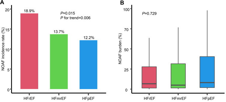
A total of 200 (14.2%) patients with HF developed post-MI NOAF. As shown in Figure 1A, NOAF incidence increased in parallel with the magnitude of impaired EF values (18.9%, 13.7% and 12.2% in HFrEF, HFmrEF, and HFpEF, respectively; p for trend = 0.006). No statistical significance was found for the distribution of NOAF burden across HF subtypes (Figure 1B). Nineteen patients performed a recurrence of AF after successful cardioversion in hospital and a total of 39 patients were still in AF at discharge. As indicated in Table 2, the rates of conversion to a sinus rhythm in HFrEF, HFmrEF and HFpEF groups were 90.3%, 93.6%, and 87.9%, respectively.
Figure 1.
NOAF incidence (A) and burden of NOAF (B) in three HF subtypes.
Abbreviations: NOAF, new onset atrial fibrillation; HF, heart failure; HFrEF, HF with reduced ejection fraction; HFmrEF, HF with mid-range ejection fraction; HFpEF, HF with preserved ejection fraction.
Long-Term Outcomes
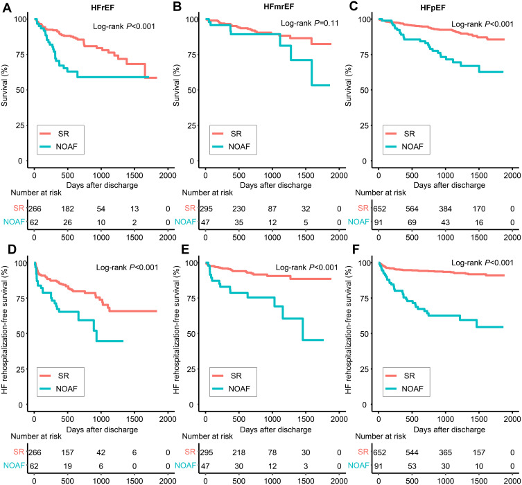
During a median follow-up of 28.5 months (IQR: 17.8–43.6), 192 (13.6%) patients died and all-cause mortality was 21.3%, 10.2% and 11.7% in HFrEF, HFmrEF and HFpEF, respectively. One hundred and ninety-five (13.8%) patients experienced HF rehospitalization. The rehospitalization rate of patients was 24.1% in HFrEF group, which reached the highest when compared to that of patients in HFmrEF (10.8%) and HFpEF (10.6%) groups, respectively. Among the patients who were still in AF at discharge, 14 patients died during the follow-up. However, the patients who died remained in AF during follow-up period were not accurately gathered since the mortality was ascertained from telephone interviews with family members who may not be able to detail patients’ situation. As demonstrated in Figure 2, patients with NOAF in HFrEF and HFpEF were associated with an elevated all-cause mortality compared to those with SR. NOAF was significantly associated with an increased all-cause mortality only in patients with HFrEF after multivariable adjustment (HR: 1.79, 95% CI: 1.03–3.12, p = 0.040) (Table 3). However, the NOAF was independently associated with the HF rehospitalization in all HF subtypes (Figure 2). This association seemed to be more significant in HFmrEF (HR: 5.08; 95% CI: 2.29–11.25) and HFpEF (HR: 2.83; 95% CI: 1.64–4.90) groups (Table 3).
Figure 2.
Long-term survival in patients with HFrEF (A), HFmrEF (B) and HFpEF (C), and HF rehospitalization-free survival in patients with HFrEF (D), HFmrEF (E) and HFpEF (F).
Abbreviations: NOAF, new onset atrial fibrillation; SR, sinus rhythm; HF, heart failure; AMI, acute myocardial infarction; HFrEF, HF with reduced ejection fraction; HFmrEF, HF with mid-range ejection fraction; HFpEF, HF with preserved ejection fraction.
Table 3.
Long-Term Outcomes
| HFrEF | HFmrEF | HFpEF | ||||
|---|---|---|---|---|---|---|
| HR | p value | HR | p value | HR | p value | |
| All-cause death | ||||||
| Unadjusted HR | 2.58 (1.56–4.26) | <0.001 | 1.88 (0.85–4.14) | 0.117 | 3.42 (2.16–5.42) | <0.001 |
| Adjusted HRa | 2.07 (1.25–3.43) | 0.005 | 0.99 (0.43–2.28) | 0.986 | 1.42 (0.84–2.40) | 0.191 |
| Adjusted HRb | 1.79 (1.03–3.12) | 0.040 | 1.61 (0.61–4.30) | 0.334 | 1.43 (0.84–2.43) | 0.183 |
| HF rehospitalization | ||||||
| Unadjusted HR | 2.23 (1.37–3.64) | 0.001 | 4.14 (2.13–8.05) | <0.001 | 6.23 (3.97–9.76) | <0.001 |
| Adjusted HRa | 1.93 (1.18–3.16) | 0.009 | 2.62 (1.27–5.39) | 0.009 | 3.93 (2.37–6.53) | <0.001 |
| Adjusted HRb | 1.93 (1.11–3.34) | 0.020 | 5.08 (2.29–11.25) | <0.001 | 2.83 (1.64–4.90) | <0.001 |
Notes: aAdjusted for GRACE risk score. bAdjusted for age, sex, hypertension, diabetes, heart failure, chronic kidney disease, ischemic stroke/TIA, vascular disease, heart rate, systolic blood pressure, Killip class, out of-hospital cardiac arrest, and medications at discharge (antiplatelet, ACEI/ARB, b-blocker, statin).
Abbreviations: HR, hazard ratio; SR sinus rhythm; NOAF, new-onset atrial fibrillation.
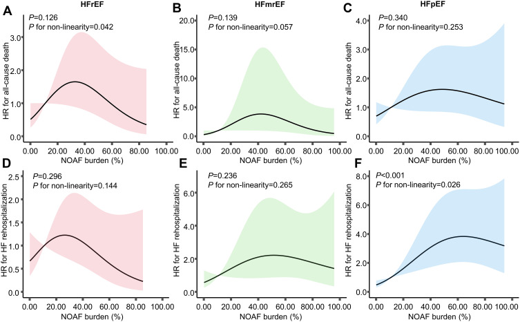
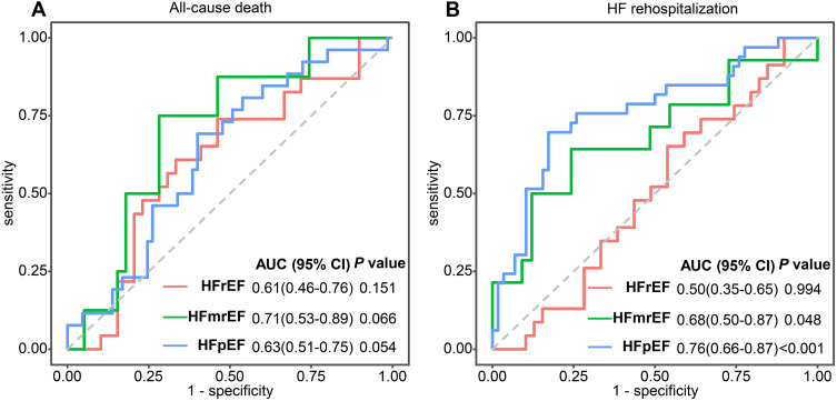
In patients with post-MI NOAF, we further investigated the prognostic impact of NOAF burden across HF subtypes. As displayed in Table 4, multivariable models revealed that NOAF burden was associated with HF rehospitalization only in HFpEF (per 10% increase, HR 1.25, 95% CI: 1.11–1.40). Furthermore, our restricted cubic spline analysis illustrated an inverted U-shaped trajectory of post-AMI NOAF burden with HF rehospitalization in HFpEF (Figure 3). Our ROC analysis showed a good predictive performance of NOAF burden for the risk of HF rehospitalization among the HFpEF individuals, as evidenced by an area under the curve of 0.76 (95% CI: 0.66–0.87), but suboptimal results among those with HFrEF and HFmrEF (Figure 4).
Table 4.
Association of NOAF Burden and Long-Term Outcomes in NOAF Patients
| HFrEF | HFmrEF | HFpEF | ||||
|---|---|---|---|---|---|---|
| HR | p value | HR | p value | HR | p value | |
| All-cause death | ||||||
| Unadjusted HR (per 10% increase) | 1.03 (0.90–1.18) | 0.653 | 1.08 (0.91–1.28) | 0.389 | 1.07 (0.95–1.19) | 0.271 |
| Adjusted HRa | 0.98 (0.84–1.14) | 0.817 | 1.06 (0.88–1.28) | 0.531 | 1.06 (0.94–1.19) | 0.345 |
| Adjusted HRb | 0.97 (0.74–1.27) | 0.839 | 0.81 (0.44–1.50)# | 0.505 | 1.08 (0.93–1.24) | 0.327 |
| HF rehospitalization | ||||||
| Unadjusted HR (per 10% increase) | 0.96 (0.82–1.12) | 0.570 | 1.10 (0.97–1.26) | 0.154 | 1.20 (1.10–1.31) | <0.001 |
| Adjusted HRa | 0.93 (0.78–1.09) | 0.364 | 1.10 (0.96–1.25) | 0.175 | 1.21 (1.10–1.32) | <0.001 |
| Adjusted HRb | 0.98 (0.76–1.27) | 0.880 | 1.19 (0.92–1.55) | 0.180 | 1.25 (1.11–1.40) | <0.001 |
Notes: #Indicated the association of NOAF burden (per 1% increase) and all-cause death. aAdjusted for GRACE risk score. bAdjusted for age, sex, hypertension, diabetes, heart failure, chronic kidney disease, ischemic stroke/TIA, vascular disease, heart rate, systolic blood pressure, Killip class, out of-hospital cardiac arrest, and medications at discharge (antiplatelet, ACEI/ARB, b-blocker, statin).
Abbreviations: HR, hazard ratio; SR sinus rhythm; NOAF, new-onset atrial fibrillation.
Figure 3.
The association of post-MI NOAF burden with all-cause death and HF rehospitalization in HFrEF, HFmrEF and HFpEF groups.
Notes: Post-MI NOAF burden referred to an in-hospital parameter and derived from continuous electrical monitoring during hospitalization. The association of post-MI NOAF burden with all-cause death in HFrEF (A), HFmrEF (B) and HFpEF (C) groups. The association of post-MI NOAF burden with HF rehospitalization in HFrEF (D), HFmrEF (E) and HFpEF (F) groups.
Abbreviations: NOAF, new onset atrial fibrillation; HF, heart failure; AMI, acute myocardial infarction; HFrEF, HF with reduced ejection fraction; HFmrEF, HF with mid-range ejection fraction; HFpEF HF with preserved ejection fraction.
Figure 4.
Receiver operating characteristic (ROC) curves for burden of NOAF for all-cause death (A) and HF rehospitalization (B) in patients with NOAF in three HF subtypes.
Abbreviations: NOAF, new onset atrial fibrillation; HF, heart failure; AMI, acute myocardial infarction; HFrEF, HF with reduced ejection fraction; HFmrEF, HF with mid-range ejection fraction; HFpEF HF with preserved ejection fraction; AUC, area under curve.
Discussion
Within a cohort of 1413 patients suffered HF during AMI hospitalization, we found that NOAF incidence increased in parallel with the magnitude of impaired LVEF values. When compared with SR, NOAF was significantly associated with an increased risk of HF rehospitalization across the three HF subtypes while a poor survival was only observed in the HFrEF individuals after accounting for potential confounders. Our exploratory analysis demonstrated that a greater burden of NOAF was associated with HF rehospitalization in HFpEF rather than in HFrEF or HFmrEF. Moreover, an inverted U-shaped trajectory of post-AMI NOAF burden with HF hospitalization was found.
In the current analysis, we found that 14.2% of the studied population experienced NOAF during the index AMI hospitalization, which is consistent with the reported prevalence of post-MI NOAF ranging from 6% to 21%.1 Moreover, NOAF was more common in HFrEF compared with that in HFmrEF and HFpEF, which is in line with prior studies where the decreased LVEF value was identified as a significant predictor of NOAF.4,7,8 A possible explanation might be that the left ventricular dysfunction could increase left atrial (LA) diameter and LA pressure, thus initiating LA remodeling and prompting the progression of AF.8
So far, there are numerous studies that have reported the significant association between AF and the long-term mortality in patients with HF, whereas its impact in different HF subtypes remains undetermined. A recent study performed on 91,360 AMI patients indicated that patients with post-MI HFrEF (HR 4.5, 95% CI 4.4–4.6) had a worst prognosis for long-term mortality compared with those with no HF and post-MI HFpEF (HR 3.3, 95% CI 3.1–3.4).20 Mentias et al also demonstrated that in AF patients HFrEF was related to a higher risk for all-cause mortality compared with patients with no HF and HFpEF.21 A similar result was received in a meta-analysis, which reported that all-cause mortality was significantly higher in AF patients with HFrEF compared to those with HFpEF (HR 1.24, 95% CI 1.12–1.36, p < 0.001).12 In contrast, McManus et al illustrated that AF patients had higher risk of death irrespective HF subtypes compared to the SR individuals.14 Conversely, some other studies suggested that AF was associated with death in HFpEF but not in HFrEF patients.22,23 Cheng et al showed that AF was related to a higher risk of death in HFpEF (HR 1.20, 95% CI 1.12–1.28) compared to those in HFrEF (HR 1.07, 95% CI 1.01–1.12).24 In our study, patients with post-MI NOAF were at a higher risk of all-cause mortality in HFrEF after full adjustment. The potential explanations for the detrimental impact of NOAF in those with HFrEF could be attributed to that HFrEF patients had poor baseline characteristics and comorbidities (Table 1), and the relatively higher incidence of NOAF in HFrEF also made it easier to observe adverse events. Further studies were warranted to explore the exact mechanisms.
As expected, we found that NOAF was an independent risk factor for HF rehospitalization in the study population after adjusting for potential confounders. Compared to the patients in HFmrEF and HFpEF groups, the patients in HFrEF group had the highest rehospitalization rate. Although there existed obvious relationship between NOAF and the elevated rehospitalization rate in HFrEF patients, the NOAF seemed to be more significantly associated with an increased risk of HF rehospitalization in patients with HFpEF and HFmrEF groups. This could be partially explained as patients with HFrEF were more likely to benefit from the use of β-blocker (69.4% vs 68.1% vs.50.5%) and antiarrhythmic drugs (21.0% vs 17.0% vs 4.4%). In addition, the detrimental impact of NOAF on the prognosis of HFrEF individuals might be attenuated to some extent since the deteriorated LVEF could per se result in poor survival.25 Former research illustrated that NOAF was related to an increased long-term risk of HF hospitalization in the whole AMI population irrespective of HF subtypes.4
AF burden has been associated with increased risks of adverse cardiovascular events among patients with AF in various settings.5,6,26,27 In this regard, to explore the relationship between the NOAF and HF more comprehensively, we further evaluated the prognostic impact of NOAF burden on HF rehospitalization in NOAF patients across HF subtypes. Multivariable-adjusted cox regression models suggested that a higher NOAF burden was independently associated with an increased risk of HF rehospitalization only in HFpEF group (per 10% increase, HR 1.25, 95% CI: 1.11–1.40), and our ROC analysis showed a good predictive performance of NOAF burden for HF rehospitalization only in HFpEF as well. The difference in the impact of NOAF burden among the three HF subgroups may be due to more strengthen pharmacological treatments being applied in patients with HFrEF and HFmrEF. Furthermore, we observed an inverted U-shaped trajectory of post-AMI NOAF burden with HF rehospitalization in HFpEF. Such a trajectory may be ascribed to the fact that paroxysmal AF episodes were more likely to be detected in the high AF burden individuals, thus leading to timely treatment. Taken together, these results suggested that more attention should be paid to patients with high NOAF burden, particularly in those with HFpEF, and a more strengthen rhythm control strategy may reduce HF rehospitalization.
Limitations
Our study had several limitations. First, our study was a retrospective, observational and sing-center study. Second, NOAF burden cannot be exactly calculated without aggressive monitoring such as an insertable cardiac monitoring system. Third, we did not monitor AF burden during the follow-up.
Conclusion
Our study demonstrated that in patients with post-MI HF, NOAF carried a worse prognosis for all-cause mortality in the HFrEF group and for HF rehospitalization in all HF subtypes. In addition, post-MI NOAF burden was significantly associated with an increased risk of HF hospitalization simply in the HFpEF individuals. Our findings suggest that in patients with HFrEF after AMI, continuous electrical monitoring and strengthen treatment should be conducted to prevent the occurrence of NOAF; while in those with HFpEF, rhythm control may be beneficial in improving their prognosis.
Funding Statement
This work was supported by the Natural Science Foundation of Shanghai [18ZR1429700], the Shanghai Sailing Program [19YF1437900], and the National Natural Science Foundation of China [81270193 and 81900385].
Ethics Statement
The study was reviewed and approved by the Ethics Committee of Shanghai Tenth People’s Hospital.
Author Contributions
All authors made a significant contribution to the work reported, whether that is in the conception, study design, execution, acquisition of data, analysis and interpretation, or in all these areas; took part in drafting, revising or critically reviewing the article; gave final approval of the version to be published; have agreed on the journal to which the article has been submitted; and agree to be accountable for all aspects of the work.
Disclosure
The authors declared that they had no conflicts of interest in relation to this article.
References
- 1.Schmitt J, Duray G, Gersh B, Hohnloser S. Atrial fibrillation in acute myocardial infarction: a systematic review of the incidence, clinical features and prognostic implications. Eur Heart J. 2009;30(9):1038–1045. doi: 10.1093/eurheartj/ehn579 [DOI] [PubMed] [Google Scholar]
- 2.Crenshaw B, Ward S, Granger C, Stebbins A, Topol E, Califf R. Atrial fibrillation in the setting of acute myocardial infarction: the GUSTO-I experience. Global utilization of Streptokinase and TPA for occluded coronary arteries. J Am Coll Cardiol. 1997;30(2):406–413. doi: 10.1016/s0735-1097(97)00194-0 [DOI] [PubMed] [Google Scholar]
- 3.Pedersen O, Bagger H, Køber L, Torp-Pedersen C. The occurrence and prognostic significance of atrial fibrillation/-flutter following acute myocardial infarction. TRACE Study group. TRAndolapril Cardiac Evalution. Eur Heart J. 1999;20(10):748–754. doi: 10.1053/euhj.1998.1352 [DOI] [PubMed] [Google Scholar]
- 4.Luo J, Xu S, Li H, et al. Long-term impact of new-onset atrial fibrillation complicating acute myocardial infarction on heart failure. ESC Heart Fail. 2020;7(5):2762–2772. doi: 10.1002/ehf2.12872 [DOI] [PMC free article] [PubMed] [Google Scholar]
- 5.Go AS, Reynolds K, Yang J, et al. Association of burden of atrial fibrillation with risk of ischemic stroke in adults with paroxysmal atrial fibrillation: the KP-RHYTHM Study. JAMA Cardiol. 2018;3(7):601–608. doi: 10.1001/jamacardio.2018.1176 [DOI] [PMC free article] [PubMed] [Google Scholar]
- 6.Luo J, Xu S, Li H, et al. Long-term impact of the burden of new-onset atrial fibrillation in patients with acute myocardial infarction: results from the NOAFCAMI-SH registry. Europace. 2021;23(2):196–204. doi: 10.1093/europace/euaa234 [DOI] [PubMed] [Google Scholar]
- 7.Cosentino N, Ballarotto M, Campodonico J, et al. Impact of glomerular filtration rate on the incidence and prognosis of new-onset atrial fibrillation in acute myocardial infarction. J Clin Med. 2020;9(5):May. doi: 10.3390/jcm9051396 [DOI] [PMC free article] [PubMed] [Google Scholar]
- 8.Zeng R, Chen M, Lian B, Liao P, Zhang M. Left ventricular ejection fraction and left atrium diameter related to new-onset atrial fibrillation following acute myocardial infarction: a systematic review and meta-analysis. Oncotarget. 2017;8(46):81137–81144. doi: 10.18632/oncotarget.20821 [DOI] [PMC free article] [PubMed] [Google Scholar]
- 9.Spencer F, Meyer T, Goldberg R, et al. Twenty year trends (1975–1995) in the incidence, in-hospital and long-term death rates associated with heart failure complicating acute myocardial infarction: a community-wide perspective. J Am Coll Cardiol. 1999;34(5):1378–1387. doi: 10.1016/s0735-1097(99)00390-3 [DOI] [PubMed] [Google Scholar]
- 10.Desta L, Jernberg T, Lofman I, et al. Incidence, temporal trends, and prognostic impact of heart failure complicating acute myocardial infarction. The SWEDEHEART Registry (Swedish Web-System for Enhancement and Development of Evidence-Based Care in Heart Disease Evaluated According to Recommended Therapies): a study of 199,851 patients admitted with index acute myocardial infarctions, 1996 to 2008. JACC Heart Fail. 2015;3(3):234–242. doi: 10.1016/j.jchf.2014.10.007 [DOI] [PubMed] [Google Scholar]
- 11.Parissis JT, Ikonomidis I, Rafouli-Stergiou P, et al. Clinical characteristics and predictors of in-hospital mortality in acute heart failure with preserved left ventricular ejection fraction. Am J Cardiol. 2011;107(1):79–84. doi: 10.1016/j.amjcard.2010.08.044 [DOI] [PubMed] [Google Scholar]
- 12.Kotecha D, Chudasama R, Lane DA, Kirchhof P, Lip GY. Atrial fibrillation and heart failure due to reduced versus preserved ejection fraction: a systematic review and meta-analysis of death and adverse outcomes. Int J Cardiol. 2016;203:660–666. doi: 10.1016/j.ijcard.2015.10.220 [DOI] [PubMed] [Google Scholar]
- 13.Olsson LG, Swedberg K, Ducharme A, et al. Atrial fibrillation and risk of clinical events in chronic heart failure with and without left ventricular systolic dysfunction: results from the Candesartan in heart failure-Assessment of reduction in mortality and morbidity (CHARM) program. J Am Coll Cardiol. 2006;47(10):1997–2004. doi: 10.1016/j.jacc.2006.01.060 [DOI] [PubMed] [Google Scholar]
- 14.McManus DD, Hsu G, Sung SH, et al. Atrial fibrillation and outcomes in heart failure with preserved versus reduced left ventricular ejection fraction. J Am Heart Assoc. 2013;2(1). doi: 10.1161/jaha.112.005694 [DOI] [PMC free article] [PubMed] [Google Scholar]
- 15.Ponikowski P, Voors AA, Anker SD, et al. 2016 ESC Guidelines for the diagnosis and treatment of acute and chronic heart failure: the Task Force for the diagnosis and treatment of acute and chronic heart failure of the European Society of Cardiology (ESC). Developed with the special contribution of the Heart Failure Association (HFA) of the ESC. Eur J Heart Fail. 2016;18(8):891–975. doi: 10.1002/ejhf.592 [DOI] [PubMed] [Google Scholar]
- 16.Bozkurt B, Coats A, Tsutsui H, et al. Universal definition and classification of heart failure: a report of the Heart Failure Society of America, Heart Failure Association of the European Society of Cardiology, Japanese Heart Failure Society and Writing Committee of the Universal Definition of Heart Failure. J Card Fail. 2021. doi: 10.1016/j.cardfail.2021.01.022 [DOI] [Google Scholar]
- 17.Januzzi J, Chen-Tournoux A, Christenson R, et al. N-terminal pro-B-type natriuretic peptide in the emergency department: the ICON-RELOADED Study. J Am Coll Cardiol. 2018;71(11):1191–1200. doi: 10.1016/j.jacc.2018.01.021 [DOI] [PubMed] [Google Scholar]
- 18.Lang R, Badano L, Mor-Avi V, et al. Recommendations for cardiac chamber quantification by echocardiography in adults: an update from the American Society of Echocardiography and the European Association of Cardiovascular Imaging. J Am Soc Echocardiogr. 2015;28(1):1–39.e14. doi: 10.1016/j.echo.2014.10.003 [DOI] [PubMed] [Google Scholar]
- 19.Hindricks G, Potpara T, Dagres N, et al. 2020 ESC Guidelines for the diagnosis and management of atrial fibrillation developed in collaboration with the European Association for Cardio-Thoracic Surgery (EACTS): the Task Force for the diagnosis and management of atrial fibrillation of the European Society of Cardiology (ESC) Developed with the special contribution of the European Heart Rhythm Association (EHRA) of the ESC. Eur Heart J. 2021;42(5):373–498. doi: 10.1093/eurheartj/ehaa612 [DOI] [PubMed] [Google Scholar]
- 20.Desta L, Jernberg T, Spaak J, Hofman-Bang C, Persson H. Heart failure with normal ejection fraction is uncommon in acute myocardial infarction settings but associated with poor outcomes: a study of 91,360 patients admitted with index myocardial infarction between 1998 and 2010. Eur J Heart Fail. 2016;18(1):46–53. doi: 10.1002/ejhf.416 [DOI] [PubMed] [Google Scholar]
- 21.Mentias A, Briasoulis A, Shantha G, Alvarez P, Vaughan-Sarrazin M. Impact of heart failure type on thromboembolic and bleeding risk in patients with atrial fibrillation on oral anticoagulation. Am J Cardiol. 2019;123(10):1649–1653. doi: 10.1016/j.amjcard.2019.02.027 [DOI] [PMC free article] [PubMed] [Google Scholar]
- 22.Linssen GC, Rienstra M, Jaarsma T, et al. Clinical and prognostic effects of atrial fibrillation in heart failure patients with reduced and preserved left ventricular ejection fraction. Eur J Heart Fail. 2011;13(10):1111–1120. doi: 10.1093/eurjhf/hfr066 [DOI] [PubMed] [Google Scholar]
- 23.Eapen ZJ, Greiner MA, Fonarow GC, et al. Associations between atrial fibrillation and early outcomes of patients with heart failure and reduced or preserved ejection fraction. Am Heart J. 2014;167(3):369–375 e2. doi: 10.1016/j.ahj.2013.12.001 [DOI] [PubMed] [Google Scholar]
- 24.Cheng M, Lu X, Huang J, Zhang J, Zhang S, Gu D. The prognostic significance of atrial fibrillation in heart failure with a preserved and reduced left ventricular function: insights from a meta-analysis. Eur J Heart Fail. 2014;16(12):1317–1322. doi: 10.1002/ejhf.187 [DOI] [PubMed] [Google Scholar]
- 25.Dries D, Exner D, Gersh B, Domanski M, Waclawiw M, Stevenson L. Atrial fibrillation is associated with an increased risk for mortality and heart failure progression in patients with asymptomatic and symptomatic left ventricular systolic dysfunction: a retrospective analysis of the SOLVD trials. J Am Coll Cardiol. 1998;32(3):695–703. doi: 10.1016/S0735-1097(98)00297-6 [DOI] [PubMed] [Google Scholar]
- 26.Piccini JP, Passman R, Turakhia M, Connolly AT, Nabutovsky Y, Varma N. Atrial fibrillation burden, progression, and the risk of death: a case-crossover analysis in patients with cardiac implantable electronic devices. Europace. 2019;21(3):404–413. doi: 10.1093/europace/euy222 [DOI] [PubMed] [Google Scholar]
- 27.Sarkar S, Koehler J, Crossley GH, et al. Burden of atrial fibrillation and poor rate control detected by continuous monitoring and the risk for heart failure hospitalization. Am Heart J. 2012;164(4):616–624. doi: 10.1016/j.ahj.2012.06.020 [DOI] [PubMed] [Google Scholar]


