
Figure 4. Cryo-EM structure of a TMEM106B protofilament highlighting the key structural features.
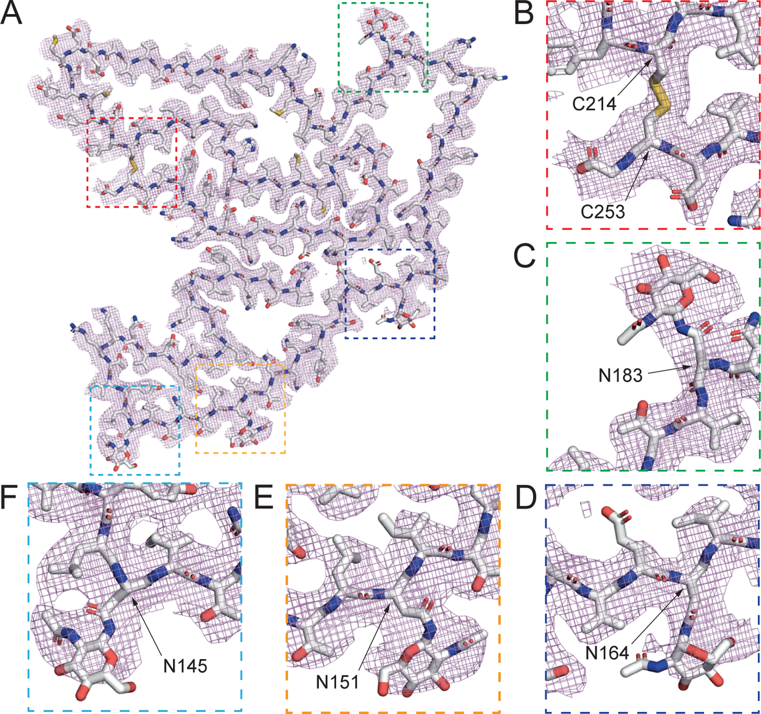
(A) Cryo-EM density (mesh) and atomic model (sticks) of a TMEM106B protofilament. (B) A disulfide bond between C214 and C253. (C) Polymorphic site T185S and glycosylated asparagine at N183. (D) Glycosylated N164, (E) N151, and (F) N145. See also Figures S4 and S5 and Table S2.