PLOS One logoLink to PLOS One
. 2022 Apr 21;17(4):e0263102. doi: 10.1371/journal.pone.0263102

Reversible domain closure modulates GlnBP ligand binding affinity

Qun Chen 1,#, Fang Li 1,#, Xiaobing Zuo 2, Jin Chen 3, Peiwu Qin 1, Chuhui Wang 1, Jin Xu 4, Danyu Yang 4, Baogang Xing 5, Ying Liu 6, Peng Jia 6, Linling Li 7, Chengming Yang 5,*, Dongmei Yu 8,*
Editor: Michael Massiah9
PMCID: PMC9022810  PMID: 35446849

Abstract

Glutamine binding protein (GlnBP) is an Escherichia Coli periplasmic binding protein, which binds and carries glutamine to the inner membrane ATP-binding cassette (ABC) transporter. GlnBP binds the ligand with affinity around 0.1μM measured by isothermal titration calorimetry (ITC) and ligand binding stabilizes protein structure shown by its increase in thermodynamic stability. However, the molecular determinant of GlnBP ligand binding is not known. Electrostatic and hydrophobic interaction between GlnBP and glutamine are critical factors. We propose that the freedome of closure movement is also vital for ligand binding. In order to approve this hypothesis, we generate a series of mutants with different linker length that has different magnitude of domain closure. Mutants show different ligand binding affinity, which indicates that the propensity of domain closure determines the ligand binding affinity. Ligand binding triggers gradual ensemble conformational change. Structural changes upon ligand binding are monitored by combination of small angle x-ray scattering (SAXS) and NMR spectroscopy. Detailed structure characterization of GlnBP contributes to a better understanding of ligand binding and provides the structural basis for biosensor design.

Introduction

In Gram-negative bacteria, periplasmic ATP-binding cassette (ABC) transporter systems are responsible for transporting a broad variety of nutrients across the cytoplasmic membrane. ABC transporter consists of three components: a periplasmic binding protein, which shuttles the ligands through the outer membranes into the periplasmic spaces; an integral membrane protein complex that provides the transmembrane pathway; and two cytoplasmic nucleotide-binding domains that provide the driving power by hydrolyzing ATP molecules. The interaction between the protein transporter and ligand activates the energy-coupling transportation pathway and results in the opening of a channel and the subsequent translocation of the ligand across the cytoplasmic membrane [1, 2].

The first periplasmic binding protein was discovered by Pardee in 1996 [3]. Since then, many periplasmic binding proteins have been characterized and their structures have been studied extensively by x-ray crystallography and solution NMR [47]. The periplasmic binding proteins of ABC transporters from Gram-negative bacteria possess a common architecture (Fig 1). Glutamine binding protein (GlnBP), ATP-binding cassette (ABC) transporter, in this study is Escherichia Coli periplasmic binding protein of L-glutamine, which binds and carries glutamine to the inner membrane. The crystal structures of ligand-free and bound GlnBP show obvious conformational change upon ligand binding by structural alignment [4, 7]. GlnBP is classified in the same Structural Classification of Proteins SCOP family (phosphate- binding protein-like) as human GluR2, but shares 17% sequence identity with GluR2. GlnBP is comprised of two domains linked by rigid β strands, whose movement induces open-to-close conformation upon ligand binding. GlnBP undergoes conformational change due to the hinge bending and twisting motion [8]. The deep central cleft between domains provides the ligand-binding site with sub-micromolar affinity. The conformational changes upon ligand binding have been studied by the combination of chemical shift perturbation and 15N-1H scalar coupling [9]. The aliphatic side chain of the glutamine is sandwiched in a hydrophobic pocket formed between Phe13 and Phe50, which has 21 van der Waals contacts with GlnBP Lys115 and His156. These interactions are unique to GlnBP among known amino acid binding proteins, which apparently contribute to the ligand binding specificity of GlnBP. The hydrogen-bond networks increase the stability of GlnBP-ligand complex [4].

Fig 1. Model of glutamine transportation and structure of GlnBP.

Fig 1

(a) Ligand-bound GlnBP interacts with membrane ABC transporter and induces the transportation of glutamine into the cytoplasm. (b) GlnBP has two domains and the interdomain rotation is demonstrated by the transition from green to red structure model while maintaining the position of another domain when glutamine binds to GlnBP. (c) The linker region of GlnBP is highlighted in red color.

Ligand-bound GlnBP triggers the conformational change of downstream membrane transporter, but not ligand free GlnBP. Ligand binding modifies GlnBP’s binding property with inner membrane transporter. GlnBP structural information is required to delineate GlnBP binding specificity and dynamic property. The use of GlnBP as glutamine biosensor has been proposed. Ligand is removed completely from GlnBP by GdnHCl and EGTA; then GlnBP is refolded on desalting column to remove GdnHCl. Ligand binding affinity is measured by isothermal titration calorimetry. However, the detailed knowledge of protein dynamics, binding affinity, and thermal stability is required for the development of sensor [8]. Here, we present the first small angle x-ray scattering study on GlnBP to monitor the gradual ensemble structural changes upon ligand binding. To map out determining factors for ligand binding, we generate a few mutants with different freedom and magnitude of domain closure. Our hypothesis is that domain closure is critical for ligand binding and this restriction of domain closure can be reversible controlled in a chemical manner. Mutants are created with site-directed mutagenesis to change the ligand affinity by introducing a linker on the opposite site of ligand binding pocket. The molecular weights of mutants are verified by mass spectrometry. We utilized different biophysical techniques to characterize the GlnBP wildtype and mutants. Thermal stability is studied with urea CD titration and Gibbs free energy is extracted from the CD urea titration curve. GlnBP conformational changes have been studied by NMR [5, 9], but the complete backbone assignment is still missing. Sedimentation velocity experiments of analytical ultracentrifuge conclude that GlnBP is monomeric and interference data shows the presence of aggregates ranging from ~0.1% at 83 μM to ~0.2% at 1480 μM. The data presented in this article is a further step towards understanding of the ligand binding, structural changes, and thermodynamics of GlnBP.

Experimental methods

GlnBP protein expression, purification, and removal of ligand

GlnBP gene is cloned into pET11a vector and the sequence is verified by DNA sequencing. Plasmid with GlnBP gene is transformed into BL21 star (DE3) competent cells for protein expression. Bacteria are cultured overnight in a 5 ml LB medium containing 100 μg/ml of ampicilin at 37°C, and then cell pellet (minimize the volume of LB medium) is transferred into 1 L M9 minimal medium (6.78 g Na2HPO4, 3.0 g KH2PO4, 1 g NaCl, 2 g Glucose, 1 g 15N NHCl4, 2 mM MgSO4, 0.2 mM CaCl2, 1x MEM vitamin mix, 1x trace element solution, 100 μg ampicilin per liter medium) at 37°C until OD600 reaches around 0.8. 1 mM IPTG is used to induce protein express for 3 hours at 37°C. Cells are harvested at 5000 g for 30 min at 4°C. The cell pellets are resuspended in the buffer (10 mM sodium acetate, pH 5.5) and broken by microfluidizer at 1200 psi. Clear supernatant is collected by centrifuge at 12,000 rpm for 30 min. Protein is purified by cation exchange (CM column) and GlnBP collections are pooled and buffer exchanged for gel filtration with 10 mM Tris and 300 mM NaCl. The fractions with GlnBP from gel filtration are pooled and exchanged to 10 mM Tris buffer with Amicon Ultra-10K (10,000 Da MWCO) device. Concentrated GlnBP run through cation exchange chromatography and elute with a linear gradient of 0–500 mM NaCl. The identity and integrity of the final protein is confirmed by SDS-PAGE. The ligand glutamine is dissociated from protein by treatment with 6 M guanidine hydrochloride at room temperature for 2 hours. Desalting column is equilibrated with 2 M and 6 M GdnHCl and GlnBP is run through the column to remove glutamine. The desalting column is thoroughly washed and equilibrated with 10 mM Tris. Guanidine is removed by running the desalting column with 10 mM Tris. 10 mM Tris (pH 6.8) buffer will be used for further NMR and ITC experiments, but 10 mM phosphate buffer will be used for CD urea titration because of the strong absorbance of Tris buffer.

Isothermal titration calorimetry

GlnBP is dialyzed into buffer A (10 mM Tris at pH 7.2). Glutamine is dissolved into buffer A. Background titration is performed to check the dilution effect of glutamine. For optimal measurement of ITC, the product of protein concentration and association constant should stay in the range between 1 to 100. The protein concentration is 10–30 μM and ligand is 10 times concentrated about 300 μM. Around 1.8 ml protein is loaded into the sample cell with long syringe and 292 μl ligand is loaded into injection needle with stirring to remove bubbles. First step injects 2 μl ligand into protein solution, and then 10 μl ligand is injected each time with 4 min delay between injections. The plot of DP (different power) with respect to the time is drawn by MicroCal software and thermal parameter and binding affinity are extracted by fitting the data.

Circular dichroism

Urea denaturation is monitored by circular dichroism (CD) with an AVIV Model 62DS spectrometer. Experiments are conducted in 10 mm cuvettes with constant 2.0 mL volume. About 5–6 μM protein in 10 mM phosphate buffer (pH 7.5) is used in all the assays. Wavelength from 190 nm to 230 nm is scanned to find appropriate wavelength to monitor the protein structural changes. The dynode is kept below 500. The urea titration is from 0 M to 8 M with 0.1 M increment for 80 shots and 30 s averaging time. The sample is stirred at 78 rpm during the process of titration. The data is analyzed by Origin. The ellipticity data, corrected for dilution, are fit to Eq (1):

y=yn+mn[urea]+(yu+mu[urea])×exp((ΔG0m[urea]RT))1+exp((ΔG0m[urea]RT)) (1)

where mn and yn are the slope and intercept, respective, for the pre-transition baseline; yu and mu are the slope and intercept for the post-transitional baseline; ΔG0 is the Gibbs energy change for unfolding in the absence of urea; m describes the sensitivity of conformational free energy to urea concentration [10].

Small angle X-ray scattering

SAXS is performed at Argonne National Laboratory sector 12-ID-B. Data are collected at room temperature on a 1024 x 1024-pixel CCD detector with sample-to-detector distance of 1 m and the transmission intensity is measured with a PIN diode beamstop. Protein solution scattering is conducted with concentration of 6.5 mg/ml in 10 mM Tris, pH 7.5. Background scattering from buffer is subtracted from all samples. The zero angle scattering intensity I(0) and overall radius of gyration Rg are obtained from a Guinier approximation to the low-q region of the scattering profiles satisfying the condition, Qmax* Rg < 1.3. The P(r) functions have the characteristic bell shape of globular complexes with well-defined maximum diameter size for all the samples prepared. I(q) is the scattered X-ray intensity per unit solid angle and q is the amplitude of the scattering vector, given by 4π(sinθ)/λ, where 2θ is the scattering angle and λ is the wavelength of the scattered X-rays (0.98 Å). ΔP(r) manifests statistical significant variation in the distribution of the frequency of vectors lengths in the P(r) functions [11].

NMR spectroscopy

NMR experiments are performed at 310 K on a Bruker Ultrashield 800 MHz and 500 MHz with a z-gradient cryoprobe. Protein samples are concentrated to 500 μM with 10 mM Tris (pH 7.5) and 7% D2O is added. All NMR spectra are processed with software package NMRPipe [12] and analyzed using NMRView [13]. Quadrature detection in the indirect dimensions is achieved either with States-TPPI or Echo/antiecho methods. Sequential backbone assignments are obtained from the following two- and three-dimensional experiments: 2D 1H-15N HSQC, 3D HNCO, HNCA, HN(CO)CACB, and HNCACB [14].

Analytical ultracentrifugation

Sedimentation-velocity experiments were performed at 20°C in a Beckman XL-I analytical ultracentrifuge using an An50Ti rotor. Aliquots of protein and reference buffer were loaded into a sedimentation-velocity cell equipped with a dual-sector charcoal-Epon centerpiece. The reference buffer used for studies of the ligand-free protein was an eluted fraction lacking protein, as determined by A280, obtained from size-exclusion chromatography of the protein sample. The reference buffer for centrifugation of CfPutA in the presence of ligands was the buffer from dialysis. Following a 2-h temperature equilibration, the sample was centrifuged at 35,000 rpm. The radial distribution of the sample was monitored with Rayleigh interference optics. Data were acquired at 2-min intervals for 300 radial scans. The data set was analyzed globally to obtain the sedimentation coefficient (c(s)) and molecular mass (c(M)) distributions using Sedfit.

Results

Generation of GlnBP mutants with different domain linker length

We create N170C (Asn 170 was mutated to Cys) mutants and introduce extra cysteine at C terminal that will form disulfide as an artificial liner, which controls the magnitude of domain closure based on our structural analysis. The N170 and C terminal residues are located at the interface between two domains. Our hypothesis is that the linkage of these two residues will perturb the domain closure that will affect ligand binding affinity. We introduce domain linker with 0 to 5 alanines that show different magnitude of restriction on domain closure. The linker composed of disulfide bond can be reversibly broken by reducing reagents.

Characterization of ligand free GlnBP

The structural and thermodynamics change of GlnBP upon ligand binding are investigated by ITC, CD, NMR, and SAXS. Protein is purified by ion exchange and gel filtration chromatography. The purity is checked by SDS-PAGE, which shows a single band at 25 KDa. Overexpressed GlnBP is a mixture of ligand-free and bound GlnBP, which could be inferred from the shoulder peak on ion exchange chromatography. The collections are pooled and treated with guanidine hydrochloride to disrupt the ligand binding pocket and release ligand. The sample is loaded into the desalting column while maintaining 2 M guanidine during the process of separation. Then, we exchange the HPLC buffer with 10 mM Tris and run the sample through desalting column again to remove guanidine (S1 Fig).

NMR assignments and ligand binding characterized by solution NMR

NMR methods have been developed to measure structural changes of GlnBP [9]. However, the complete backbone assignment of ligand-free GlnBP is still missing and 92% backbone assignment is achieved in this study. Assignment starts from residues with unique chemical shifts like alanine (average 19.11 ppm for Cβ), serine and threonine (average 63.74 ppm and 69.62 ppm for Cβ respectively), and glycine (average 45.38 ppm for Cα) (S2 Fig). Connectivity between residues was established initially using MARS [15] in an iterative manner, adding or adjusting assignments manually. Structural changes of GlnBP upon ligand binding are shown by dramatic change in heteronuclear single quantum correlation spectroscopy (HSQC) spectra of ligand-free and bound GlnBP (BMRB ID: 10171). Residues that are far from ligand binding sites also show chemical shift perturbation, which means that the binding event not only change the structure of binding pocket but change the whole protein structure (Fig 2A–2B).

Fig 2.

Fig 2

(a) Overlay of HSQC spectra for ligand-bound and free GlnBP. The red spectrum represents ligand-bound GlnBP and the blue one is GlnBP in ligand-free form; (b) HSQC of ligand-free GlnBP with the assignments.

Gradual structural changes of GlnBP after ligand binding

Sedimentation velocity of analytical ultracentrifuge is used to check the aggregates of GlnBP at different concentrations. GlnBP is primarily monomeric, but inference data shows the presence of aggregates and the amount of aggregate appears to depend on the loading concentration from 0.1% at 83 μM to 2% at 1480 μM. SAXS is sensitive to tertiary and quarterly structural changes that is suitable for monitoring the conformational changes in ligand binding. The protein concentration for SAXS is 197 μM in 10 mM Tris, which corresponds to 4.9 mg/ml. Guinier analysis is used to determine the overall radius of gyration, Rg, and the concentration-normalized forward scattering intensity, I(0)/c, which are functions of spatial size and molecular mass respectively. The Guinier plots are linear at the low q-range at all concentrations, indicating that GlnBP has a well-defined size under aqueous condition and radiation-induced aggregation is not an issue. Rg and I(0)/c, determined from linear fitting to the data over the q range satisfying the q * Rg < 1.3 condition (q = 0.0146 to 0.0416 Å-1), display very little concentration dependence, indicating that no change in oligomeric state occurs over the investigated concentration range (Fig 3A and 3B). Structural changes of GlnBP are also shown by SAXS ΔP(r) curves. With the increase in ligand concentration, the gradual changes are shown on ΔP(r) curves (Fig 3C and 3D). The gradual changes indicate the gradual occupation of binding pockets after increase in ligand concentration. The conformational changes are not abrupt and the magnitude of changes correlate with ligand binding.

Fig 3. Small angle X-ray scattering of GlnBP.

Fig 3

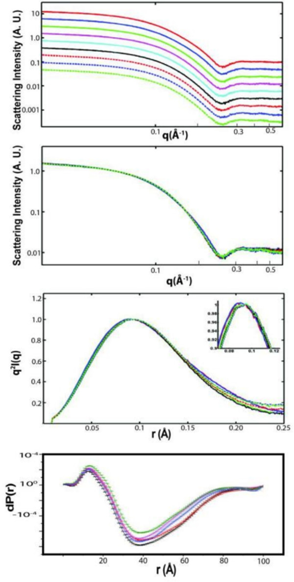
(a) Offset of I(q) vs q plot for GlnBP with different concentrations of ligand. Red line is 400 μM and green line is 0 μM. The increment is 50 μM from 0 to 400 μM; (b) Overlay of I(q) vs q plots for GlnBP with different concentrations of ligand; (c) Pair distance distribution function P(r) plot of GlnBP with different concentrations of ligands; (d) ΔP(r) shows the difference of conformational changes when different concentrations of ligand were titrated into the GlnBP protein solution.

Thermal stability of GlnBP in ligand free and bound state

Thermo stability is measured with urea CD titration (Fig 4A–4B). The difference of Gibbs free energy between ligand-free and bound GlnBP is 59.82 kJ/mol and the energy of hydrogen bond is 5–30 kJ/mol, which indicates that ligand binding introduces extra hydrogen bonds and van der Walls interactions that account for the energy difference. Tertiary structure changes dramatically but secondary structures are maintained, which explain the higher energy level of ligand-free GlnBP. GlnBP is rigid with [10] 15N NOE values around 0.9 except the N and C terminals and some residues that show slightly higher flexibility (data not shown).

Fig 4. CD Urea titration.

Fig 4

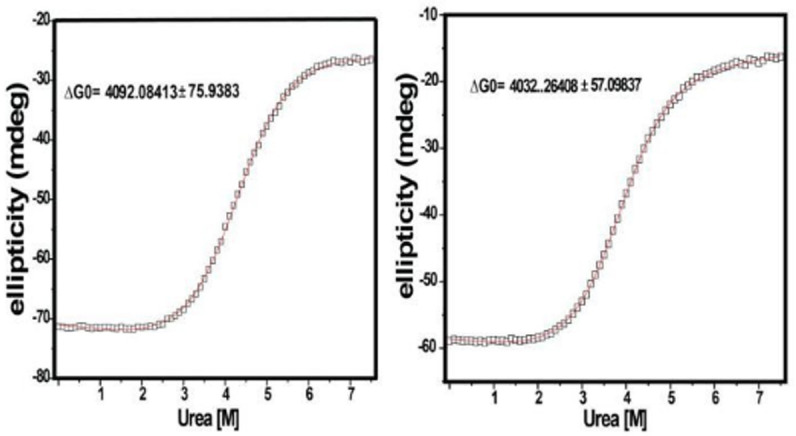
(a) 10 M urea was titrated into 5 M ligand bound and (b) ligand free GlnBP solution.

Domain closure affects GlnBP ligand binding affinity

The ligand binding affinity is measured with ITC for wild-type and mutants of GlnBP. Mutants with different ligand binding affinity are created by introducing a polypeptide linker composed of different numbers of alanine residue from 0 to 5 residues. The smaller side chain of alanine reduces the possibility of creating steric hindrance. The length of the linker between N170C (Asn 170 was mutated to Cys) and C terminal cysteine controls the magnitude of domain closure based on our structural analysis. The formation of disulfide bond is verified by Ellman’s assay and mass spectrometry. The formation of disulfide bond decreases molecular weight by 2 due to the formation of new polypeptide bond, which can be verified by the molecular weight of GlnBP mutant with four alanine liner. Reduction of disulfide bond changes the molecular weight from 25043 to 25041 (S3 Fig). The correlation between domain closure and ligand binding is observed since shorter linker mutants have a lower binding affinity, which indicates that ligand binding triggers the protein conformational change and induces domain closure to create a perfect binding site for ligand. The mutants with artificial linker can be reversed by reducing reagent. The presence of linker inhibits domain closure and reduces ligand binding affinity. Breakage of linker restore the ligand binding comparable to wild type protein (Fig 5, S4 Fig).

Fig 5. ITC of ligand binding.

Fig 5

(a) ITC for wild-type GlnBP; (b) ITC for GlnBP 2A mutant and (c) with the addition of reducing reagents (DTT); (d) ITC for GlnBP 3A mutant and (e) with the addition of reducing reagents (DTT).

Discussion

GlnBP is a soluble glutamine binding protein swimming in the periplasmic space and escorts free glutamine into membrane transporter. Ligand binding to GlnBP triggers a large-scale conformational change and two domains close to pack the glutamine into the ligand binding pocket. The ligand binding affinity determines the efficiency of transportation in vivo and the application of GlnBP in the field of biosensors. Dynamics of GlnBP have been characterized by NMR [5] and molecular dynamics simulation [16]. However, the correlation of conformation change and ligand binding affinity remains elusive. We use NMR, SAXS, and ITC to characterize the ligand binding of GlnBP and corresponding structural changes. The complementary nature of these techniques provides biophysical insights of molecular binding. We generate GlnBP mutants with different domain linker length to investigate the perturbation of domain closure capability on protein ligand binding. We find that mutants of GlnBP with different domain closure capability show different ligand binding property. Longer linker allows the free domain closure that has the highest ligand binding affinity. Shorter domain linker blocks domain closure that has the lowest binding affinity. Breakage of disulfide bond release the restriction on domain closure and restore ligand binding affinity. We discover a reversible way to control domain closure and ligand binding. The study shows the correlation between ligand binding affinity and domain closure, which is the major conformational change upon ligand binding and determinant for ligand binding.

The structure of GlnBP is strikingly similar to the ligand binding core (S1S2) of ionotropic glutamate receptor. Ionotropic glutamate receptors (iGluR) are major excitatory receptors in the vertebrate central nervous system. The ligand-binding core of iGluR is formed by polypeptide fragments S1 and S2, which could be coupled with a 13-residue liner to form a new construct S1S2. S1S2 has the same binding property as wild-type protein. The homotetrameric structure of intact iGluR was resolved recently and it demonstrates that S1S2 construct has identical structure as the ligand-binding core of intact receptor. Structural and dynamic information obtained from GlnBP could shed light on the function of iGluR and provides hints about the structural conservation even if their sequences have diverged dramatically. Potential drug development can target the domain closure to perturb the ligand binding, which provide theoretical foundation for targeted therapeutics.

Supporting information

S1 Fig. Purification of GlnBP and removal of Gln ligand through desalting column.

(TIF)

S2 Fig. Representative 3D NMR for GlnBP backbone chemical shift assignment.

(TIF)

S3 Fig. Mass spectrometer characterization of GlnBP in intact and reduce format.

The corresponding molecular weight is 25041 and 25043.

(TIF)

S4 Fig. ITC of GlnBP with 4 and 5 alanine linkers.

(TIF)

Acknowledgments

The use of the Advanced Photon Source, an Office of Science User Facility operated for the U.S. Department of Energy (DOE) Office of Science by Argonne National Laboratory, was supported by the U.S. DOE.

Data Availability

The data are available through the BMRB Data Bank, ID number: 50310.

Funding Statement

Prof. Peiwu Qin received funding from the National Natural Science Foundation of China (grant numbers 31970742, 81972854, 81802626, http://www.nsfc.gov.cn/english/site_1/index.html), the Science, Technology and Innovation Commission of Shenzhen Municipality (grant number JSGG20191129110812, http://stic.sz.gov.cn/), and the Southern University of Science and Technology Hospital Dean Research Fund (2020-A4). The funders had no role in study design, data collection and analysis, decision to publish, or preparation of the manuscript.

References

  • 1.Grunau A, Geraki K, Grossmann JG, Gutierrez A. Conformational dynamics and the energetics of protein—ligand interactions: role of interdomain loop in human cytochrome P450 reductase. Biochemistry. 2007;46(28):8244–55. Epub 2007/06/22. doi: 10.1021/bi700596s . [DOI] [PubMed] [Google Scholar]
  • 2.Hu Y, Fan CP, Fu G, Zhu D, Jin Q, Wang DC. Crystal structure of a glutamate/aspartate binding protein complexed with a glutamate molecule: structural basis of ligand specificity at atomic resolution. Journal of molecular biology. 2008;382(1):99–111. Epub 2008/07/22. doi: 10.1016/j.jmb.2008.06.091 . [DOI] [PubMed] [Google Scholar]
  • 3.Linton KJ. Structure and function of ABC transporters. Physiology (Bethesda). 2007;22:122–30. Epub 2007/04/11. doi: 10.1152/physiol.00046.2006 . [DOI] [PubMed] [Google Scholar]
  • 4.Sun YJ, Rose J, Wang BC, Hsiao CD. The structure of glutamine-binding protein complexed with glutamine at 1.94 A resolution: comparisons with other amino acid binding proteins. Journal of molecular biology. 1998;278(1):219–29. Epub 1998/05/26. doi: 10.1006/jmbi.1998.1675 . [DOI] [PubMed] [Google Scholar]
  • 5.Bermejo GA, Strub MP, Ho C, Tjandra N. Ligand-free open-closed transitions of periplasmic binding proteins: the case of glutamine-binding protein. Biochemistry. 2010;49(9):1893–902. Epub 2010/02/10. doi: 10.1021/bi902045p ; PubMed Central PMCID: PMC2831130. [DOI] [PMC free article] [PubMed] [Google Scholar]
  • 6.Yu J, Simplaceanu V, Tjandra NL, Cottam PF, Lukin JA, Ho C. 1H, 13C, and 15N NMR backbone assignments and chemical-shift-derived secondary structure of glutamine-binding protein of Escherichia coli. Journal of biomolecular NMR. 1997;9(2):167–80. Epub 1997/02/01. doi: 10.1023/a:1018606304131 . [DOI] [PubMed] [Google Scholar]
  • 7.Hsiao CD, Sun YJ, Rose J, Wang BC. The crystal structure of glutamine-binding protein from Escherichia coli. Journal of molecular biology. 1996;262(2):225–42. Epub 1996/09/20. doi: 10.1006/jmbi.1996.0509 . [DOI] [PubMed] [Google Scholar]
  • 8.Pang A, Arinaminpathy Y, Sansom MS, Biggin PC. Interdomain dynamics and ligand binding: molecular dynamics simulations of glutamine binding protein. FEBS letters. 2003;550(1–3):168–74. Epub 2003/08/26. doi: 10.1016/s0014-5793(03)00866-4 . [DOI] [PubMed] [Google Scholar]
  • 9.Ma J, Gruschus JM, Tjandra N. 15N-1H scalar coupling perturbation: an additional probe for measuring structural changes due to ligand binding. Journal of the American Chemical Society. 2009;131(29):9884–5. Epub 2009/07/08. doi: 10.1021/ja903552q . [DOI] [PMC free article] [PubMed] [Google Scholar]
  • 10.Henzl MT, Ndubuka K. Low-affinity signature of the rat beta-parvalbumin CD site. Evidence for remote determinants. Biochemistry. 2007;46(1):23–35. Epub 2007/01/03. doi: 10.1021/bi061421h . [DOI] [PubMed] [Google Scholar]
  • 11.Fenton AW, Williams R, Trewhella J. Changes in small-angle X-ray scattering parameters observed upon binding of ligand to rabbit muscle pyruvate kinase are not correlated with allosteric transitions. Biochemistry. 2010;49(33):7202–9. Epub 2010/08/18. doi: 10.1021/bi100147w ; PubMed Central PMCID: PMC2924747. [DOI] [PMC free article] [PubMed] [Google Scholar]
  • 12.Delaglio F, Grzesiek S, Vuister GW, Zhu G, Pfeifer J, Bax A. NMRPipe: a multidimensional spectral processing system based on UNIX pipes. Journal of biomolecular NMR. 1995;6(3):277–93. Epub 1995/11/01. doi: 10.1007/BF00197809 . [DOI] [PubMed] [Google Scholar]
  • 13.Johnson BA. Using NMRView to visualize and analyze the NMR spectra of macromolecules. Methods Mol Biol. 2004;278:313–52. Epub 2004/08/20. doi: 10.1385/1-59259-809-9:313 . [DOI] [PubMed] [Google Scholar]
  • 14.Ferentz AE, Wagner G. NMR spectroscopy: a multifaceted approach to macromolecular structure. Quarterly reviews of biophysics. 2000;33(1):29–65. Epub 2000/11/15. doi: 10.1017/s0033583500003589 . [DOI] [PubMed] [Google Scholar]
  • 15.Jung YS, Zweckstetter M. Mars—robust automatic backbone assignment of proteins. Journal of biomolecular NMR. 2004;30(1):11–23. Epub 2004/09/29. doi: 10.1023/B:JNMR.0000042954.99056.ad . [DOI] [PubMed] [Google Scholar]
  • 16.Loeffler HH, Kitao A. Collective dynamics of periplasmic glutamine binding protein upon domain closure. Biophysical journal. 2009;97(9):2541–9. Epub 2009/11/04. doi: 10.1016/j.bpj.2009.08.019 ; PubMed Central PMCID: PMC2770614. [DOI] [PMC free article] [PubMed] [Google Scholar]

Decision Letter 0

Michael Massiah

Transfer Alert

This paper was transferred from another journal. As a result, its full editorial history (including decision letters, peer reviews and author responses) may not be present.

29 Jan 2021

PONE-D-20-37100

Combination of SAXS and NMR to monitor the conformation changes of E. Coli glutaminine binding protein upon ligand binding

PLOS ONE

Dear Dr. Yu,

Thank you for submitting your manuscript to PLOS ONE. Let me apologize for the slowness of the review process. The holiday season and COVID19 has affected most schedules.  After careful consideration, we feel that it has merit but does not fully meet PLOS ONE’s publication criteria as it currently stands. As you can see, one reviewer had fair but strong objections to the manuscript.  Despite their recommendation, I will recommend major revision because if these objections can be properly addressed, it would make the manuscript stronger. Also the other reviewer was less critical but you can see if their review that some of their concerns also resonant with comments of the other reviewer. Therefore, we invite you to submit a revised version of the manuscript that addresses the points raised during the review process.

Please submit your revised manuscript by Mar 13 2021 11:59PM. If you will need more time than this to complete your revisions, please reply to this message or contact the journal office at plosone@plos.org. When you're ready to submit your revision, log on to https://www.editorialmanager.com/pone/ and select the 'Submissions Needing Revision' folder to locate your manuscript file.

Please include the following items when submitting your revised manuscript:

  • A rebuttal letter that responds to each point raised by the academic editor and reviewer(s). You should upload this letter as a separate file labeled 'Response to Reviewers'.

  • A marked-up copy of your manuscript that highlights changes made to the original version. You should upload this as a separate file labeled 'Revised Manuscript with Track Changes'.

  • An unmarked version of your revised paper without tracked changes. You should upload this as a separate file labeled 'Manuscript'.

If you would like to make changes to your financial disclosure, please include your updated statement in your cover letter. Guidelines for resubmitting your figure files are available below the reviewer comments at the end of this letter.

If applicable, we recommend that you deposit your laboratory protocols in protocols.io to enhance the reproducibility of your results. Protocols.io assigns your protocol its own identifier (DOI) so that it can be cited independently in the future. For instructions see: http://journals.plos.org/plosone/s/submission-guidelines#loc-laboratory-protocols

We look forward to receiving your revised manuscript.

Kind regards,

Michael Massiah

Academic Editor

PLOS ONE

Journal Requirements:

When submitting your revision, we need you to address these additional requirements.

1. Please ensure that your manuscript meets PLOS ONE's style requirements, including those for file naming. The PLOS ONE style templates can be found at

https://journals.plos.org/plosone/s/file?id=wjVg/PLOSOne_formatting_sample_main_body.pdf and

https://journals.plos.org/plosone/s/file?id=ba62/PLOSOne_formatting_sample_title_authors_affiliations.pdf

2. Thank you for stating the following in the Acknowledgments Section of your manuscript:

"This work was funded by National Natural Science Foundation of China (31970742, 81972854, 81802626), Science,

Technology and Innovation Commission of Shenzhen Municipality (JSGG20191129110812), and Southern

University of Science and Technology Hospital Dean Research Fund (2020-A4)."

We note that you have provided funding information that is not currently declared in your Funding Statement. However, funding information should not appear in the Acknowledgments section or other areas of your manuscript. We will only publish funding information present in the Funding Statement section of the online submission form.

Please remove any funding-related text from the manuscript and let us know how you would like to update your Funding Statement. Currently, your Funding Statement reads as follows:

"Prof. Peiwu Qin received National Natural Science Foundation of China (31970742, 81972854, 81802626, http://www.nsfc.gov.cn/english/site_1/index.html), and Science, Technology and Innovation Commission of Shenzhen Municipality (JSGG20191129110812, http://stic.sz.gov.cn/). The funders had no role in study design, data collection and analysis, decision to publish, or preparation of the manuscript."

Please include your amended statements within your cover letter; we will change the online submission form on your behalf.

3. We note that you have stated that you will provide repository information for your data at acceptance. Should your manuscript be accepted for publication, we will hold it until you provide the relevant accession numbers or DOIs necessary to access your data. If you wish to make changes to your Data Availability statement, please describe these changes in your cover letter and we will update your Data Availability statement to reflect the information you provide.

[Note: HTML markup is below. Please do not edit.]

Reviewers' comments:

Reviewer's Responses to Questions

Comments to the Author

1. Is the manuscript technically sound, and do the data support the conclusions?

The manuscript must describe a technically sound piece of scientific research with data that supports the conclusions. Experiments must have been conducted rigorously, with appropriate controls, replication, and sample sizes. The conclusions must be drawn appropriately based on the data presented.

Reviewer #1: Yes

Reviewer #2: No

**********

2. Has the statistical analysis been performed appropriately and rigorously?

Reviewer #1: Yes

Reviewer #2: No

**********

3. Have the authors made all data underlying the findings in their manuscript fully available?

The PLOS Data policy requires authors to make all data underlying the findings described in their manuscript fully available without restriction, with rare exception (please refer to the Data Availability Statement in the manuscript PDF file). The data should be provided as part of the manuscript or its supporting information, or deposited to a public repository. For example, in addition to summary statistics, the data points behind means, medians and variance measures should be available. If there are restrictions on publicly sharing data—e.g. participant privacy or use of data from a third party—those must be specified.

Reviewer #1: No

Reviewer #2: No

**********

4. Is the manuscript presented in an intelligible fashion and written in standard English?

PLOS ONE does not copyedit accepted manuscripts, so the language in submitted articles must be clear, correct, and unambiguous. Any typographical or grammatical errors should be corrected at revision, so please note any specific errors here.

Reviewer #1: Yes

Reviewer #2: No

**********

5. Review Comments to the Author

Please use the space provided to explain your answers to the questions above. You may also include additional comments for the author, including concerns about dual publication, research ethics, or publication ethics. (Please upload your review as an attachment if it exceeds 20,000 characters)

Reviewer #1: The authors have investigated the configurational modifications of the glutamine binding protein (GlnBP) upon ligand binding by NMR and SAXS in order to determine the correlation between ligand binding affinity and domain closure. Dynamics of the hinge region, located between the two domains that constitute the protein, induce open-to-close conformation upon ligand binding and can be determined by variation of ligand concentration and mutants with linkers of different lengths. Although a complete structure determination of the conformational variations has not been achieved, the authors present a relevant structural characterization that contributes to the knowledge in the corresponding research field.

This article is of interest, well-written, and the experiments seem to be well-conducted, with few issues that need to be addressed that are listed below.

- The authors could use SAXS analysis to obtain envelope models for the different domain closure configurations and complement NMR data. Different methodologies for protein structure determination by NMR and SAXS conjugation can be implemented, as described at Rodriguez-Zamora, P. "Conjugation of NMR and SAXS for flexible and multidomain protein structure determination: from sample preparation to model refinement." Progress in biophysics and molecular biology 150 (2020): 140-144. If such data is available it should be included in the article, otherwise it would be recommended to mention the possibility of an NMR-SAXS conjugated analysis as a perspective for this work.

- A figure showing the protein hinge section and the linker introduction is required to illustrate the region of interest.

- Figure 5 is missing a color code that clarifies the results obtained by SAXS.

- Few grammar errors need to be corrected throughout the manuscript. A PDF copy with some of those errors highlighted is attached.

Reviewer #2: The manuscript by Chen et al. described the structure changes of addition mutant of E. coli. Glutamine binding protein (GlnBP) upon ligand (glutamine) binding. The experimental methods of ITC, NMR, SAXS etc. were applied. The conclusion was the change of binding affinity was associated with quaternary structure change (inter-domain) caused by the lengthened linker mutant. While the inter-domain reorganization may contribute to the binding affinity changes, the contribution is one of many factors that could affect binding affinity. Potentially, electrostatic and hydrophobic interactions between protein side chains and Gln should dominate the interaction, not discussed in the manuscript. The authors don’t seem to have a clear experimental design or conclusion. In general, for a study like this, the hypotheses should be raised at the beginning, followed by experimental demonstration, result interpretation and final conclusion. Now, the manuscript lacks coherent reasoning or conclusion. In the end, there is no take home message from the study. The manuscript is highly immature and should not be published in the current format. Major issues are listed below.

1. The result did not provide any quantitative correlation between ITC results and any others like SAXS or NMR. Therefore, the science advance on this binding topic is minimal.

2. The scattering of “Results” part was not acceptable, where sections were missing. The authors appeared to be random in narratively describing the results. Normally, sections of mutation selection, structure changes, correlation between xxx and yyy etc, should be in “Results”. For a biophysics study like this, a table of GlnBP protein of WT, mutant 1, mutant 2 … should be presented. This should be followed by structural characterization (NMR., SAXS, SV-AUC) at free and binding states of each protein. The authors should then infer from analytical results on the structural change of GlnBP at secondary, tertiary, quaternary structure levels. For example, NMR chemical shift could be sensitive to secondary and tertiary structure, relaxation could be sensitive to quaternary and oligomerization, SAXS could be sensitive to quaternary or domain structure, SV-AUC is for oligomerization. All of these standard reasonings are missing in the manuscript.

3. The 1st paragraph in “Results” belongs to method description.

4. Page 2 Paragraph 1, the statement “two domain linked by a flexible “hinge” region” was not correct. The 2 domains were linked by 2 rigid beta-strands per crystal structure.

5. The method section missed the description of SV-AUC method.

**********

6. PLOS authors have the option to publish the peer review history of their article (what does this mean?). If published, this will include your full peer review and any attached files.

If you choose “no”, your identity will remain anonymous but your review may still be made public.

Do you want your identity to be public for this peer review? For information about this choice, including consent withdrawal, please see our Privacy Policy.

Reviewer #1: No

Reviewer #2: No

[NOTE: If reviewer comments were submitted as an attachment file, they will be attached to this email and accessible via the submission site. Please log into your account, locate the manuscript record, and check for the action link "View Attachments". If this link does not appear, there are no attachment files.]

While revising your submission, please upload your figure files to the Preflight Analysis and Conversion Engine (PACE) digital diagnostic tool, https://pacev2.apexcovantage.com/. PACE helps ensure that figures meet PLOS requirements. To use PACE, you must first register as a user. Registration is free. Then, login and navigate to the UPLOAD tab, where you will find detailed instructions on how to use the tool. If you encounter any issues or have any questions when using PACE, please email PLOS at figures@plos.org. Please note that Supporting Information files do not need this step.

Attachment

Submitted filename: Review_Combination of SAXS and NMR to monitor the conformation changes of E.pdf

Attachment

Submitted filename: PONE-D-20-37100_reviewer.pdf

Attachment

Submitted filename: review.docx

PLoS One. 2022 Apr 21;17(4):e0263102. doi: 10.1371/journal.pone.0263102.r002

Author response to Decision Letter 0


24 Aug 2021

Reviewer #1: The authors have investigated the configurational modifications of the glutamine binding protein (GlnBP) upon ligand binding by NMR and SAXS in order to determine the correlation between ligand binding affinity and domain closure. Dynamics of the hinge region, located between the two domains that constitute the protein, induce open-to-close conformation upon ligand binding and can be determined by variation of ligand concentration and mutants with linkers of different lengths. Although a complete structure determination of the conformational variations has not been achieved, the authors present a relevant structural characterization that contributes to the knowledge in the corresponding research field.

This article is of interest, well-written, and the experiments seem to be well-conducted, with few issues that need to be addressed that are listed below.

1- The authors could use SAXS analysis to obtain envelope models for the different domain closure configurations and complement NMR data. Different methodologies for protein structure determination by NMR and SAXS conjugation can be implemented, as described at Rodriguez-Zamora, P. "Conjugation of NMR and SAXS for flexible and multidomain protein structure determination: from sample preparation to model refinement." Progress in biophysics and molecular biology 150 (2020): 140-144. If such data is available it should be included in the article, otherwise it would be recommended to mention the possibility of an NMR-SAXS conjugated analysis as a perspective for this work.

We thank reviewer for the valuable suggestions. We conducted SAXS experiments at Argonne National Laboratory and the data was analyzed with the assistance of the staff scientist at Argonne. Both the ligand bound and free GlnBP have been solved by X-ray crystallography. Thus, we feel it is not necessary to get the shape of molecules since atomic structure is available. SAXS is capable of monitoring the gradual structural changes upon ligand binding, which is not possible by x-ray diffraction. The structural changes of GlnBP were confirmed and monitored from two complementary techniques, which is enough for our purpose to claim the gradual domain closure upon ligand binding.

2- A figure showing the protein hinge section and the linker introduction is required to illustrate the region of interest.

For the GlnBP structure, we add figure 1c to illustrate the linker region of interest. The green indicates the large domain of GlnBP, the magenta represents the small domain, and the hinges we are interested (residues 85–89 and residues 181–185) is highlighted in red. We introduce the linker between position N170 and C terminal labeled with cyan color.

Red line is 400 μM and green line is 0 μM. The increment is 50 μM from 0 to 400 μM.

- Few grammar errors need to be corrected throughout the manuscript. A PDF copy with some of those errors highlighted is attached.

We thank reviewer for pinpointing the grammar errors and we correct them.

Reviewer #2: The manuscript by Chen et al. described the structure changes of addition mutant of E. coli. Glutamine binding protein (GlnBP) upon ligand (glutamine) binding. The experimental methods of ITC, NMR, SAXS etc. were applied. The conclusion was the change of binding affinity was associated with quaternary structure change (inter-domain) caused by the lengthened linker mutant. While the inter-domain reorganization may contribute to the binding affinity changes, the contribution is one of many factors that could affect binding affinity. Potentially, electrostatic and hydrophobic interactions between protein side chains and Gln should dominate the interaction, not discussed in the manuscript. The authors don’t seem to have a clear experimental design or conclusion. In general, for a study like this, the hypotheses should be raised at the beginning, followed by experimental demonstration, result interpretation and final conclusion. Now, the manuscript lacks coherent reasoning or conclusion. In the end, there is no take home message from the study. The manuscript is highly immature and should not be published in the current format. Major issues are listed below.

We thank reviewer for the valuable comments. We did make a hypothesis that domain closure is critical for ligand binding at the beginning. In order to approve this hypothesis, we create a series of artificial linker to change the domain closure. Then, we study how the ligand binding is affected by these mutants. The reviewer is absolutely correct that electrostatic and hydrophobic interactions between side chain and Gln influence ligand binding. However, this has been demonstrated in many ligand binding proteins. Domain closure or linker affect ligand binding, which has never been investigated to our knowledge. We maintain the protein sequence to exclude the possibility that electrostatic or hydrophobic interaction determines ligand binding.

We add a few sentences in the discussion to highlight the significance of this study. We use NMR, SAXS, and ITC to characterize the ligand binding of GlnBP and corresponding structural changes. The complementary nature of these techniques provide biophysical insights of molecular binding. We generate GlnBP mutants with different domain linker length to investigate the perturbation of domain closure capability on protein ligand binding. We find that mutants of GlnBP with different domain closure capability show different ligand binding property. Longer linker allows the free domain closure that has the highest ligand binding affinity. Shorter domain linker blocks domain closure that has the lowest binding affinity. Breakage of disulfide bond releases the restriction on domain closure and restore ligand binding affinity. We discover a reversible way to control domain closure and ligand binding. The study shows the correlation between ligand binding affinity and domain closure for the first time, which is the major conformational change upon ligand binding and determinant for ligand binding.

1. The result did not provide any quantitative correlation between ITC results and any others like SAXS or NMR. Therefore, the science advance on this binding topic is minimal.

ITC, NMR, and SAXS study different properties of protein complex. ITC studies the ligand binding affinity that shows different linker length perturbs the ligand binding affinity. Then, we use SAXS to show that ligand binding gradually trigger domain closure and protein conformation changes. The results from three techniques are complementary and complete to characterize the ligand binding. SAXS and NMR both provide aqueous structural information and we have demonstrated the ligand binding induced conformation changes from these two techniques. Since crystal structures of GlnBP in ligand free and bound state are available, we skip the generation of protein shape by SAXS and NMR structure determination.

2. The scattering of “Results” part was not acceptable, where sections were missing. The authors appeared to be random in narratively describing the results. Normally, sections of mutation selection, structure changes, correlation between xxx and yyy etc, should be in “Results”. For a biophysics study like this, a table of GlnBP protein of WT, mutant 1, mutant 2 … should be presented. This should be followed by structural characterization (NMR., SAXS, SV-AUC) at free and binding states of each protein. The authors should then infer from analytical results on the structural change of GlnBP at secondary, tertiary, quaternary structure levels. For example, NMR chemical shift could be sensitive to secondary and tertiary structure, relaxation could be sensitive to quaternary and oligomerization, SAXS could be sensitive to quaternary or domain structure, SV-AUC is for oligomerization. All of these standard reasonings are missing in the manuscript.

We thank reviewer for this valuable comment. We add section titles in the results part. We add one more section to introduce the design logic behind the mutants with different length of domain linker as the following:

Generation of GlnBP mutants with different domain linker length

We create N170C (Asn 170 was mutated to Cys) mutant and introduce extra cysteine at C terminal that will form disulfide as an artificial liner, which controls the magnitude of domain closure based on our structural analysis. The N170 and C terminal residues are located at the interface between two domains. Our hypothesis is that the linkage of these two residues will perturb the domain closure that will affect ligand binding affinity. We introduce domain linker with 0 to 5 alanine that show different magnitude of restriction on domain closure. The linker composed of disulfide bond can be reversibly broken by reducing reagents.

We add a few sentences to each section of results to give detail explanation about the physical and biological meaning of the data.

We agree with the reviewer that correlation between xxx and yyy etc, should be in “Results”. We show the results in a reversible assay. The mutants we generate can introduce a reversible linker. The ligand binding affinity is perturbed and restored after domain linker formation or breakage. Reversibility is a stronger evidence than correlation analysis. The biophysical measurements of diverse GlnBP mutants are not complete between the structural changes are minimal and ligand binding is the most important property we are interested.

3. The 1st paragraph in “Results” belongs to method description.

The first paragraph illustrates how we prepare and characterize ligand free GlnBP since GlnBP from cell lysate contain both ligand free and bound GlnBP. We treat the purified sample with detergent and remove the ligand. If the sample is a mixture of ligand free and bound format, the following binding assay will have great measurement errors.

4. Page 2 Paragraph 1, the statement “two domain linked by a flexible “hinge” region” was not correct.

We changed to “The 2 domains were linked by 2 rigid β-strands per crystal structure.”

5. The method section missed the description of SV-AUC method.

We thank reviewer for pointing this out. We add one paragraph to describe the SV-AUC method as the following:

Analytical ultracentrifugation

Sedimentation-velocity experiments were performed at 20 °C in a Beckman XL-I analytical ultracentrifuge using an An50Ti rotor. Aliquots of protein and reference buffer were loaded into a sedimentation-velocity cell equipped with a dual-sector charcoal-Epon centerpiece. The reference buffer used for studies of the ligand-free protein was an eluted fraction lacking protein, as determined by A280, obtained from size-exclusion chromatography of the protein sample. The reference buffer for centrifugation of CfPutA in the presence of ligands was the buffer from dialysis. Following a 2-h temperature equilibration, the sample was centrifuged at 35,000 rpm. The radial distribution of the sample was monitored with Rayleigh interference optics. Data were acquired at 2-min intervals for 300 radial scans. The data set was analyzed globally to obtain the sedimentation coefficient (c(s)) and molecular mass (c(M)) distributions using Sedfit.

Attachment

Submitted filename: Response to the Reviewers-only some parts-v3.docx

Decision Letter 1

Michael Massiah

16 Sep 2021

PONE-D-20-37100R1Combination of SAXS and NMR to monitor the conformation changes of E. Coli glutaminine binding protein upon ligand bindingPLOS ONE

Dear Dr. Yu,

Thank you for submitting your manuscript to PLOS ONE. As you can see, one of the reviewers still has serious concerns, some of which appears to not be address in this revision. Can you please address of these concerns. Thanks. After careful consideration, we feel that it has merit but does not fully meet PLOS ONE’s publication criteria as it currently stands. Therefore, we invite you to submit a revised version of the manuscript that addresses the points raised during the review process.

Please submit your revised manuscript by Oct 31 2021 11:59PM. If you will need more time than this to complete your revisions, please reply to this message or contact the journal office at plosone@plos.org. When you're ready to submit your revision, log on to https://www.editorialmanager.com/pone/ and select the 'Submissions Needing Revision' folder to locate your manuscript file.

Please include the following items when submitting your revised manuscript:

  • A rebuttal letter that responds to each point raised by the academic editor and reviewer(s). You should upload this letter as a separate file labeled 'Response to Reviewers'.

  • A marked-up copy of your manuscript that highlights changes made to the original version. You should upload this as a separate file labeled 'Revised Manuscript with Track Changes'.

  • An unmarked version of your revised paper without tracked changes. You should upload this as a separate file labeled 'Manuscript'.

If you would like to make changes to your financial disclosure, please include your updated statement in your cover letter. Guidelines for resubmitting your figure files are available below the reviewer comments at the end of this letter.

If applicable, we recommend that you deposit your laboratory protocols in protocols.io to enhance the reproducibility of your results. Protocols.io assigns your protocol its own identifier (DOI) so that it can be cited independently in the future. For instructions see: https://journals.plos.org/plosone/s/submission-guidelines#loc-laboratory-protocols. Additionally, PLOS ONE offers an option for publishing peer-reviewed Lab Protocol articles, which describe protocols hosted on protocols.io. Read more information on sharing protocols at https://plos.org/protocols?utm_medium=editorial-email&utm_source=authorletters&utm_campaign=protocols.

We look forward to receiving your revised manuscript.

Kind regards,

Michael Massiah

Academic Editor

PLOS ONE

Journal Requirements:

Please review your reference list to ensure that it is complete and correct. If you have cited papers that have been retracted, please include the rationale for doing so in the manuscript text, or remove these references and replace them with relevant current references. Any changes to the reference list should be mentioned in the rebuttal letter that accompanies your revised manuscript. If you need to cite a retracted article, indicate the article’s retracted status in the References list and also include a citation and full reference for the retraction notice.

[Note: HTML markup is below. Please do not edit.]

Reviewers' comments:

Reviewer's Responses to Questions

Comments to the Author

1. If the authors have adequately addressed your comments raised in a previous round of review and you feel that this manuscript is now acceptable for publication, you may indicate that here to bypass the “Comments to the Author” section, enter your conflict of interest statement in the “Confidential to Editor” section, and submit your "Accept" recommendation.

Reviewer #1: All comments have been addressed

Reviewer #2: (No Response)

**********

2. Is the manuscript technically sound, and do the data support the conclusions?

The manuscript must describe a technically sound piece of scientific research with data that supports the conclusions. Experiments must have been conducted rigorously, with appropriate controls, replication, and sample sizes. The conclusions must be drawn appropriately based on the data presented.

Reviewer #1: Partly

Reviewer #2: No

**********

3. Has the statistical analysis been performed appropriately and rigorously?

Reviewer #1: N/A

Reviewer #2: No

**********

4. Have the authors made all data underlying the findings in their manuscript fully available?

The PLOS Data policy requires authors to make all data underlying the findings described in their manuscript fully available without restriction, with rare exception (please refer to the Data Availability Statement in the manuscript PDF file). The data should be provided as part of the manuscript or its supporting information, or deposited to a public repository. For example, in addition to summary statistics, the data points behind means, medians and variance measures should be available. If there are restrictions on publicly sharing data—e.g. participant privacy or use of data from a third party—those must be specified.

Reviewer #1: Yes

Reviewer #2: No

**********

5. Is the manuscript presented in an intelligible fashion and written in standard English?

PLOS ONE does not copyedit accepted manuscripts, so the language in submitted articles must be clear, correct, and unambiguous. Any typographical or grammatical errors should be corrected at revision, so please note any specific errors here.

Reviewer #1: Yes

Reviewer #2: No

**********

6. Review Comments to the Author

Please use the space provided to explain your answers to the questions above. You may also include additional comments for the author, including concerns about dual publication, research ethics, or publication ethics. (Please upload your review as an attachment if it exceeds 20,000 characters)

Reviewer #1: (No Response)

Reviewer #2: The manuscript by Chen et al. remained immature after the revision. Though a series of artificial linker mutants were generated, the mutant protein was only subject to ITC study, not NMR, SAXS or AUC. The NMR study on the same WT protein and ligand has been extensively studied by others before. The authors’ current studies were isolated on different proteins, and lacked quantitative results or correlation. This type of scattering in study was not reflected in “title”, which implied NMR and SAXS were applied on mutants, actually not. A table listing protein variants, methods and results were suggested to the authors, but authors did not take the suggestion. The main conclusion of longer linker introducing tighter binding between protein and ligand was commonly assumed in protein biochemistry because of less steric restriction. The study using ITC only verifies the common knowledge. A scientific approach should drive from the avenue of free energy landscape to interpret results and give more quantitative insight to molecular interaction. Due to the less quantitation and correlation, the manuscript did not deliver any robust new findings that merit the publication.

**********

7. PLOS authors have the option to publish the peer review history of their article (what does this mean?). If published, this will include your full peer review and any attached files.

If you choose “no”, your identity will remain anonymous but your review may still be made public.

Do you want your identity to be public for this peer review? For information about this choice, including consent withdrawal, please see our Privacy Policy.

Reviewer #1: No

Reviewer #2: No

[NOTE: If reviewer comments were submitted as an attachment file, they will be attached to this email and accessible via the submission site. Please log into your account, locate the manuscript record, and check for the action link "View Attachments". If this link does not appear, there are no attachment files.]

While revising your submission, please upload your figure files to the Preflight Analysis and Conversion Engine (PACE) digital diagnostic tool, https://pacev2.apexcovantage.com/. PACE helps ensure that figures meet PLOS requirements. To use PACE, you must first register as a user. Registration is free. Then, login and navigate to the UPLOAD tab, where you will find detailed instructions on how to use the tool. If you encounter any issues or have any questions when using PACE, please email PLOS at figures@plos.org. Please note that Supporting Information files do not need this step.

PLoS One. 2022 Apr 21;17(4):e0263102. doi: 10.1371/journal.pone.0263102.r004

Author response to Decision Letter 1


13 Nov 2021

We addressed the first reviewer’s comments, and the following is our response to the second reviewer’s concerns. We really thank reviewer for above points, in our abstract, we want to approve hypothesis that the freedom of closure is vital for ligand binding, and our experiments approved this hypothesis. We change the manuscript title to “Reversible domain closure modulates GlnBP ligand binding affinity” that summarize the chief finding more appropriately. The mutants are variants of GlnBP with different linker length from 05 alanine residues. Thus, we think it is not necessary to list these simple mutants with one more table. The reviewer comments that “The study using ITC only verifies the common knowledge”. Our manuscript is the first study to investigate the correlation between domain closure and ligand binding. ITC for ligand binding is a common knowledge. The usage of ITC in characterization of domain closure is novel. We thank review for the suggestion about evergy landscape. Our focus is the relationship between domain closure and ligand binding affinity. Gibbs energy changes is a concrete demonstration or explaination of our conclusion. ITC and SAXS are direct measurements to approve the relationship between domain opening and ligand binding.

Attachment

Submitted filename: Response to Reviewers.pdf

Decision Letter 2

Michael Massiah

13 Jan 2022

Reversible domain closure modulates GlnBP ligand binding affinity

PONE-D-20-37100R2

Dear Dr. Yu,

We’re pleased to inform you that your manuscript has been judged scientifically suitable for publication and will be formally accepted for publication once it meets all outstanding technical requirements.

Within one week, you’ll receive an e-mail detailing the required amendments. When these have been addressed, you’ll receive a formal acceptance letter and your manuscript will be scheduled for publication.

An invoice for payment will follow shortly after the formal acceptance. To ensure an efficient process, please log into Editorial Manager at http://www.editorialmanager.com/pone/, click the 'Update My Information' link at the top of the page, and double check that your user information is up-to-date. If you have any billing related questions, please contact our Author Billing department directly at authorbilling@plos.org.

If your institution or institutions have a press office, please notify them about your upcoming paper to help maximize its impact. If they’ll be preparing press materials, please inform our press team as soon as possible -- no later than 48 hours after receiving the formal acceptance. Your manuscript will remain under strict press embargo until 2 pm Eastern Time on the date of publication. For more information, please contact onepress@plos.org.

Kind regards,

Michael Massiah

Academic Editor

PLOS ONE

Additional Editor Comments (optional):

Reviewers' comments:

Acceptance letter

Michael Massiah

12 Apr 2022

PONE-D-20-37100R2

Reversible domain closure modulates GlnBP ligand binding affinity

Dear Dr. Yu:

I'm pleased to inform you that your manuscript has been deemed suitable for publication in PLOS ONE. Congratulations! Your manuscript is now with our production department.

If your institution or institutions have a press office, please let them know about your upcoming paper now to help maximize its impact. If they'll be preparing press materials, please inform our press team within the next 48 hours. Your manuscript will remain under strict press embargo until 2 pm Eastern Time on the date of publication. For more information please contact onepress@plos.org.

If we can help with anything else, please email us at plosone@plos.org.

Thank you for submitting your work to PLOS ONE and supporting open access.

Kind regards,

PLOS ONE Editorial Office Staff

on behalf of

Dr. Michael Massiah

Academic Editor

PLOS ONE

Associated Data

    This section collects any data citations, data availability statements, or supplementary materials included in this article.

    Supplementary Materials

    S1 Fig. Purification of GlnBP and removal of Gln ligand through desalting column.

    (TIF)

    S2 Fig. Representative 3D NMR for GlnBP backbone chemical shift assignment.

    (TIF)

    S3 Fig. Mass spectrometer characterization of GlnBP in intact and reduce format.

    The corresponding molecular weight is 25041 and 25043.

    (TIF)

    S4 Fig. ITC of GlnBP with 4 and 5 alanine linkers.

    (TIF)

    Attachment

    Submitted filename: Review_Combination of SAXS and NMR to monitor the conformation changes of E.pdf

    Attachment

    Submitted filename: PONE-D-20-37100_reviewer.pdf

    Attachment

    Submitted filename: review.docx

    Attachment

    Submitted filename: Response to the Reviewers-only some parts-v3.docx

    Attachment

    Submitted filename: Response to Reviewers.pdf

    Data Availability Statement

    The data are available through the BMRB Data Bank, ID number: 50310.


    Articles from PLoS ONE are provided here courtesy of PLOS

    RESOURCES